Podcast
Questions and Answers
What is the primary function of the musculoskeletal system?
What is the primary function of the musculoskeletal system?
- Filtering waste products from the blood.
- Producing hormones that regulate body functions.
- Supporting the body, protecting organs, and enabling movement. (correct)
- Transporting oxygen and nutrients throughout the body.
Which type of muscle tissue is responsible for voluntary movements?
Which type of muscle tissue is responsible for voluntary movements?
- Smooth muscle
- Visceral muscle
- Cardiac muscle
- Skeletal muscle (correct)
Where does hematopoiesis primarily occur?
Where does hematopoiesis primarily occur?
- In the kidneys.
- In the bone marrow. (correct)
- In the liver.
- In the spleen.
What is the role of ligaments in the musculoskeletal system?
What is the role of ligaments in the musculoskeletal system?
Which type of bone provides broad surfaces for muscle attachment and protection of internal organs?
Which type of bone provides broad surfaces for muscle attachment and protection of internal organs?
What is the function of articular cartilage?
What is the function of articular cartilage?
What is the periosteum?
What is the periosteum?
Which division of the skeleton contributes to the formation of body cavities and protects internal organs?
Which division of the skeleton contributes to the formation of body cavities and protects internal organs?
What is the function of sutures in the skull?
What is the function of sutures in the skull?
What is the mastoid process?
What is the mastoid process?
What is the role of fontanels in an infant's skull?
What is the role of fontanels in an infant's skull?
What is the term for the paired upper jawbones that are fused in the midline?
What is the term for the paired upper jawbones that are fused in the midline?
The paranasal sinuses are lined with what specific type of epithelium?
The paranasal sinuses are lined with what specific type of epithelium?
What bones make up the pectoral girdle?
What bones make up the pectoral girdle?
What is the primary function of the pectoral girdle?
What is the primary function of the pectoral girdle?
Which bone articulates with the humerus at the elbow?
Which bone articulates with the humerus at the elbow?
What is the acetabulum?
What is the acetabulum?
Which bone features the symphysis pubis located anteriorly?
Which bone features the symphysis pubis located anteriorly?
The intervertebral discs are composed of:
The intervertebral discs are composed of:
What is the largest, longest, and strongest bone in the body?
What is the largest, longest, and strongest bone in the body?
Which term refers to joints that permit free movement?
Which term refers to joints that permit free movement?
What is the joint capsule?
What is the joint capsule?
What provides additional strength to the joint capsule?
What provides additional strength to the joint capsule?
What is the role of osteoblasts?
What is the role of osteoblasts?
Myel/o is a combining form that means:
Myel/o is a combining form that means:
Which medical specialist would focus on disorders and treatment of straightening children's bones?
Which medical specialist would focus on disorders and treatment of straightening children's bones?
A bone tumor would be described using which medical term?
A bone tumor would be described using which medical term?
The term 'scoliosis' refers to
The term 'scoliosis' refers to
Pain in the lumbar region of the back would be described using which medical term?
Pain in the lumbar region of the back would be described using which medical term?
What type of injury describes the tearing of a ligament
What type of injury describes the tearing of a ligament
What is the medical term for surgical puncture to remove fluid from the joint space?
What is the medical term for surgical puncture to remove fluid from the joint space?
A bone graft describes
A bone graft describes
Which class of drugs suppress the immune system when treating rheumatoid arthritis?
Which class of drugs suppress the immune system when treating rheumatoid arthritis?
Analgesics are usually prescribed to:
Analgesics are usually prescribed to:
Flashcards
Musculoskeletal System
Musculoskeletal System
Includes muscles, bones, joints, and connective tissues for support and movement.
Muscle Tissue
Muscle Tissue
Contractile cells or fibers that provide movement of an organ or body part.
Skeletal Muscles
Skeletal Muscles
Muscles whose action is under voluntary control, such as those moving the eyeballs, tongue, and bones.
Cardiac Muscle
Cardiac Muscle
Signup and view all the flashcards
Smooth Muscles
Smooth Muscles
Signup and view all the flashcards
Appendage
Appendage
Signup and view all the flashcards
Articulation
Articulation
Signup and view all the flashcards
Cancellous
Cancellous
Signup and view all the flashcards
Cruciate Ligaments
Cruciate Ligaments
Signup and view all the flashcards
Hematopoiesis
Hematopoiesis
Signup and view all the flashcards
Adduction
Adduction
Signup and view all the flashcards
Abduction
Abduction
Signup and view all the flashcards
Flexion
Flexion
Signup and view all the flashcards
Extension
Extension
Signup and view all the flashcards
Fleshy Attachments
Fleshy Attachments
Signup and view all the flashcards
Fibrous Attachments
Fibrous Attachments
Signup and view all the flashcards
Aponeurosis
Aponeurosis
Signup and view all the flashcards
Ligaments
Ligaments
Signup and view all the flashcards
Bones
Bones
Signup and view all the flashcards
Diaphysis
Diaphysis
Signup and view all the flashcards
Medullary Cavity
Medullary Cavity
Signup and view all the flashcards
Epiphysis
Epiphysis
Signup and view all the flashcards
Articular Cartilage
Articular Cartilage
Signup and view all the flashcards
Periosteum
Periosteum
Signup and view all the flashcards
Flat Bones
Flat Bones
Signup and view all the flashcards
Long bones
Long bones
Signup and view all the flashcards
Surface Features of Bones
Surface Features of Bones
Signup and view all the flashcards
Skeletal System Divisions
Skeletal System Divisions
Signup and view all the flashcards
Skull
Skull
Signup and view all the flashcards
Suture
Suture
Signup and view all the flashcards
Cranial Bones
Cranial Bones
Signup and view all the flashcards
Frontal Bone
Frontal Bone
Signup and view all the flashcards
Parietal Bones
Parietal Bones
Signup and view all the flashcards
Study Notes
- The musculoskeletal system enables support and movement of body parts and organs.
- It is comprised of muscles, bones, joints, and related structures, like tendons and connective tissues.
Muscles
- Muscle tissue produces organ/body part movement with contractile cells or fibers.
- Muscles contribute to posture, produce body heat, act as protective coverings for internal organs, and make up most of the body's bulk
- Muscles can be excited by stimuli, contact, relax, and return to regular size/shape.
- Whether attached to bones, internal organs, or blood vessels, muscles' primary function is movement.
- Apparent muscle motion involves walking and talking; less apparent motions include food passage/elimination through the digestive system, blood propulsion through arteries, and bladder contraction to eliminate urine.
- There are three types of muscle tissue: skeletal, cardiac, and smooth.
- Skeletal muscles, also called voluntary or striated muscles, are under voluntary control and move eyeballs, tongue, and bones.
- Cardiac muscle is only in the heart, is recognized for its branched interconnections, makes up most of the heart wall, and shares similarities with skeletal and smooth muscles.
- Like skeletal muscle, it is striated; like smooth muscle, it produces rhythmic involuntary contractions.
- Smooth muscles, also called involuntary or visceral muscles, are involuntary, found mainly in visceral organs, artery and respiratory passage walls, and urinary and reproductive ducts.
- Smooth muscles contract using the autonomic (involuntary) nervous system.
Body Movements Produced by Muscle Action
- Adduction: moves closer to the midline
- Abduction: moves away from the midline
- Flexion: decreases the angle of a joint
- Extension: increases the angle of a joint
- Rotation: moves a bone around its own axis
- Pronation: turns the palm down
- Supination: turns the palm up
- Inversion: moves the sole of the foot inward
- Eversion: moves the sole of the foot outward
- Dorsiflexion: elevates the foot
- Plantar flexion: lowers the foot (points the toes)
Attachments
- Muscles attach to bones using fleshy or fibrous attachments.
- Fleshy attachments occur when muscle fibers arise directly from the bone.
- Distributing force across wide areas, they are weaker than fibrous attachments.
- Fibrous attachments occur when connective tissue converges at the muscle's end to become continuous and indistinguishable from the periosteum.
- Aponeurosis fibrous attachments span a large bone area.
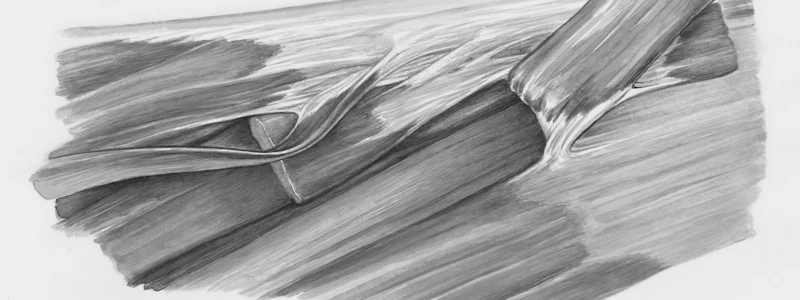
- Tendons are connective tissue fibers that form a cord or strap.
- Tendons localize a great deal of force in a bone's small area.
- Ligaments use flexible bands of fibrous tissue that resist strains; they hold bones close together in a synovial joint.
- Cruciate ligaments in the knee help prevent anterior-posterior displacement of articular surfaces and secure articulating bones when standing.
- Fleshy attachments occur when muscle fibers arise directly from the bone.
Bones
- Bones provide the body's framework, protect internal organs, store calcium and other minerals, and produce blood cells within bone marrow (hematopoiesis).
- Along with soft tissue, bones enclose and protect most vital organs; the skull's bones protect the brain, and the rib cage protects the heart and lungs.
- The skeletal system facilitates movement, providing attachment points for muscles, tendons, and ligaments.
- Bone marrow is responsible for hematopoiesis, producing millions of blood cells to replace those that are destroyed continuously.
- Bones behave as a mineral storehouse, mostly for phosphorus and calcium.
- For a mineral deficiency, like calcium during pregnancy, bodies withdraw it from bones where dietary supply is insufficient.
Bone Types
- There are four principal types of bone: short, irregular, flat, and long.
- Short bones are somewhat cube-shaped, consisting of a spongy bone core (cancellous bone) and a thin surface layer of compact bone.
- Short bones include the bones of the ankles, wrists, and toes.
- Irregular bones cannot be classified as short or long due to their complex shapes.
- These include vertebrae and the middle ear bones.
- Flat bones offer broad surfaces for muscular attachment or protection for internal organs like the skull's bones, shoulder blades, and sternum.
- Long bones are in the appendages/extremities of the body, such as the legs, arms, and fingers.
- Parts of a long bone include the diaphysis, compact bone, medullary cavity, distal epiphysis, proximal epiphysis, articular cartilage, spongy bone, and periosteum.
- The diaphysis is the bone's shaft or long, main portion and consists of compact bone.
- Compact bone forms a cylinder and surrounds a central canal called the medullary cavity (marrow cavity), which contains fatty yellow marrow comprised mostly of fat cells and a few scattered blood cells.
- Distal and proximal epiphyses are bulbous-shaped ends that provide space for muscle and ligament attachments near joints.
- Epiphyses have bulbous shapes that give space for muscular and ligament attachments near the joints and consist largely of
- Porous spongy bone surrounded by a compact bone layer.
- Within spongy bone is red bone marrow, which is richly supplied with blood and consists of immature and mature blood cells in varying development stages.
- In adults, red bone marrow is responsible for production of red blood cells (erythropoiesis), formation of white blood cells (leukopoiesis), and platelets.
- The periosteum uses a dense, white, fibrous membrane that covers bones remaining surface and contains numerous blood and lymph vessels and nerves.
- Bones that lose periosteal covering due to injury can scale or die, but it otherwise aids repairs and nutrition
- In growing bones, the inner layer contains bone-forming cells called osteoblasts.
- The periosteum provides a means for bone repair and general bone nutrition because blood vessels and osteoblasts are there and acts as an attachment point for muscles, ligaments, and tendons.
- Parts of a long bone include the diaphysis, compact bone, medullary cavity, distal epiphysis, proximal epiphysis, articular cartilage, spongy bone, and periosteum.
- Short bones are somewhat cube-shaped, consisting of a spongy bone core (cancellous bone) and a thin surface layer of compact bone.
Surface Features of Bones
- Bones consist of projections, depressions, and openings that are rarely smooth.
- These provide sites for muscle and ligament attachments, as well as pathways and openings for blood vessels, nerves, and ducts.
- Projections are evident in bones, where some serve as articulation points, and can be rounded, sharp, or narrow and have a large edge.
Bone Surface Types
- Nonarticulating surfaces are attached to a trochanter, very large irregularly shaped process, found only on the femur.
- Sites of muscle and ligament attachments present tubercles, with small, rounded processes present on the femur.
- There are tuberosities in large, rounded processes, like the humerus' tuberosity.
- Articulating surfaces contain condyles (rounded, articulating knobs) and heads (prominent, rounded, articulating ends of bone).
- These are located examples are located on the humerus' condyle and femur's head.
- Depressions and openings include Foramen ( the skull nerve opening), Fissure (The sphenoid bone slit), Meatus (The temporal bone passage), and Sinus (frontal duct cavity).
Divisions of the Skeletal System
- The skeletal system in a human adult is made of 206 individual bones, separated into axial and appendicular skeletons.
Axial Skeleton
- The axial skeleton has skull, rib cage, vertebral column, contributing to body cavity formation and protecting the brain, spinal cord, and organs.
Skull
- The skull's bony makeup consists of cranial and facial bones, which join by sutures.
- Sutures are the junction lines between 2 skull connecting bones and are usually immovable.
- Eight bones, known as the cranium/skull, enclose/protect the brain and hearing/equilibrium organs.
- Cranial bones connected to muscles provide head movements, chewing, and facial expressions.
- An infant's skull contains an unossified membrane (soft spot, incomplete bone formation) between the cranial bones is called the fontanel.
- Pulses of blood vessels felt under skin.
- Fontanels allow fetal bone movement through the birth canal to fuse and become immobile in early childhood.
- Frontal Bone: Forms the skull's anterior portion (forehead) and bony cavity roofs for eyeballs.
- Parietal Bone: Located at each skull side just behind the frontal bone and forms the upper sides and cranium’s roof.
- Coronal Suture: Where Parietal bone meets the frontal bone.
- Occipital Bone: Forms the back and base of the skull with a spinal cord opening in its base.
- Temporal Bones: Located on each skull side; forms part of lower cranium.
- Has a complex shape with cavities/recesses associated with hearing organs.
- Projects down to form the mastoid process, which allows attachments for neck muscles.
- Sphenoid Bone: Located at the skull base's middle part to have a central wedge to join other cranial bones.
- Ethmoid Bone: Forms most of bony area between the nasal cavity and eye orbits, and it is light and spongy.
- All facial bones besides the mandible (lower jaw) are joined by sutures and are immovable.
- Movement needed for chewing (mastication): The (9) maxillae, paired-upper jaw bones, are fused in the midline by a suture, and make up the upper jaw and hard palate (mouth roof).
- Cleft palate results during improper maxillary fusion before birth.
- Sockets in The maxillae (singular, maxilla) and mandible are for tooth roots.
- Nasal Bones: Lie side-by-side, fused medially for nose shape and bridge.
- Lacrimal Bones: Located at each eye corner.
- These thin, small bones unite to form the groove for lacrimal sac and canals through which tear ducts pass in nasal cavity.
- Zygomatic Bones: Located on face side below the eyes; cheekbones.
- Vomer: a thin, single bone forms the lower section of the nasal septum.
- Paranasal sinuses, such as that of the frontal, ethmoidal, sphenoidal, and maxillary sinuses, function as cavities within the cranial and facial bones, opening into the nasal cavities and lined with ciliary epithelium.
- Difficulty draining sinuses causes a feeling of congestion during upper respiratory infections (URI) or allergies.
- The internal organs of the chest (thorax), are enclosed and protected by a bony rib cage with 12 pairs of ribs attached to the spine.
- First seven form True Ribs attach directly to the sternum with a costal cartilage strip.
- The costal cartilage doesn't directly fasten to the sternum in the next five, they are known as false ribs.
- The last two, a type of false rib, are not connected to the sternum and attach to thoracic vertebrae (floating ribs).
- The vertebral column has 26 bones called vertebrae (singular, vertebra).
- It provides support, a bony canal for the spinal cord supports, resilient and balanced thanks to spinal curves.
- The cervical and lumbar regions curve forward and the thoracic and sacral regions curve backward.
- Abnormal curves are due to congenital defect, poor posture, or bone disease.
- The column has five regions, named from location within the spine column.
- The seven cervical vertebrae form the neck framework with the first cervical vertebra, the atlas, supports the skull.
- the second cervical vertebra, the axis, rotates the skull.
- 12 Thoracic, support the chest and articulate for ribs.
- The next five, Lumbar, are placed in the lower back, hold the most weight.
- Sacral has five fused bones in sacrum portion of vertebral column.
- Coccyx has tail of vertebral for three to five fragmented bones.
- The column has five regions, named from location within the spine column.
- The vertebrae separates round structures found between structure called (intevertebral discs), fibrous substance and the fluid mass on inside.
- If it has pressure on the nerve root where nerves exit from the back it cause it commonly called hernia nucleus polyposus.
Studying That Suits You
Use AI to generate personalized quizzes and flashcards to suit your learning preferences.


