Podcast
Questions and Answers
What is the function of the epicardium?
What is the function of the epicardium?
- Lines the internal cavity of the heart
- Receives oxygenated blood from the pulmonary veins
- Contributes to the heart's pumping action
- Serves to lubricate and protect the outer section of the heart (correct)
Which chamber of the heart receives deoxygenated blood from systemic veins?
Which chamber of the heart receives deoxygenated blood from systemic veins?
- Right atrium (correct)
- Left ventricle
- Right ventricle
- Left atrium
What is the main function of the endocardium?
What is the main function of the endocardium?
- Forcefully pumps blood out of the heart
- Lines the internal cavity of the heart
- Protects the outer section of the heart
- Provides a smooth surface for blood to flow against (correct)
Which layer of the heart wall is responsible for the heart's pumping action?
Which layer of the heart wall is responsible for the heart's pumping action?
Where is two-thirds of the heart's mass located in relation to the body?
Where is two-thirds of the heart's mass located in relation to the body?
What encloses and protects the heart?
What encloses and protects the heart?
What is the function of the atrioventricular valves in the heart?
What is the function of the atrioventricular valves in the heart?
Which valve is responsible for maintaining blood flow from the right ventricle to the pulmonary trunk?
Which valve is responsible for maintaining blood flow from the right ventricle to the pulmonary trunk?
What is the primary function of the coronary arteries in the heart?
What is the primary function of the coronary arteries in the heart?
What is the purpose of the parietal layer of the pericardium?
What is the purpose of the parietal layer of the pericardium?
Which layer of the heart wall is responsible for lubricating and protecting the outer section of the heart?
Which layer of the heart wall is responsible for lubricating and protecting the outer section of the heart?
How many layers make up the heart wall, and what are they?
How many layers make up the heart wall, and what are they?
Flashcards are hidden until you start studying
Study Notes
Heart Structure
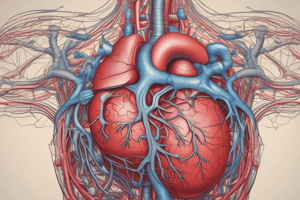
The heart is a four-chambered muscular organ that plays a crucial role in pumping blood throughout the body. It is roughly the size and shape of a man's closed fist, with two-thirds of its mass located on the left side of the body. The heart is enclosed in a protective sac called the pericardium, which is lined with the parietal layers of a serous membrane.
Layers of the Heart Wall
The heart wall is composed of three layers: the outermost layer, the epicardium; the middle layer, the myocardium; and the innermost layer, the endocardium. The epicardium is a thin-layered membrane that serves to lubricate and protect the outer section of the heart. The myocardium is a layer of muscle tissue that contributes to the thickness and is responsible for the heart's pumping action. The endocardium is the innermost layer, which lines the internal cavity of the heart and provides a smooth surface for blood to flow against.
Chambers of the Heart
The heart's internal cavity is divided into four chambers: the right atrium, left atrium, right ventricle, and left ventricle. The right atrium and ventricle make up the right side of the heart, while the left atrium and ventricle make up the left side. The right atrium receives deoxygenated blood from systemic veins, while the left atrium receives oxygenated blood from the pulmonary veins. The right and left ventricles are thick-walled chambers that forcefully pump blood out of the heart.
Valves of the Heart
The heart has two types of valves that keep the blood flowing in the correct direction: atrioventricular valves (also called cuspid valves) and semilunar valves. The right atrioventricular valve is the tricuspid valve, while the left atrioventricular valve is the bicuspid, or mitral, valve. The valve between the right ventricle and pulmonary trunk is the pulmonary semilunar valve, and the valve between the left ventricle and the aorta is the aortic semilunar valve.
Blood Supply to the Myocardium
The myocardium, the working muscle of the heart, requires a constant supply of oxygen and nutrients to function efficiently. This demand is met by the heart's own circulation system, known as coronary circulation. The right and left coronary arteries, branches of the ascending aorta, supply blood to the walls of the myocardium. After passing through capillaries in the myocardium, the blood enters a system of cardiac (coronary) veins, most of which drain into the coronary sinus, which opens into the right atrium.
External Structure of Heart
The pericardium, a fibrous sac, surrounds the heart and protects it from surrounding organs. The pericardium has two layers: the visceral layer, which directly covers the outside of the heart, and the parietal layer, which forms a sac around the outer region of the heart and contains the fluid in the pericardial cavity.
Blood Flow through the Heart
The heart functions as two pumps, one on the right and one on the left, working simultaneously. Blood flows from the right atrium to the right ventricle, then is pumped to the lungs to receive oxygen. From the lungs, the blood flows to the left atrium, then to the left ventricle, which pumps the oxygenated blood to the systemic circulation.
Structure of the Heart Wall
The heart wall is made up of three layers: the epicardium, myocardium, and endocardium. The epicardium is the outermost layer, which serves to lubricate and protect the outer section of the heart. The myocardium is a layer of muscle tissue that contributes to the thickness and is responsible for the heart's pumping action. The endocardium is the innermost layer, which lines the internal cavity of the heart and provides a smooth surface for blood to flow against.
In summary, the human heart is a four-chambered muscular organ enclosed in a protective pericardial sac. It has three layers of tissue forming the heart wall: the outermost epicardium, the middle myocardium, and the innermost endocardium. The heart is divided into four chambers: the right atrium, left atrium, right ventricle, and left ventricle. The heart has two types of valves: atrioventricular valves and semilunar valves, which keep the blood flowing in the correct direction. The heart's myocardium receives a continuous supply of oxygen and nutrients through the coronary circulation. The heart functions as two pumps, one on the right and one on the left, working simultaneously to pump blood throughout the body.
Studying That Suits You
Use AI to generate personalized quizzes and flashcards to suit your learning preferences.


