Podcast
Questions and Answers
Which of the following accurately describes the location of the oesophagogastric junction in relation to the vertebral column?
Which of the following accurately describes the location of the oesophagogastric junction in relation to the vertebral column?
- Lies to the right of the T11 vertebra.
- Lies to the left of the T12 vertebra.
- Lies to the left of the T11 vertebra. (correct)
- Lies to the right of the T12 vertebra.
What is the primary function of the diaphragmatic musculature immediately superior to the Z-line in the oesophagus?
What is the primary function of the diaphragmatic musculature immediately superior to the Z-line in the oesophagus?
- To act as a physiological sphincter, contracting and relaxing to prevent reflux. (correct)
- To provide structural support to the esophageal hiatus.
- To secrete mucous to protect the esophageal lining.
- To facilitate the swallowing process through peristaltic movements.
A patient presents with chronic reflux leading to metaplastic changes in the mucosa of the oesophagus. Which condition is most likely indicated by these changes?
A patient presents with chronic reflux leading to metaplastic changes in the mucosa of the oesophagus. Which condition is most likely indicated by these changes?
- Barrett's oesophagus (correct)
- Oesophageal varices
- Hiatal hernia
- Achalasia
Which of the following best describes the primary role of the pyloric sphincter?
Which of the following best describes the primary role of the pyloric sphincter?
A patient's pyloric sphincter is overly tight, restricting the movement of food. Which condition is the patient most likely experiencing?
A patient's pyloric sphincter is overly tight, restricting the movement of food. Which condition is the patient most likely experiencing?
Which of the following is a function of the gastric canal?
Which of the following is a function of the gastric canal?
In which histological zone of the gastric mucosa are G-cells, responsible for gastrin production, primarily located?
In which histological zone of the gastric mucosa are G-cells, responsible for gastrin production, primarily located?
A patient is diagnosed with a peptic ulcer in the stomach's antrum. What is a common characteristic of ulcers in this region?
A patient is diagnosed with a peptic ulcer in the stomach's antrum. What is a common characteristic of ulcers in this region?
Gastric ulcers on the posterior wall of the stomach are most likely to penetrate which of the following structures, leading to bleeding?
Gastric ulcers on the posterior wall of the stomach are most likely to penetrate which of the following structures, leading to bleeding?
What is the primary attachment point of the lesser omentum?
What is the primary attachment point of the lesser omentum?
Through which anatomical structure do the greater and lesser sacs of the peritoneum communicate?
Through which anatomical structure do the greater and lesser sacs of the peritoneum communicate?
The celiac trunk gives rise to which set of arteries that supply the stomach?
The celiac trunk gives rise to which set of arteries that supply the stomach?
Which vein does the Superior Mesenteric Vein join to form the Hepatic Portal Vein of the stomach?
Which vein does the Superior Mesenteric Vein join to form the Hepatic Portal Vein of the stomach?
Which nerve provides sympathetic innervation to the stomach via the celiac plexus?
Which nerve provides sympathetic innervation to the stomach via the celiac plexus?
What is the function of the vagus nerve innervation to the stomach?
What is the function of the vagus nerve innervation to the stomach?
What is a common cause of acute gastritis?
What is a common cause of acute gastritis?
What is a frequent cause of chronic gastritis?
What is a frequent cause of chronic gastritis?
Which condition may directly result from ulceration into the splenic artery?
Which condition may directly result from ulceration into the splenic artery?
What is the approximate length of the oesophagus?
What is the approximate length of the oesophagus?
A patient presents with a hiatal hernia where the cardia and parts of the fundus have protruded into the mediastinum. What symptom is the patient most likely to experience?
A patient presents with a hiatal hernia where the cardia and parts of the fundus have protruded into the mediastinum. What symptom is the patient most likely to experience?
According to the provided text, how many parts does the stomach have?
According to the provided text, how many parts does the stomach have?
Which of the following best describes the structure of the greater omentum?
Which of the following best describes the structure of the greater omentum?
Which of the following structures surrounds the oesophagus, before following it through the diaphragm into the abdomen?
Which of the following structures surrounds the oesophagus, before following it through the diaphragm into the abdomen?
In the context of a hiatal hernia, which anatomical change contributes to the occurrence of the hernia?
In the context of a hiatal hernia, which anatomical change contributes to the occurrence of the hernia?
Which of the following is a characteristic of gastric ulcers, where the pain is more pronounced?
Which of the following is a characteristic of gastric ulcers, where the pain is more pronounced?
Flashcards
Oesophagus
Oesophagus
A fibromuscular tube that extends from the pharynx to the stomach, approximately 25cm in length.
Oesophageal Strictures
Oesophageal Strictures
Narrowing of the oesophagus at three points: the cervical, thoracic, and oesophageal regions.
Oesophageal Hiatus
Oesophageal Hiatus
The point where the oesophagus passes through the diaphragm, located at the T10 vertebral level.
Hiatus Hernia
Hiatus Hernia
Signup and view all the flashcards
Stomach
Stomach
Signup and view all the flashcards
Rugae
Rugae
Signup and view all the flashcards
Gastric Canal
Gastric Canal
Signup and view all the flashcards
Oesophagogastric Junction
Oesophagogastric Junction
Signup and view all the flashcards
Z-Line
Z-Line
Signup and view all the flashcards
Pyloric Sphincter
Pyloric Sphincter
Signup and view all the flashcards
Rugae
Rugae
Signup and view all the flashcards
Chronic Reflux
Chronic Reflux
Signup and view all the flashcards
Greater Omentum
Greater Omentum
Signup and view all the flashcards
Lesser Omentum
Lesser Omentum
Signup and view all the flashcards
Epiploic Foramen
Epiploic Foramen
Signup and view all the flashcards
Coeliac Trunk
Coeliac Trunk
Signup and view all the flashcards
Anterior Vagus Nerve
Anterior Vagus Nerve
Signup and view all the flashcards
Posterior Vagus Nerve
Posterior Vagus Nerve
Signup and view all the flashcards
Gastritis
Gastritis
Signup and view all the flashcards
Greater Splanchnic Nerve
Greater Splanchnic Nerve
Signup and view all the flashcards
Study Notes
- The lecture is about the anatomy of the stomach
- The objectives are to describe the gross structure, parts, and curvatures of the stomach, the structure and function of the oesophageal/gastric sphincter and the pyloric sphincter, the macro and microscopic structure of the gastric mucosa Also, be able to describe and identify the attachments of the lesser and greater omentum, curvatures of the stomach and its regions (fundus, body, pyloric antrum& pylorus), the coeliac trunk and its main branches (left gastric, splenic and common hepatic), and the blood supply (arteries&veins) of stomach the enterance of the lesser sac (epiploic foramen)
Oesophagus
- A fibromuscular tube that extends from the pharynx to the stomach and is roughly 25cm in length
- Has three main strictures: cervical, thoracic, and oesophageal
- The oesophageal constrictor occurs where the oesophagus passes through the diaphragm at the oesophageal hiatus (T10)
- The anterior and posterior vagal trunks join the oesophageal plexus around the oesophagus, and then follow it enter the abdomen.
Clinical Considerations: Hiatus Hernia
- A protrusion of part of the stomach into the mediastinum through the oesophageal hiatus of the diaphragm
- Hernias occur due to weakening in the diaphragm muscle causing the widening of the oesophageal hiatus, allowing stomach parts to move upwards
- It can be either the fundus alone (usually no regurgitation) or the cardia and parts of the fundus (some regurgitation may occur)
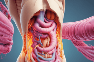
General Anatomy of Stomach
- The stomach is a J-shaped organ with 5 parts and a greater and lesser curvature
- The parts include the cardia, fundus, body, antrum, and pylorus
- The stomach lies in the epigastric (E), umbilical (U), and left hypochondrium (LH) regions
- The size and position of the stomach depends on body shape, degree of distension, and posture
Gastric Mucosa
- These are images of a Gastric Mucosa Endoscopy
Oesophageal/gastric sphincter and the pyloric sphincter
- Sphincters are present at the entry and exit points of the stomach
Inferior Oesophageal Sphincter
- The oesophagogastric junction lies to the left of the T11 vertebra on the horizontal plane that passes through the tip of the xiphoid process
- The Z-Line is where the mucosa changes from oesophageal to gastric
- Immediately superior to the Z-line, the diaphragmatic musculature forms the oesophageal hiatus, functioning as a physiological sphincter that contracts and relaxes thus preventing the reflux
Reflux of Gastric Contents
- Reflux into the oesophagus is common
- Frequent reflux or deficient clearance may cause "heartburn" and/or inflammation with ulceration
- Chronic reflux can lead to metaplastic changes in the mucosa (Barrett's oesophagus)
Pyloric Sphincter
- At the pyloric end of the stomach, the circular muscle coat is thickened
- This thickening produces the pyloric sphincter
- The pyloric sphincter controls the discharge of stomach contents through the pyloric orifice into the duodenum
Macro and Microscopic Structure of the Gastric Mucosa
- Rugae: Longitudinal folds of gastric mucosa
- Gastric canal: Forms temporarily between the gastric folds along the lesser curvature to allow saliva and other fluids to pass to the pylorus
- The Gastric mucosa has 3 histologically zones:
- Cardia: Neck cells produce mucus
- Fundus and Body: Neck cells produce mucus, Parietal cells secrete acid, and Chief cells secrete pepsinogen
- Pyloric: Neck cells secrete mucus, G-Cells secrete gastrin
Peptic Ulcers
- An ulcer is an erosion or loss of continuity
- Commonly occurs in the antrum and along the lesser curvature of the stomach
- Perforation of ulcers leads to the spillage of gastric contents into the peritoneal cavity, affecting abdominal structures, such as the pancreas and associated blood vessels
Gastric Ulcers
- With gastric ulcers there is pain on eating, whereas with duodenal ulcers there is pain when hungry
- Posterior wall ulcers penetrate the gastroduodenal artery and bleed
- Anterior wall ulcers penetrate the peritoneum and perforate into the peritoneal cavity
Greater Omentum
- A four-layered peritoneal fold that hangs down like an apron from the Greater Curve of the Stomach
- It folds back and attaches to the anterior surface of the Transverse Colon and its mesentery
Lesser Omentum
- A double-layered peritoneal fold that connects the Lesser Curvature of the Stomach and the Proximal part of the Duodenum to the Liver
- Connects the stomach to the portal triad
Epiploic Foramen
- The greater and lesser sacs communicate through the omental foramen (epiploic foramen), an opening posterior to the free edge of the lesser omentum
- Locate the omental foramen by running a finger along the gall bladder to free the edge of the lesser omentum; admits 2 fingers.
Stomach – Arterial Blood Supply
- The celiac trunk originates from the Abdominal Aorta and gives rise to:
- Left Gastric, Splenic and Common Hepatic Arteries (Lesser Curvature)
- Coeliac Trunk → Left Gastric
- Coeliac Trunk → Common Hepatic, Right Gastric (Greater Curvature)
- Coeliac Trunk → Splenic, Left Gastro-Omental (epiploic)
- Coeliac Trunk → Common Hepatic → Gastroduodenal, Right Gastro-Omental (Fundus and Body)
- Coeliac Trunk → Splenic, Posterior Gastric / Small Gastric arteries (5-7)
Stomach – Venous Drainage
- Left Gastric Vein & Right Gastric Vein follow the course of the LT & RT Gastric Artery and drain into the Hepatic Portal Vein
- Short Gastric Vein follows the course of the Short Gastric Artery and drains into the Splenic Vein, which joins the SMV to form the Hepatic Portal Vein
- Left Gastro-Omental Vein follows the course of the Left Gastro-Omental Artery, drains into the Splenic Vein, which joins the SMV to form the Hepatic Portal Vein Right Gastro-Omental Vein follows the course of the Right Gastro-Omental Artery and drains into the Superior Mesenteric Vein, which joins the Splenic Vein to form the Hepatic Portal Vein
Nerve Supply of the Stomach
- Sympathetic nerve supply from T6 to T10 segments is via the Greater Splanchnic Nerve to the Celiac plexus
- The left & right vagus nerves enter the abdomen through the oesophageal hiatus (T10 vertebral level)
- Anterior vagus supplies the anterior surface the stomach (cardia&lesser curvature)and gives off a hepatic branch of (liver, gall bladder & the pylorus)
- Posterior vagus gives off a celiac branch (which passes to the celiac plexus) & a posterior branch to the posterior surface of the stomach
- Crow's feet innervation to antropyloric area
- Two main nerve trunks branch into multiple nerves at incisura forming crow's foot, which innervates at antrum (preserved in highly selective vagotomy)
- Vagus innervation constitutes both the motor and secretory nerve supply of the stomach
Inflammation of the stomach (Gastritis)
- Can be acute, and caused by NSAIDS / Alcohol
- Results in exfoliation of the surface epithelial cells and decreased secretion of protective mucus
- Can be chronic, and caused by infection with the bacteria Helicobacter Pylori
- Inflammatory changes in the mucosa result in atrophy and epithelial metaplasia (may develop into carcinoma)
- Posterior gastric cancer or ulcer may erode the pancreas and lead to back pain
- Ulceration into Splenic artery may result in severe haemorrhage
Studying That Suits You
Use AI to generate personalized quizzes and flashcards to suit your learning preferences.