Podcast
Questions and Answers
What are the three functional units of the small bowel?
What are the three functional units of the small bowel?
The three functional units are the duodenum, jejunum, and ileum.
What is the average length of the jejunum and ileum combined?
What is the average length of the jejunum and ileum combined?
The average length is 6 meters.
How do the valvulae conniventes change from the jejunum to the ileum?
How do the valvulae conniventes change from the jejunum to the ileum?
They are large and thick in the jejunum and decrease in size to disappear entirely in the distal ileum.
Where is the jejunum primarily located within the abdomen?
Where is the jejunum primarily located within the abdomen?
What characteristic is observed regarding the wall thickness of the jejunum and ileum?
What characteristic is observed regarding the wall thickness of the jejunum and ileum?
What happens to the density of valvulae as one moves from the jejunum to the distal ileum?
What happens to the density of valvulae as one moves from the jejunum to the distal ileum?
What is the function of the ileocecal valve?
What is the function of the ileocecal valve?
How long is the large intestine, and what does it encompass?
How long is the large intestine, and what does it encompass?
Where does the appendix attach to the large intestine?
Where does the appendix attach to the large intestine?
What distinguishes the transverse colon from the other parts of the large intestine?
What distinguishes the transverse colon from the other parts of the large intestine?
Flashcards
Small Bowel Location
Small Bowel Location
The section of the bowel between the stomach and colon, comprised of the duodenum, jejunum, and ileum.
Jejunum and Ileum Length
Jejunum and Ileum Length
The combined length of the jejunum and ileum typically ranges from 3 to 10 meters, averaging 6 meters.
Small Bowel Attachment
Small Bowel Attachment
The jejunum and ileum are connected to the mesentery, a membrane-like structure.
Valvulae Conniventes
Valvulae Conniventes
Signup and view all the flashcards
Small Bowel X-Ray Appearance
Small Bowel X-Ray Appearance
Signup and view all the flashcards
Jejunum vs. Ileum - Length
Jejunum vs. Ileum - Length
Signup and view all the flashcards
Jejunum vs. Ileum - Diameter
Jejunum vs. Ileum - Diameter
Signup and view all the flashcards
Jejunum vs. Ileum - Wall Thickness
Jejunum vs. Ileum - Wall Thickness
Signup and view all the flashcards
Jejunum vs. Ileum - Location
Jejunum vs. Ileum - Location
Signup and view all the flashcards
Small Bowel Follow-Through (SBFT)
Small Bowel Follow-Through (SBFT)
Signup and view all the flashcards
Small Bowel Density
Small Bowel Density
Signup and view all the flashcards
Small Bowel Exam
Small Bowel Exam
Signup and view all the flashcards
Large Bowel Length
Large Bowel Length
Signup and view all the flashcards
Taenia Coli
Taenia Coli
Signup and view all the flashcards
Haustra
Haustra
Signup and view all the flashcards
Cecum
Cecum
Signup and view all the flashcards
Ileocecal Valve
Ileocecal Valve
Signup and view all the flashcards
Appendix
Appendix
Signup and view all the flashcards
Ascending Colon
Ascending Colon
Signup and view all the flashcards
Transverse Colon
Transverse Colon
Signup and view all the flashcards
Descending Colon
Descending Colon
Signup and view all the flashcards
Sigmoid Colon
Sigmoid Colon
Signup and view all the flashcards
Rectum
Rectum
Signup and view all the flashcards
Anal Canal
Anal Canal
Signup and view all the flashcards
Radiographic Features of Colon
Radiographic Features of Colon
Signup and view all the flashcards
Study Notes
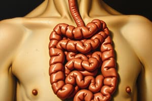
Small Bowel
- Divided into three sections: duodenum, jejunum, and ileum
- Average length of the jejunum and ileum is 6 meters, ranging from 3 to 10 meters
- Attached to mesentery; the root of the mesentery extends from the duodeno-jejunal junction (left of L2 vertebrae) to the right sacroiliac joint
- Mucosal folds called valvulae conniventes are present, largest in the jejunum and decrease in size distally in the ileum, disappearing in the distal ileal loops.
Radiographic Features (Abdominal X-ray)
- Air/fluid levels are often seen, up to five on an erect abdominal X-ray
- The small bowel generally lies centrally
- Valvulae conniventes are thin, circular folds of mucosa that can be seen passing across the full width of the lumen on an X-ray
Jejunum (Comparison with Ileum)
- Shorter, occupying the proximal 2/5 of the small intestine
- Wider (3-3.5 cm)
- Thicker walls
- Located in the left upper abdomen
- Thicker and more prominent valvulae conniventes
Ileum (Comparison with Jejunum)
- Longer, occupying the distal 3/5 of the small intestine
- Narrower (2.5 cm)
- Thinner walls
- Located in the right iliac fossa
- Thinner and less prominent valvulae conniventes
Barium Studies of Small Bowel Loops
- Small Bowel Follow-Through (SBFT): Performed after an upper GI fluoroscopic study; contrast column moves from stomach and duodenum into the small bowel. Can be done without upper GI first. This shows mucosal detail of Jejunum has high density, with density reduction in the proximal ileum.
- Small Bowel Enema: Naso-jejunal tube is passed to the duodenojejunal flexure and barium is passed directly into the small intestine; good for double contrast images.
Large Bowel
- Approximately 1.5 meters long
- Outer longitudinal muscle layers (taeniae coli) are shorter than the bowel, causing sacculations (haustra)
- Divided into cecum, appendix, ascending colon, hepatic flexure, transverse colon, splenic flexure, descending colon, sigmoid colon, and rectum
- Cecum: The starting point, a blind pouch of the ascending colon
- Appendix: A thin tube attached to the cecum
- Ascending Colon: Runs superiorly on the right side of the abdomen from the right iliac fossa to the right lobe of the liver
- Transverse Colon: Most mobile and longest part; found between the right and left colic flexures
- Descending Colon: Extends from the splenic flexure to the sigmoid colon
- Sigmoid Colon: S-shaped loop linking the descending colon to the rectum
- Rectum: From the sigmoid colon to the anal canal, occupying the sacrococcygeal curvature
- Anal Canal: A narrow muscular canal, directed almost perpendicular to the rectum
Radiographic Features (Abdominal X-ray - Large Bowel)
- Retroperitoneal structures of the colon (ascending, descending, and rectum) are relatively constant in position
- The cecum is often the widest part and can vary in position
- Haustra (sacculations) are characteristic of the colon and are not present in the rectum
- Normal large bowel appearance has peripheral position, presence of haustra, and presence of feces.
Studying That Suits You
Use AI to generate personalized quizzes and flashcards to suit your learning preferences.