Podcast
Questions and Answers
What is the correct order of the four stages in skin cell formation?
What is the correct order of the four stages in skin cell formation?
- Keratinization, cell division, desquamation, cell differentiation
- Desquamation, cell differentiation, keratinization, cell division
- Cell division, cell differentiation, keratinization, desquamation (correct)
- Cell differentiation, keratinization, desquamation, cell division
Which of the following is NOT a primary skin lesion?
Which of the following is NOT a primary skin lesion?
- Papule
- Scale (correct)
- Wheal
- Macule
Which layer of the skin contains the sweat glands and nerve endings?
Which layer of the skin contains the sweat glands and nerve endings?
- Dermis (correct)
- Subcutaneous layer
- Epidermis
- Stratum corneum
What is the term used to describe skin lesions that are raised and palpable?
What is the term used to describe skin lesions that are raised and palpable?
Which of the following is NOT a characteristic of skin lesions used for accurate description?
Which of the following is NOT a characteristic of skin lesions used for accurate description?
What is the difference between a nodule and a tumor?
What is the difference between a nodule and a tumor?
Which of these is NOT a condition discussed under the category of papulo-squamous diseases?
Which of these is NOT a condition discussed under the category of papulo-squamous diseases?
Which of these skin lesions is NOT classified as a secondary lesion?
Which of these skin lesions is NOT classified as a secondary lesion?
What is the term used to describe a flat, non-palpable skin lesion that is less than 1 cm in diameter?
What is the term used to describe a flat, non-palpable skin lesion that is less than 1 cm in diameter?
Which type of skin lesion is characterized by a circumscribed superficial cavity filled with purulent exudate?
Which type of skin lesion is characterized by a circumscribed superficial cavity filled with purulent exudate?
What is the term used to describe a flat or barely elevated plaque that is often associated with conditions like atopic dermatitis?
What is the term used to describe a flat or barely elevated plaque that is often associated with conditions like atopic dermatitis?
What is the term used to describe a palpable, solid, fatty or cystic lesion that is larger than 1 cm in diameter?
What is the term used to describe a palpable, solid, fatty or cystic lesion that is larger than 1 cm in diameter?
What is the term used to describe a dried serum, blood, or exudate on the skin?
What is the term used to describe a dried serum, blood, or exudate on the skin?
Which type of skin lesion is characterized by an epidermis defect that heals without a scar?
Which type of skin lesion is characterized by an epidermis defect that heals without a scar?
What is the term used to describe a defect in the dermis or deeper layers of skin that heals with a scar?
What is the term used to describe a defect in the dermis or deeper layers of skin that heals with a scar?
What term describes a fibrous tissue replacement, often visible after a skin injury?
What term describes a fibrous tissue replacement, often visible after a skin injury?
What does the term 'confluence' refer to, when describing skin lesions?
What does the term 'confluence' refer to, when describing skin lesions?
Which of the following is NOT considered a characteristic of palpation when evaluating skin lesions?
Which of the following is NOT considered a characteristic of palpation when evaluating skin lesions?
What are the two main categories of lesion arrangement?
What are the two main categories of lesion arrangement?
What does 'margination' refer to, when describing skin lesions?
What does 'margination' refer to, when describing skin lesions?
Which of the following is used to describe the type of skin lesion?
Which of the following is used to describe the type of skin lesion?
What is the term for the extent and distribution of a skin lesion?
What is the term for the extent and distribution of a skin lesion?
When assessing skin lesions, what does the 'single vs multiple' category refer to?
When assessing skin lesions, what does the 'single vs multiple' category refer to?
Which of the following characteristics is assessed during palpation?
Which of the following characteristics is assessed during palpation?
Which of these is NOT a common site for Lichen Simplex Chronicus?
Which of these is NOT a common site for Lichen Simplex Chronicus?
Which of the following is associated with a coin-shaped plaque of grouped small papules and vesicles on an erythematous base?
Which of the following is associated with a coin-shaped plaque of grouped small papules and vesicles on an erythematous base?
Which of the following conditions is often associated with venous insufficiency?
Which of the following conditions is often associated with venous insufficiency?
Which of the following treatment options is commonly used for both Nummular Eczema and Stasis Dermatitis?
Which of the following treatment options is commonly used for both Nummular Eczema and Stasis Dermatitis?
Which of the following factors can elicit or exacerbate Atopic Dermatitis?
Which of the following factors can elicit or exacerbate Atopic Dermatitis?
Which of the following conditions is often seen in immunosuppressed individuals, such as those with HIV or Parkinson's Disease?
Which of the following conditions is often seen in immunosuppressed individuals, such as those with HIV or Parkinson's Disease?
What is the common term used to describe the itch-scratch cycle often seen in Atopic Dermatitis?
What is the common term used to describe the itch-scratch cycle often seen in Atopic Dermatitis?
Which of these options is a common treatment for severe cases of Contact Dermatitis?
Which of these options is a common treatment for severe cases of Contact Dermatitis?
Which of the following conditions is NOT typically associated with a chronic or relapsing course?
Which of the following conditions is NOT typically associated with a chronic or relapsing course?
Which of these conditions is often associated with a personal or family history of allergies, such as allergic rhinitis or asthma?
Which of these conditions is often associated with a personal or family history of allergies, such as allergic rhinitis or asthma?
Which of these medications may be prescribed for disabling oral lesions in Erythema Multiforme?
Which of these medications may be prescribed for disabling oral lesions in Erythema Multiforme?
What is the primary treatment approach for Stevens-Johnson Syndrome (SJS) and Toxic Epidermal Necrolysis (TEN)?
What is the primary treatment approach for Stevens-Johnson Syndrome (SJS) and Toxic Epidermal Necrolysis (TEN)?
What distinguishes Stevens-Johnson Syndrome (SJS) from Toxic Epidermal Necrolysis (TEN)?
What distinguishes Stevens-Johnson Syndrome (SJS) from Toxic Epidermal Necrolysis (TEN)?
What is a common symptom experienced by patients with Stevens-Johnson Syndrome (SJS) and Toxic Epidermal Necrolysis (TEN) early in the disease course?
What is a common symptom experienced by patients with Stevens-Johnson Syndrome (SJS) and Toxic Epidermal Necrolysis (TEN) early in the disease course?
Which of the following is NOT a potential trigger for Stevens-Johnson Syndrome (SJS) and Toxic Epidermal Necrolysis (TEN)?
Which of the following is NOT a potential trigger for Stevens-Johnson Syndrome (SJS) and Toxic Epidermal Necrolysis (TEN)?
What is Nikolsky's sign and what does it indicate?
What is Nikolsky's sign and what does it indicate?
Which of these is NOT a characteristic of Erythema Multiforme (EM)?
Which of these is NOT a characteristic of Erythema Multiforme (EM)?
What is the key characteristic that must be present for a diagnosis of acne vulgaris?
What is the key characteristic that must be present for a diagnosis of acne vulgaris?
Which of these medications can be effective in treating moderate acne vulgaris, but should be used with caution during pregnancy?
Which of these medications can be effective in treating moderate acne vulgaris, but should be used with caution during pregnancy?
Which of the following is a potential side effect of benzoyl peroxide gel?
Which of the following is a potential side effect of benzoyl peroxide gel?
Which of the following medications is NOT typically used in the treatment of acne vulgaris?
Which of the following medications is NOT typically used in the treatment of acne vulgaris?
What is the primary reason for the occurrence of comedones in acne vulgaris?
What is the primary reason for the occurrence of comedones in acne vulgaris?
Which of these factors is NOT a known contributor to the pathogenesis of acne vulgaris?
Which of these factors is NOT a known contributor to the pathogenesis of acne vulgaris?
Which of the following is a common presentation of pemphigus?
Which of the following is a common presentation of pemphigus?
Which of the following is a characteristic of pemphigoid, but NOT of pemphigus?
Which of the following is a characteristic of pemphigoid, but NOT of pemphigus?
Which of the following statements accurately describes the Nikolsky sign?
Which of the following statements accurately describes the Nikolsky sign?
Which of the following is NOT a characteristic of toxic epidermal necrolysis (TEN)?
Which of the following is NOT a characteristic of toxic epidermal necrolysis (TEN)?
Flashcards
Papulo-squamous diseases
Papulo-squamous diseases
Skin disorders characterized by papules and plaques such as eczema, psoriasis.
Desquamation
Desquamation
The process of shedding dead skin cells from the outer layer of skin.
Vesicular bullae
Vesicular bullae
Fluid-filled blisters that can occur in conditions like pemphigus and pemphigoid.
Acneiform lesions
Acneiform lesions
Signup and view all the flashcards
Primary skin lesions
Primary skin lesions
Signup and view all the flashcards
Secondary skin lesions
Secondary skin lesions
Signup and view all the flashcards
Epidermal layers
Epidermal layers
Signup and view all the flashcards
Skin cell formation steps
Skin cell formation steps
Signup and view all the flashcards
CLAMPS TN
CLAMPS TN
Signup and view all the flashcards
Macule
Macule
Signup and view all the flashcards
Papule
Papule
Signup and view all the flashcards
Pustule
Pustule
Signup and view all the flashcards
Plaque
Plaque
Signup and view all the flashcards
Nodule
Nodule
Signup and view all the flashcards
Wheal
Wheal
Signup and view all the flashcards
Vesicle/Bulla
Vesicle/Bulla
Signup and view all the flashcards
Location/Distribution
Location/Distribution
Signup and view all the flashcards
Arrangement
Arrangement
Signup and view all the flashcards
Margination
Margination
Signup and view all the flashcards
Palpation
Palpation
Signup and view all the flashcards
Shape
Shape
Signup and view all the flashcards
Type of lesion
Type of lesion
Signup and view all the flashcards
Single vs Multiple lesions
Single vs Multiple lesions
Signup and view all the flashcards
Color
Color
Signup and view all the flashcards
Erythema Multiforme
Erythema Multiforme
Signup and view all the flashcards
Stevens-Johnson Syndrome (SJS)
Stevens-Johnson Syndrome (SJS)
Signup and view all the flashcards
Toxic Epidermal Necrolysis (TEN)
Toxic Epidermal Necrolysis (TEN)
Signup and view all the flashcards
Clinical Diagnosis
Clinical Diagnosis
Signup and view all the flashcards
Biopsy
Biopsy
Signup and view all the flashcards
Nikolsky Sign
Nikolsky Sign
Signup and view all the flashcards
Mucous Membrane Involvement
Mucous Membrane Involvement
Signup and view all the flashcards
Continuous Antiviral Therapy
Continuous Antiviral Therapy
Signup and view all the flashcards
Lichen Simplex Chronicus
Lichen Simplex Chronicus
Signup and view all the flashcards
Symptoms of Lichen Simplex Chronicus
Symptoms of Lichen Simplex Chronicus
Signup and view all the flashcards
Numular Eczema
Numular Eczema
Signup and view all the flashcards
Acute vs Chronic Contact Dermatitis
Acute vs Chronic Contact Dermatitis
Signup and view all the flashcards
Symptoms of Stasis Dermatitis
Symptoms of Stasis Dermatitis
Signup and view all the flashcards
Atopic Dermatitis
Atopic Dermatitis
Signup and view all the flashcards
Triggers of Atopic Dermatitis
Triggers of Atopic Dermatitis
Signup and view all the flashcards
Seborrheic Dermatitis
Seborrheic Dermatitis
Signup and view all the flashcards
Treatment for Lichen Simplex Chronicus
Treatment for Lichen Simplex Chronicus
Signup and view all the flashcards
Common Areas for Contact Dermatitis
Common Areas for Contact Dermatitis
Signup and view all the flashcards
Pemphigus
Pemphigus
Signup and view all the flashcards
Bullous pemphigoid
Bullous pemphigoid
Signup and view all the flashcards
Acne vulgaris
Acne vulgaris
Signup and view all the flashcards
Comedones
Comedones
Signup and view all the flashcards
Flare-ups
Flare-ups
Signup and view all the flashcards
Propionibacterium acnes
Propionibacterium acnes
Signup and view all the flashcards
Treatment for mild acne
Treatment for mild acne
Signup and view all the flashcards
Moderate acne treatment
Moderate acne treatment
Signup and view all the flashcards
Follicular keratinization
Follicular keratinization
Signup and view all the flashcards
Study Notes
Dermatology 1
- The course is Dermatology 1, taught by Professor Jacobus, MSBS, PA-C.
- The course is offered at South College.
Topics
- Papulo-squamous diseases: Dermatitis, eczema, drug eruptions, lichen planus, pityriasis rosea, and psoriasis.
- Desquamation: Erythema multiforme, Stevens-Johnson syndrome, and toxic epidermal necrolysis.
- Vesicular bullae: Pemphigoid and pemphigus.
- Acneiform lesions: Acne vulgaris, rosacea, and folliculitis.
Instructional Objectives
- Identify and describe the etiology, epidemiology, clinical features, differential diagnosis, and management of selected skin disorders.
- Identify and accurately describe skin lesions using standard terms:
- Number: single, multiple
- Pigmentation/color: white, flesh-colored, pink, pearly, erythematous, tan-brown, salmon, black, purple, violaceous, and yellow
- Shape and arrangement: annular, round/discoid, linear, oval, iris/target, zosteriform, serpiginous, stellate, reticulate, and morbilliform
- Texture: consistency, mobility, temperature, tenderness, depth
- Borders/margins: well-defined, ill-defined
- Type: Primary lesions (macule, tumor, patch, wheal, papule, vesicle, plaque, bulla, nodule, pustule, cyst, and telangiectasia) or secondary lesions (crust, fissure, scale, ulcer, lichenified, keloid, erosion, hypertrophic scar, atrophy, and excoriation)
- Arrangement/location/distribution: localized, regional, and generalized
- Associated changes
Skin layers
- The layers of the skin include the epidermis, dermis, and subcutaneous tissue.
- Epidermal layers include the stratum corneum, granular layer, spinous layer, and basal layer.
Skin cell formation
- Keratinocytes divide in the deepest (basal) layer
- Cells move up the dermis, changing shape and composition
- Cells secrete keratin proteins and lipids to form a protective matrix
- Outermost skin cells die and shed.
Skin Exam
- Be thorough; examine patients in gowns.
- Use good lighting; consider magnification.
- Check scalp, palms, soles, and nails.
- Document any findings in detail, photograph, and monitor changes over time.
Approach to Diagnosis
- Use the CLAMPS TN method: color, location/distribution, arrangement, margination, palpation, shape, type, and number.
Types of Skin Lesions (examples)
- Crust
- Cyst
- Macule
- Papule
- Pustule
- Ulcer
- Vesicle
- Wheal
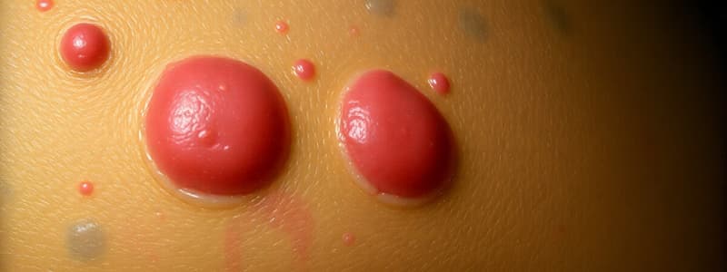
Macule
- Non-palpable
- <1 cm diameter
- Varied pigmentation from surrounding skin
- No elevation or depression
- Patch: macule > 1 cm diameter
Papule
- Palpable
- <1 cm diameter
- Isolated or grouped
- Pustule: small, circumscribed papule containing purulent material
Pustule
- Circumscribed superficial cavity with purulent exudate.
- Exudate can be white, yellow, greenish-yellow, or hemorrhagic.
Plaque
- Plateau-like elevation
- Lichenification - less defined large plaque (thickened, rough skin)
- Patch – flat or barely elevated plaque
Nodule
- Palpable, solid, fatty or cystic
- Round or ellipsoidal
- Larger than a papule
- Tumor: nodule > 2 cm
Wheal
- Irregularly-shaped, elevated, edematous
- Erythematous or paler than surrounding skin
- Well-demarcated borders
- Disappears within 24-48 hours
Vesicle/Bulla
- Blister
- Vesicle <0.5cm; Bulla >0.5 cm
- Well-defined
- Thin roof
- Serum and blood
Secondary Skin Lesions
- Crust-dried serum, blood, or exudate
- Scales-flakes
- Erosion - epidermis defect (heals without scar)
- Ulcer - defect in dermis or deeper (heals with scar)
- Scar - fibrous tissue replacement
- Atrophy - diminution of some or all layers of skin
History
- Demographics: age, race, sex, occupation, hobbies.
- Chemical/toxin exposure?
- Constitutional symptoms (acute vs chronic).
- History of skin lesions - OLD CARTS.
HX of Skin Lesion (OLD CARTS)
- When did lesion appear (first noticed)?
- Where did lesion appear (site of onset)?
- Does it come and go, or is it constant?
- Does it itch, hurt, or bleed?
- How has it spread (pattern/evolution)?
- How have individual lesions changed?
- What are provocative factors?
- What are previous treatments (topical, systemic)?
Papulosquamous Disease
- Eczema/ Dermatitis
- Drug eruptions
- Lichen planus
- Pityriasis rosea
- Psoriasis
Eczema/Dermatitis
- Dyshidrotic eczema
- Lichen simplex chronicus
- Nummular eczema
- Contact dermatitis
- Stasis dermatitis
- Atopic dermatitis
- Seborrheic dermatitis
- Perioral dermatitis
Desquamation
-
Erythema multiforme
-
Stevens-Johnson Syndrome (SJS)
-
Toxic Epidermal Necrolysis (TEN)
-
Erythema multiforme - presentation, history taking, target lesions
• Stevens-Johnson Syndrome (SJS)/Toxic Epidermal Necrolysis (TEN) – acute, life-threatening mucocutaneous reaction, necrosis & detachment of epidermis, differentiating SJS from TEN, rare, present, idiopathic or drug-induced, higher incidence in HIV & active cancer, F>M.
• Nikolsky sign-ability of sloughing to expand.
Vesiculobullous Disease
-
Bullous pemhigoid- autoimmune, elderly patients, large, tense bullae
-
Pemphigus- chronic or acute, bullous autoimmune disease, adults 40-60, predilection scalp, face, chest, axillae, groin, umbilicus. painful. PE- vesicles and bullae, easily rupture, flaccid and weeping, Nikolsky sign
-
Acne & Related Disorders - acne vulgaris, rosacea, hidradenitis suppurativa
-
Acne vulgaris - inflammation of pilosebaceous units, comes (open = blackheads, closed= whiteheads) ,can result in pits, depressions, scars or hyperpigmentation, more severe in males than females;less often in Asians; cystic acne can be familial.
-
Factors in pathogenesis- follicular keratinization, androgens, Propionibacterium acnes
-
Contributing factors - meds (lithium, isoniazid), steroids, OCP, androgens; stress; occlusion/pressure; cosmetics; pomade; sweat, worse in winter, comedones necessary diagnosis; labs- none, course clears by early 20's, flares with winter or menses- treatment, removal of plugs, topical antibiotics, topical retinoids; combination therapy works best.
-
Treatment- moderate: same + PO antibiotics (minocycline, doxycycline, tapered to 50mg/d as acne lessens.
Rosacea
• Chronic inflammation of facial pilosebaceous units. • 30-50 y/o, F>M. • Episodic erythema, flushing & blushing, stages I, II, & III, persistent erythema, telangiectasias, papules, tiny pustules, nodules. • Note, no comedones • Triggered by hot liquids, spicy foods, alcohol/wine, aged cheese, exposure to sun & heat, stress • Duration: days, weeks, months • Late stage: rhinophyma • Treatment: reduce or eliminate alcohol/caffeine, topical antibiotics, PO antibiotics better than topical, PO isotretinoin for severe disease, surgery for rhinophyma/telangiectasias (lasers)
Folliculitis
• Inflammation or infection of superficial hair follicles. • Perifollicular papules and/or pustules with surrounding erythema; hair bearing skin; often pruritic • More common in males • Risk factors: prolonged antibiotic use, topical corticosteroids, hot tubs • Etiology: most common staph aureus; hot tub-->> pseudomonas auruginosa. •Treatment: Benzoyl peroxide wash (bleaches) topical mupirocin, clindamycin, erythromycin. If no improvement -- >PO Cephalexin.
Hidradenitis Suppurativa
• Chronic, suppurative • Apocrine gland skin. • Axillae, inguinocrural, anogenital, inframammary, rarely scalp • F (axillae)>M (anogenital) • FHx: NC acne & HS • Unknown etiology • Risk factors: obesity, smoking, genetic predisp. to acne. • Lesions: tender open & double comedones, red nodules/abscesses, sinus tracts, “bridge” scars, hypertrophic & keloidal scars, contractures. • Pathogenesis: follicle plugging > dilated follicle & apocrine duct inflammation > bacterial growth > extension > ulceration, fibrosis, sinus tracts > scarring. • Treatment (combination): intralesional steroids, then I&D abscess, prednisone for severe pain, & inflammation, surgery, PO antibiotics, Isotretinoin, adalimumab
Studying That Suits You
Use AI to generate personalized quizzes and flashcards to suit your learning preferences.


