Podcast
Questions and Answers
What is the primary function of alveolar pores in the respiratory system?
What is the primary function of alveolar pores in the respiratory system?
- They provide nutritional support to alveolar cells.
- They allow communication between alveoli. (correct)
- They facilitate gas exchange.
- They support structural integrity of alveoli.
Which cell type acts as the first line of defense in the lungs?
Which cell type acts as the first line of defense in the lungs?
- Dust cells (correct)
- Type I alveolar cells
- Interstitial fibroblasts
- Endothelial cells
What type of particles do dust cells primarily phagocytose?
What type of particles do dust cells primarily phagocytose?
- Dust and coal particles (correct)
- Intravenous pathogens
- Oxygen molecules
- Excessive surfactant
What feature is characteristic of the cytoplasm of dust cells in smokers?
What feature is characteristic of the cytoplasm of dust cells in smokers?
Which of the following correctly describes the fate of dust cells after phagocytosing particles?
Which of the following correctly describes the fate of dust cells after phagocytosing particles?
What are the two main types of cells lining the alveoli?
What are the two main types of cells lining the alveoli?
What is the main function of Type 2 pneumocytes?
What is the main function of Type 2 pneumocytes?
Which structures make up the interalveolar septum?
Which structures make up the interalveolar septum?
How do Type 1 pneumocytes primarily contribute to gas exchange?
How do Type 1 pneumocytes primarily contribute to gas exchange?
What is a defining characteristic of Type 2 pneumocytes as observed under an electron microscope?
What is a defining characteristic of Type 2 pneumocytes as observed under an electron microscope?
What role do interalveolar septa play in the structure of the alveoli?
What role do interalveolar septa play in the structure of the alveoli?
What percentage of the alveolar lining epithelium is made up of Type 2 pneumocytes?
What percentage of the alveolar lining epithelium is made up of Type 2 pneumocytes?
What prevents alveolar collapse during respiration?
What prevents alveolar collapse during respiration?
What is the primary histological structure that separates the air in the alveoli from the blood in the capillaries?
What is the primary histological structure that separates the air in the alveoli from the blood in the capillaries?
Which statement best describes the structure of the interalveolar septa?
Which statement best describes the structure of the interalveolar septa?
What characteristic of the cytoplasm of heart failure cells is notable?
What characteristic of the cytoplasm of heart failure cells is notable?
What is the ultimate fate of dust cells after they have phagocytosed foreign particles?
What is the ultimate fate of dust cells after they have phagocytosed foreign particles?
Which type of cells contributes to the structure of the interalveolar septum, in addition to the alveolar epithelial cells?
Which type of cells contributes to the structure of the interalveolar septum, in addition to the alveolar epithelial cells?
Which statement accurately describes Type 1 pneumocytes?
Which statement accurately describes Type 1 pneumocytes?
What is the primary role of pulmonary surfactant?
What is the primary role of pulmonary surfactant?
What type of cells are Type 2 pneumocytes classified as?
What type of cells are Type 2 pneumocytes classified as?
Which component surrounds the terminal bronchioles?
Which component surrounds the terminal bronchioles?
How do Type 2 pneumocytes differ from Type 1 pneumocytes in terms of percentage of lining epithelium?
How do Type 2 pneumocytes differ from Type 1 pneumocytes in terms of percentage of lining epithelium?
What feature distinguishes Type 2 pneumocytes from Type 1 pneumocytes at the cellular level?
What feature distinguishes Type 2 pneumocytes from Type 1 pneumocytes at the cellular level?
What are the alveolar sacs comprised of?
What are the alveolar sacs comprised of?
Which of the following best describes the structure of the interalveolar septum?
Which of the following best describes the structure of the interalveolar septum?
Flashcards
Respiratory Bronchioles
Respiratory Bronchioles
Narrow tubes where alveoli open into the wall, and are part of the functional and structural unit of the lungs.
Alveolar Sac
Alveolar Sac
A group of alveoli that share a common central space.
Type 1 Pneumocytes
Type 1 Pneumocytes
Flat, squamous cells that form the majority of the alveolar lining, crucial for gas exchange.
Type 2 Pneumocytes
Type 2 Pneumocytes
Signup and view all the flashcards
Pulmonary Surfactant
Pulmonary Surfactant
Signup and view all the flashcards
Interalveolar septum
Interalveolar septum
Signup and view all the flashcards
Alveoli
Alveoli
Signup and view all the flashcards
Terminal Bronchioles
Terminal Bronchioles
Signup and view all the flashcards
Alveolar capillary membrane
Alveolar capillary membrane
Signup and view all the flashcards
Alveolar pores
Alveolar pores
Signup and view all the flashcards
Dust cells
Dust cells
Signup and view all the flashcards
Heart failure cells
Heart failure cells
Signup and view all the flashcards
Interalveolar septum
Interalveolar septum
Signup and view all the flashcards
Type I alveolar cells
Type I alveolar cells
Signup and view all the flashcards
Type 1 Pneumocytes
Type 1 Pneumocytes
Signup and view all the flashcards
Type 2 Pneumocytes
Type 2 Pneumocytes
Signup and view all the flashcards
Pulmonary Surfactant
Pulmonary Surfactant
Signup and view all the flashcards
Alveoli
Alveoli
Signup and view all the flashcards
Alveolar Sac
Alveolar Sac
Signup and view all the flashcards
Interalveolar Septum
Interalveolar Septum
Signup and view all the flashcards
Terminal bronchioles
Terminal bronchioles
Signup and view all the flashcards
Respiratory bronchioles
Respiratory bronchioles
Signup and view all the flashcards
Alveolar capillary membrane
Alveolar capillary membrane
Signup and view all the flashcards
Alveolar pores
Alveolar pores
Signup and view all the flashcards
Dust cells
Dust cells
Signup and view all the flashcards
Heart failure cells
Heart failure cells
Signup and view all the flashcards
Interalveolar septum
Interalveolar septum
Signup and view all the flashcards
Type I alveolar cells
Type I alveolar cells
Signup and view all the flashcards
Study Notes
Respiratory System (Histology)
- Respiratory Bronchioles: Arise from terminal bronchioles.
- Lined By: Ciliated cubical cells (in some areas, non-ciliated cubical in smaller respiratory bronchioles).
- Surrounded By: Connective tissue with elastic fibers and smooth muscle fibers.
- Wall Is: Alveoli that open directly into the lumen.
Alveolar Ducts
- Definition: Narrow tubes where alveoli open into their wall.
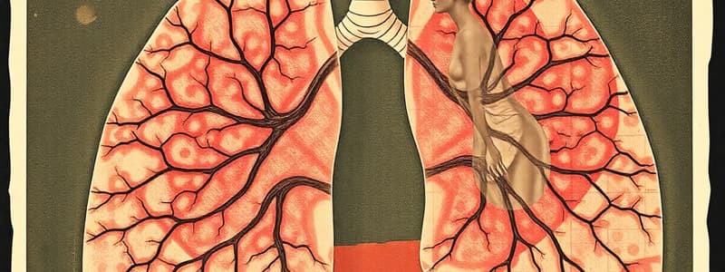
Pulmonary Alveoli
- Definition: Minute air spaces; the functional and structural unit of the lung. Groups of alveoli that open into a common central space are called alveolar sacs.
- Structure:
- Alveolar epithelium (Type 1 pneumocytes, Type 2 pneumocytes)
- Interalveolar septum
- Alveolar pores
Type 1 Pneumocytes
- Number: 95% of lining epithelium.
- LM: Flat squamous cells, less than 0.2 µm thick, with slightly thickened areas containing nuclei.
- EM: Attached to each other by occluding (tight) junctions, have thin basal lamina; perinuclear cytoplasm contains small Golgi, few mitochondria, and rough endoplasmic reticulum (rER) while cytoplasm of the thin portion is devoid of organelles.
- Function: Provide a very thin membrane for gas exchange.
Type 2 Pneumocytes
- Number: 5% of lining epithelium.
- Characteristics: Cuboidal cells with central nuclei located at the angles of interalveolar septa.
- EM: Attached to Type 1 cells by occluding junctions. Cytoplasm contains many mitochondria, rER, prominent Golgi complex and electron-dense lamellar bodies rich in phospholipids.
- Function: Act as progenitor cells for both Type 1 and Type 2 pneumocytes; secrete pulmonary surfactant.
Pulmonary Surfactant
- Definition: A mixture of phospholipids that lines the inner aspect of alveoli.
- Function: Decreases surface tension, preventing alveolar collapse.
Blood Air Barrier
- Definition: The wall separating air in alveoli from blood in capillaries.
- Structure: Includes Type 1 alveolar cells, fused basal laminae of alveolar and endothelial cells of capillaries, and endothelial cells of capillaries.
Alveolar Pores (Pores of Kohn)
- Description: Holes in the interalveolar septa that connect the alveoli.
- Importance: Facilitate communication and equalization of pressure between alveoli.
Alveolar Macrophages
-
Definition: Mononuclear phagocytes in the lungs, representing the first line of defense.
-
Site: Interalveolar septum, on luminal surface of alveoli.
-
Origin: Blood monocytes.
-
Shape: Branched cells with pseudopodia.
-
Cytoplasm: Vacuolated, contains many lysosomes.
-
Types:
- Dust cells: Phagocytose dust or coal particles. In smokers, cytoplasm is crowded with large irregular heterogeneous electron-dense bodies.
- Heart failure cells: Phagocytose extravasated red blood cells (RBCs). In heart failure, cytoplasm contains many vacuoles with hemosiderin granules.
-
Fate: Migrate to bronchi via ciliary action, swallowed and expelled in sputum.
Interalveolar Septum
- Structure:
- Alveolar epithelium on either side.
- Capillary network (pulmonary capillary bed).
- Supporting network of reticular and elastic fibers and connective tissue cells (septal cells, mast cells, lymphocytes, white blood cells [WBCs]).
- Basement membranes of alveolar epithelium and capillary beds.
- Additional Note: The two basement membranes often fuse to form the alveolar-capillary membrane.
Studying That Suits You
Use AI to generate personalized quizzes and flashcards to suit your learning preferences.
Related Documents
Description
Test your knowledge on the histological structure of the respiratory system. This quiz covers respiratory bronchioles, alveolar ducts, and the different types of pneumocytes. Understand the key features and functional significance of the lung's microscopic anatomy.


