Podcast
Questions and Answers
What type of cells forms the majority of the alveolar epithelium?
What type of cells forms the majority of the alveolar epithelium?
- Type II alveolar cells
- Cuboidal cells
- Polyhedral cells
- Type I alveolar cells (correct)
What is the main function of Type II alveolar cells?
What is the main function of Type II alveolar cells?
- To provide minimum thickness for diffusion of gases
- To form the blood-air barrier
- To regulate elasticity of the interalveolar septum
- To secrete surfactant and reduce the surface tension of alveoli (correct)
What type of fibers are present in the interalveolar septum?
What type of fibers are present in the interalveolar septum?
- Reticular fibers and cartilaginous fibers
- Collagen fibers and keratin fibers
- Elastic fibers and reticular fibers (correct)
- Elastic fibers and smooth muscle fibers
What is the characteristic feature of the alveoli?
What is the characteristic feature of the alveoli?
What is the function of the interalveolar septum?
What is the function of the interalveolar septum?
What is the primary function of the conducting system in the respiratory system?
What is the primary function of the conducting system in the respiratory system?
What is the shape of the cartilage rings found in the trachea?
What is the shape of the cartilage rings found in the trachea?
What is the function of the goblet cells found in the epithelium of the trachea?
What is the function of the goblet cells found in the epithelium of the trachea?
How many lobes does the right lung have?
How many lobes does the right lung have?
What is the characteristic of the mucosa in the bronchioles?
What is the characteristic of the mucosa in the bronchioles?
Flashcards are hidden until you start studying
Study Notes
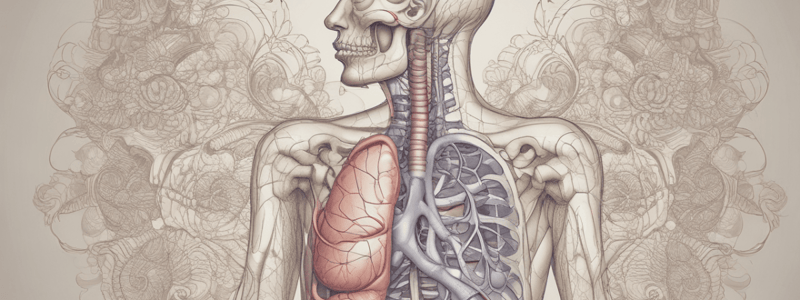
Divisions of Respiratory System
- The respiratory system has two divisions: the conducting system and the respiratory portion.
- The conducting system conducts, filters, and conditions inspired air before it reaches the lung.
- The respiratory portion is responsible for the exchange of gases.
Trachea
- The trachea is approximately 12 cm long and extends from the larynx above to the bifurcation into two bronchi in the lower end.
- The trachea is kept patent with 20 C-shaped, incomplete rings of hyaline cartilage.
Microscopic Structure of the Trachea
- Mucosa (epithelium and lamina propria):
- Epithelium: pseudostratified columnar ciliated cells with goblet cells.
- Goblet cells secrete mucus.
- Cilia move up dust particles.
- Lamina propria: rich in elastic fibers.
- Submucosa:
- Connective tissue with mixed mucoserous glands.
- Elastic membrane separating it from the lamina propria.
- The fibrocartilagenous layer:
- Connective tissue with incomplete C-shaped hyaline cartilage.
- Deficient posteriorly towards the esophagus.
- Smooth muscle fibers between the cartilage ends.
Bronchi
- Extrapulmonary bronchi are similar in structure to the trachea.
- The right lung has 3 lobes, while the left lung has 2 lobes.
Microscopic Structure of the Lung
- Intrapulmonary bronchus:
- Mucosa: folded.
- Submucosa: continuous thin sheath of smooth muscle.
- Fibrocartilagenous layer: multiple plates of cartilage and glands.
Bronchioles
- Mucosa:
- Large bronchioles: simple columnar ciliated epithelium with goblet cells.
- Small bronchioles: simple cuboidal ciliated epithelium with few Clara cells, no goblet cells.
- Muscle layer: well-developed smooth muscle layer.
- C.T layer: no cartilage or glands.
Respiratory Bronchiole
- Lined with cuboidal cells.
- Numerous alveoli open into it, and a few alveoli open into them.
Alveoli
- Polyhedral sacs with one wall deficient.
- Lined with alveolar epithelium.
- Separated by interalveolar septa, which contain:
- Elastic fibers.
- Reticular fibers.
- Pulmonary alveolar phagocytic cells.
- Blood capillaries.
Alveolar Epithelium
- Type I alveolar cells:
- Flattened cells (97% of alveolar surface).
- Small amount of cytoplasm.
- Function: provide minimum thickness for diffusion of gases and share in blood-air barrier.
- Type II alveolar cells:
- Large cuboidal or rounded cells (3% of alveolar epithelium).
- Apical surface contains microvilli.
- Vesicular nucleus.
- Cell organoids.
- Function: secrete surfactant, which reduces the surface tension of alveoli, preventing them from collapsing at the end of expiration.
Studying That Suits You
Use AI to generate personalized quizzes and flashcards to suit your learning preferences.