Podcast
Questions and Answers
What is the recommended hand position to avoid rotation of the radius and ulna during a forearm x-ray?
What is the recommended hand position to avoid rotation of the radius and ulna during a forearm x-ray?
- Neutral
- Prone
- Pronated
- Supinated (correct)
Which of the following is included in the collimation criteria for a forearm x-ray?
Which of the following is included in the collimation criteria for a forearm x-ray?
- The elbow joint only
- Both wrist and elbow joints (correct)
- Only the proximal wrist joint
- Just the distal humerus
What is the appropriate tube angle for a forearm x-ray?
What is the appropriate tube angle for a forearm x-ray?
- 45 degrees lateral
- 20 degrees cephalad
- Straight tube (correct)
- 30 degrees caudad
What is the required distance for a forearm x-ray?
What is the required distance for a forearm x-ray?
Which positioning technique is used for a lateral forearm x-ray?
Which positioning technique is used for a lateral forearm x-ray?
What characteristic indicates an adequately visualized image density in a forearm x-ray?
What characteristic indicates an adequately visualized image density in a forearm x-ray?
What must be ensured regarding the alignment of the forearm during a forearm x-ray?
What must be ensured regarding the alignment of the forearm during a forearm x-ray?
What artifact must be removed from the field of view before conducting a forearm x-ray?
What artifact must be removed from the field of view before conducting a forearm x-ray?
What is the tube angle for the Sesamoids Tangential View?
What is the tube angle for the Sesamoids Tangential View?
Where should the central ray be centered for the Foot Dorso-Plantar View?
Where should the central ray be centered for the Foot Dorso-Plantar View?
Which positioning technique is used for the Sesamoids Tangential View?
Which positioning technique is used for the Sesamoids Tangential View?
What is the recommended collimation for the Foot Dorso-Plantar View?
What is the recommended collimation for the Foot Dorso-Plantar View?
Which of the following is NOT a criterion for the Sesamoids Tangential View?
Which of the following is NOT a criterion for the Sesamoids Tangential View?
What is the kVp range specified for both the Sesamoids and Foot Dorso-Plantar Views?
What is the kVp range specified for both the Sesamoids and Foot Dorso-Plantar Views?
In the Foot Dorso-Plantar View, what should be the posture of the patient?
In the Foot Dorso-Plantar View, what should be the posture of the patient?
What is the primary instruction to the patient during the imaging for both views?
What is the primary instruction to the patient during the imaging for both views?
Which anatomical structures are visible in the lateral profile of the image?
Which anatomical structures are visible in the lateral profile of the image?
What is the purpose of including side markers in the image?
What is the purpose of including side markers in the image?
In what settings is this imaging typically requested?
In what settings is this imaging typically requested?
Which of the following describe the visual quality of the image?
Which of the following describe the visual quality of the image?
What is the correct label for the described image?
What is the correct label for the described image?
What is the optimal angle for the tube during Rosenberg imaging?
What is the optimal angle for the tube during Rosenberg imaging?
Which criteria is essential for achieving a true lateral view in Horizontal Beam Lateral Knee View imaging?
Which criteria is essential for achieving a true lateral view in Horizontal Beam Lateral Knee View imaging?
What is the recommended kVp for the Horizontal Beam Lateral Knee View?
What is the recommended kVp for the Horizontal Beam Lateral Knee View?
When performing a PA Rosenberg view, which anatomy must be included in collimation?
When performing a PA Rosenberg view, which anatomy must be included in collimation?
What should be the patient’s position for a Horizontal Beam Lateral Knee View?
What should be the patient’s position for a Horizontal Beam Lateral Knee View?
Which of the following is NOT a requirement for the PA Rosenberg criteria?
Which of the following is NOT a requirement for the PA Rosenberg criteria?
What is the purpose of the side marker in imaging?
What is the purpose of the side marker in imaging?
Which of the following describes the central ray direction for Horizontal Beam Lateral Knee View?
Which of the following describes the central ray direction for Horizontal Beam Lateral Knee View?
What is the correct angle for the central ray in the axial lateral elbow view?
What is the correct angle for the central ray in the axial lateral elbow view?
Which of the following should be included in the collimation for the toes (dorso-plantar)?
Which of the following should be included in the collimation for the toes (dorso-plantar)?
What should be seen in the axial lateral elbow view regarding the anatomy?
What should be seen in the axial lateral elbow view regarding the anatomy?
What is the recommended kVp for the toes (dorso-plantar) view?
What is the recommended kVp for the toes (dorso-plantar) view?
What is the proper posture for the patient during the axial lateral elbow imaging?
What is the proper posture for the patient during the axial lateral elbow imaging?
Which of the following practices should be followed for patient preparation before taking the radiograph?
Which of the following practices should be followed for patient preparation before taking the radiograph?
In the axial lateral elbow criteria, how should the density of the image be evaluated?
In the axial lateral elbow criteria, how should the density of the image be evaluated?
What is the recommended distance for the tube during the toe imaging?
What is the recommended distance for the tube during the toe imaging?
Study Notes
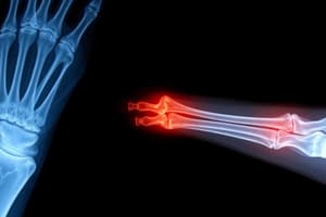
AP Forearm Positioning
- Hand supinated to prevent rotation of radius and ulna
- Straight tube angle
- Central ray perpendicular to mid radius/ulna
- Distance: 100-110cm
- Collimation: Include all skin edges, both wrist and elbow joints
- kVp: 50-60kVp
- mAs: 5mAs
- Grid: No
- Patient preparation: Remove artifacts (watches and jewelry)
AP Forearm Criteria
- Collimation: Include all anatomy, skin edges
- Alignment: Slight superimposition of radial head, neck and tuberosity over proximal ulna. Partially open elbow joint if the shoulder was placed in the same plane as the forearm.
- Anatomy: Entire Forearm, including wrist and distal humerus
- Density: Cortical outline and bony trabecular pattern visible
- Contrast: Soft tissue and bony interfaces visualized
- Markers: Side marker evident
- Identification: Appropriate labeling or anonymized
Lateral Forearm Positioning
- Patient seated with legs at right angle to table
- Entire Arm should be in the same plane
- Flex elbow 90 degrees and place medial aspect of forearm on image receptor
- Straight tube angle
- Central ray perpendicular to mid radius/ulna
- Distance: 100-110cm
- Collimation: Include all skin edges, both wrist and elbow joints
- kVp: 50-60kVp
- mAs: 5 mAs
- Grid: No
- Patient preparation: Remove artifacts (watches and jewelry)
Lateral Forearm Criteria
- Collimation: Include all anatomy, skin edges
- Alignment: Slight superimposition of radial head, neck and tuberosity over proximal ulna. Partially open elbow joint if the shoulder was placed in the same plane as the forearm.
- Anatomy: Entire forearm, including wrist and distal humerus in true lateral position. Superimposition of radius and ulna at distal end.
- Markers: Side marker evident
- Identification: Appropriate labeling or anonymized
Axial Lateral Elbow (Radial Head Trauma) Positioning
- Patient seated with arm bent at 90 degrees
- Hand pronated
- Elbow flexed at 90 degrees
- Hand pronated
- Tube angle 45 degrees towards patient
- Central Ray 45 degrees angled toward chest at mid elbow joint
- Distance: 100-110cm
- Collimation: Include skin edges, 5cm distal humerus, proximal forearm
- kVp: 55-65kVp
- mAs: 4-6 mAs
- Grid: No
- Patient preparation: Remove artifacts (clothing)
Axial Lateral Elbow (Radial Head Trauma) Criteria
- Collimation: Include all anatomy, skin edges
- Alignment: Elbow flexed at 90 degrees
- Anatomy: Radial head and neck free of superimposition, projected away from ulna. Capitellum seen.
- Density: Cortical outline and bony trabecular pattern visible
- Contrast: Soft tissue and bony interfaces visualized
- Markers: Side marker evident
- Identification: Appropriate labeling or anonymized
Toes (Dorso-Plantar) Positioning
- Patient seated on table
- Knee bent and place foot on image receptor
- Tube angle: 15 degrees angled towards heel
- Central Ray: perpendicular to third metatarso-phalangeal joint (MTP)
- Distance: 100-110cm
- Collimation: Include skin edges and adjacent toes.
- kVp: 55 kVp
- mAs: 3 mAs
- Grid: No
- Patient preparation: Remove artifacts (shoes, socks)
Toes (Dorso-Plantar) Criteria
- Collimation: Include all anatomy, skin edges and adjacent toes.
- Alignment: Toes separated without rotation of phalanges, concavity demonstrated on both sides of metatarsal shafts.
- Markers: Side marker evident
- Identification: Appropriate labeling or anonymized
Sesamoids Tangential View Positioning
- Patient lying prone on table or seated
- Foot positioned with first toe flexed and ball of foot perpendicular to image plate. Patient can remain seated with foot flexed and tape tensioning the toes back.
- Straight angle tube
- Central Ray: Centered on second metatarsal
- Distance: 100-110cm
- Collimation: Include skin edges, metatarsal head and sesamoids
- kVp: 50-55kVp
- mAs: 2-4 mAs
- Grid: No
- Patient preparation: Remove artifacts (shoes, socks)
Sesamoids Tangential View Criteria
- Collimation: Include all anatomy, skin edges
- Alignment: Open joint spaces between sesamoids and first metatarsal
- Anatomy: Both sesamoids seen in profile without superimposition
- Density: Cortical outline and bony trabecular pattern visible
- Contrast: Soft tissue and bony interfaces visualized
- Markers: Side marker evident
- Identification: Appropriate labeling or anonymized
Foot Dorso-Plantar View Positioning
- Patient seated on table
- Foot positioned flat on image plate
- Tube angle: 10 degrees towards heel
- Central Ray: Centered on base of third metatarsal
- Distance: 100-110cm
- Collimation: Include all anatomy of interest
- kVp: 50-55kVp
- mAs: 3-4 mAs
- Grid: No
- Patient preparation: Remove artifacts (shoes, socks)
Foot Dorso-Plantar View Criteria
- Collimation: Include all anatomy, skin edges
- Alignment: No rotation of foot, long axis of image plate aligned with long axis of foot.
- Anatomy: Toes to tarsals included, slight overlap of 2nd-5th metatarsal bases.
- Density: Cortical outline and bony trabecular pattern visible
- Contrast: Soft tissue and bony interfaces visualized
- Markers: Side marker evident
- Identification: Appropriate labeling or anonymized
PA Rosenberg Positioning:
- Tube angle: Parallel to tibial plateau, appx. 10 degrees caudal angle
- Central Ray: Centered to midline of knee at level of joint space
- Distance: 100-110cm
- Collimation: Include all anatomy of interest
- kVp: 60-70kVp
- mAs: 12-16 mAs
- Grid: Yes
- Patient preparation: Comfortable position, quick exposure
PA Rosenberg Criteria
- Collimation: Include all anatomy and skin edges
- Alignment: No rotation of femur or tibia. Tibial plateau in profile.
- Anatomy: Distal femur, proximal tibia and fibula. Open femoro-tibial joint spaces, intercondylar fossa visualized.
- Density: Cortical outline and bony trabecular pattern visible
- Contrast: Soft tissue and bony interfaces visualized
- Markers: Side marker evident, labeled “Wt Bearing Rosenberg”
- Identification: Appropriate labeling or anonymized
Horizontal Beam Lateral Knee View Positioning:
- Patient supine on bed, knee extended
- Performed medio-laterally with image receptor resting between patient’s knees.
- Tube angle: Perpendicular to long axis of knee.
- Central Ray: Angled appx 5 degrees caudally at distal portion of lateral condyle, superimposing the femoral condyles with medial femoral condyle appearing larger.
- Distance: 100-110cm
- Collimation: Include all anatomy of interest
- kVp: 60
- mAs: 12 mAs (adjust based on knee thickness)
- Grid: No
Horizontal Beam Lateral Criteria:
- Collimation: Include all anatomy, skin edges
- Alignment: Superimposed femoral condyles, true lateral. Patella in profile and open femoro-patellar joint space. Tibial plateau in profile.
- Anatomy: Distal femur, proximal tibia and fibula in lateral profile.
- Density: Cortical outline and bony trabecular pattern visible
- Contrast: Soft tissue and bony interfaces visualized
- Markers: Side marker evident, labeled “Horizontal Beam Lateral”
- Identification: Appropriate labeling or anonymized
Studying That Suits You
Use AI to generate personalized quizzes and flashcards to suit your learning preferences.
Related Documents
Description
This quiz focuses on the essential positioning techniques and criteria for imaging the forearm in radiography. It covers both AP and lateral views, emphasizing parameters like collimation, alignment, anatomy, and density. Test your knowledge on the crucial factors for successful forearm imaging.