Podcast
Questions and Answers
What is the recommended collimation for a forearm x-ray?
What is the recommended collimation for a forearm x-ray?
- Collimate only the lateral border closest to the elbow
- Collimate only the lateral border closest to the wrist
- Collimate the entire arm from shoulder to wrist
- Collimate both lateral borders to the actual forearm area (correct)
What is the minimum source-to-image distance (SID) for a forearm x-ray?
What is the minimum source-to-image distance (SID) for a forearm x-ray?
- 40 inches (102 cm) (correct)
- 20 inches (51 cm)
- 30 inches (76 cm)
- 60 inches (152 cm)
How should the patient be positioned for a lateral forearm x-ray?
How should the patient be positioned for a lateral forearm x-ray?
- Seat patient with shoulder raised above the table
- Seat patient with wrist flexed 90°
- Seat patient at end of table, with elbow flexed 90° (correct)
- Seat patient with elbow fully extended
What is the ideal direction of the central ray (CR) for a lateral forearm x-ray?
What is the ideal direction of the central ray (CR) for a lateral forearm x-ray?
What is the recommended minimum source-to-image receptor distance (SID) for this imaging procedure?
What is the recommended minimum source-to-image receptor distance (SID) for this imaging procedure?
For analog imaging, what is the recommended kilovoltage (kV) range?
For analog imaging, what is the recommended kilovoltage (kV) range?
How should the patient be positioned for the wrist imaging?
How should the patient be positioned for the wrist imaging?
What is the recommended central ray (CR) angle for the PA axial wrist (scaphoid) with ulnar deviation?
What is the recommended central ray (CR) angle for the PA axial wrist (scaphoid) with ulnar deviation?
Which anatomy should be clearly demonstrated in the wrist imaging?
Which anatomy should be clearly demonstrated in the wrist imaging?
What is the purpose of collimation in this imaging procedure?
What is the purpose of collimation in this imaging procedure?
What might obscure fractures of the scaphoid require in this imaging procedure?
What might obscure fractures of the scaphoid require in this imaging procedure?
What should be evident in the wrist imaging to show ulnar deviation?
What should be evident in the wrist imaging to show ulnar deviation?
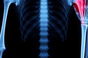
What are the clinical indications for forearm imaging?
What are the clinical indications for forearm imaging?
What is the recommended collimation for forearm imaging?
What is the recommended collimation for forearm imaging?
What projection is used for calcification or pathology of the dorsal aspect of carpal bones?
What projection is used for calcification or pathology of the dorsal aspect of carpal bones?
What is the purpose of PA and PA axial scaphoid with ulnar deviation?
What is the purpose of PA and PA axial scaphoid with ulnar deviation?
What may be required for nondisplaced fractures of the scaphoid?
What may be required for nondisplaced fractures of the scaphoid?
What should be used to protect radiosensitive tissues outside the region of interest during upper limb imaging?
What should be used to protect radiosensitive tissues outside the region of interest during upper limb imaging?
What is the recommended patient positioning for forearm imaging?
What is the recommended patient positioning for forearm imaging?
What is the purpose of the carpal bridge tangential projection?
What is the purpose of the carpal bridge tangential projection?
What is the recommended collimation to avoid cutting off anatomy at either joint during forearm imaging?
What is the recommended collimation to avoid cutting off anatomy at either joint during forearm imaging?
What is the purpose of PA and PA axial scaphoid with ulnar deviation?
What is the purpose of PA and PA axial scaphoid with ulnar deviation?
What may be required for nondisplaced fractures of the scaphoid?
What may be required for nondisplaced fractures of the scaphoid?
What should be used to protect radiosensitive tissues outside the region of interest during upper limb imaging?
What should be used to protect radiosensitive tissues outside the region of interest during upper limb imaging?
Flashcards are hidden until you start studying
Study Notes
Radiographic Imaging of Upper Limb
- Forearm routine involves AP and lateral projections to demonstrate radius, ulna, carpal bones, elbow, and humerus, with specific positioning and exposure instructions
- Clinical indications for forearm imaging include fractures, dislocations, and pathologic processes like osteomyelitis or arthritis
- Technical factors for forearm imaging include minimum SID, IR size, and kV range for analog and digital systems
- Patient positioning for forearm imaging includes fully extending the arm, dropping the shoulder, and aligning the forearm with the IR
- Collimation for forearm imaging should be focused on the actual forearm area with minimal collimation at both ends to avoid cutting off anatomy at either joint
- Carpal bridge tangential projection is used for calcification or pathology of the dorsal aspect of carpal bones, with specific technical factors and patient positioning
- Evaluation criteria for carpal bridge tangential projection include the visualization of specific carpal bones and the position and exposure requirements
- PA and PA axial scaphoid with ulnar deviation is used for possible scaphoid fractures, with a warning not to attempt the position before ruling out possible fractures
- Nondisplaced fractures of the scaphoid may require additional projections or a CT scan of the wrist
- The text provides specific technical factors and patient positioning for wrist imaging, including scaphoid views, radial deviation, and carpal canal imaging
- Shielding should be used to protect radiosensitive tissues outside the region of interest during upper limb imaging
- The text emphasizes the importance of considering beam divergence and ensuring the inclusion of a minimum distance distal to wrist and elbow joints on the image receptor.
Studying That Suits You
Use AI to generate personalized quizzes and flashcards to suit your learning preferences.