Podcast
Questions and Answers
In the femur AP position, the patient is placed in a lateral decubitus position.
In the femur AP position, the patient is placed in a lateral decubitus position.
False (B)
The central ray is projected perpendicularly towards the femur in the femur AP position.
The central ray is projected perpendicularly towards the femur in the femur AP position.
True (A)
The IR size for femur AP is 10*12 inch.
The IR size for femur AP is 10*12 inch.
False (B)
In the lateral femur position, the knee of the filming side is flexed 90 degrees.
In the lateral femur position, the knee of the filming side is flexed 90 degrees.
The patella should be shown in the entire image in the lateral femur position.
The patella should be shown in the entire image in the lateral femur position.
The femur lateral oblique position requires the patient to be placed in a lateral decubitus position.
The femur lateral oblique position requires the patient to be placed in a lateral decubitus position.
The lesser trochanter of the femur should be shown completely in the femur lateral oblique position.
The lesser trochanter of the femur should be shown completely in the femur lateral oblique position.
The femur AP position is used to show the distal portion of the femur only.
The femur AP position is used to show the distal portion of the femur only.
Flashcards are hidden until you start studying
Study Notes
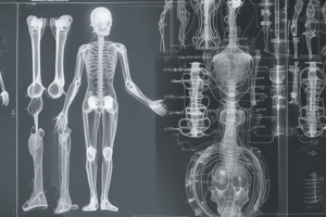
Femur Radiographic Techniques
Femur Positions
- Four types of femur positions: Femur AP, Femur Lateral, Femur Medial Oblique, and Femur Lateral Oblique
Femur AP
- Positioning: Patient in supine position, femur in center of image receptor (IR), leg rotated 5 degrees medially
- Anatomy: Includes knee and femur in true anterior-posterior projection image
- Technical Factors:
- IR size: 14*17 inch
- SID: 100cm
- Central ray: Perpendicular to femur
- Respiration: Suspended
- Collimation: Minimum of knee joint cavity and distal and proximal portions of femur
- Evaluation:
- Femur must not be in rotation, but show in true anterior-posterior projection image
- Femoral condyles should be shown symmetrically about patella
Lateral Femur
- Positioning:
- Patient in lateral decubitus or supine position
- Flex knee of filming side 45 degrees, bilateral condyles of femur perpendicular to IR
- Rotate to the back of opposite leg to avoid overlapping
- Place bottom of IR 5cm from the knee
- Evaluation:
- Patella should be shown in half-side image
- Neck of femur and greater trochanter must overlap completely
- Technical Factors:
- Same as Femur AP position
Femur Lateral Oblique
- Positioning:
- Patient in supine position
- Place femur in center of IR
- Rotate knee and femur 45 degrees laterally
- Evaluation:
- Lesser trochanter of femur should be shown separated in half-side image
Studying That Suits You
Use AI to generate personalized quizzes and flashcards to suit your learning preferences.