Podcast
Questions and Answers
What is the term used to describe the shape distortion that occurs when the object is not parallel to the image receptor?
What is the term used to describe the shape distortion that occurs when the object is not parallel to the image receptor?
- Unsharpness
- Focal spot
- Foreshortening (correct)
- Elongation
What is the result of the x-ray beam being oriented at right angles to the object but not to the image receptor?
What is the result of the x-ray beam being oriented at right angles to the object but not to the image receptor?
- Foreshortening of the image
- Elongation of the image (correct)
- Unsharpness of the image
- No distortion of the image
Why is it important to position the image receptor parallel to the long axis of the object?
Why is it important to position the image receptor parallel to the long axis of the object?
- To minimize shape distortion (correct)
- To reduce unsharpness
- To decrease the x-ray beam intensity
- To increase the focal spot
What is the result of the central ray not being directed at right angles to the object and image receptor?
What is the result of the central ray not being directed at right angles to the object and image receptor?
What is the optimal orientation of the x-ray beam to the object and image receptor?
What is the optimal orientation of the x-ray beam to the object and image receptor?
What type of views are most prone to image shape distortion?
What type of views are most prone to image shape distortion?
What is the result of the object being parallel to the image receptor, but the central ray not being directed at right angles to each?
What is the result of the object being parallel to the image receptor, but the central ray not being directed at right angles to each?
What is the reason for the distortion of the resultant image?
What is the reason for the distortion of the resultant image?
What is the purpose of orienting the central ray perpendicular to the object and image receptor?
What is the purpose of orienting the central ray perpendicular to the object and image receptor?
Which roots appear disproportionately longer in the image due to angulation?
Which roots appear disproportionately longer in the image due to angulation?
What is the effect of increasing the distance between the focal spot and the object?
What is the effect of increasing the distance between the focal spot and the object?
What is the result of decreasing the distance between the object and the image receptor?
What is the result of decreasing the distance between the object and the image receptor?
What is the relationship between the distance of the object from the focal spot and the image sharpness?
What is the relationship between the distance of the object from the focal spot and the image sharpness?
What is the effect of angulation on the image of the roots?
What is the effect of angulation on the image of the roots?
Which of the following statements is true about the image formation?
Which of the following statements is true about the image formation?
What is the purpose of adjusting the distance between the focal spot and the object?
What is the purpose of adjusting the distance between the focal spot and the object?
What is the primary benefit of the paralleling technique in intraoral radiography?
What is the primary benefit of the paralleling technique in intraoral radiography?
Why is it necessary to position the image receptor away from the teeth in the paralleling technique?
Why is it necessary to position the image receptor away from the teeth in the paralleling technique?
What is the purpose of using image receptor holders in the paralleling technique?
What is the purpose of using image receptor holders in the paralleling technique?
What is the main challenge in deriving three-dimensional information from radiographs in clinical practice?
What is the main challenge in deriving three-dimensional information from radiographs in clinical practice?
What type of radiograph is commonly used to determine the mediolateral position of an object or feature?
What type of radiograph is commonly used to determine the mediolateral position of an object or feature?
Why is it important to use multiple radiographic views to determine the location of an object or feature?
Why is it important to use multiple radiographic views to determine the location of an object or feature?
What is the main advantage of using the paralleling technique in intraoral radiography?
What is the main advantage of using the paralleling technique in intraoral radiography?
What is the purpose of using radiographs in clinical practice?
What is the purpose of using radiographs in clinical practice?
What happens when the object in question appears to move in the same direction with respect to the reference structures as does the x-ray tube?
What happens when the object in question appears to move in the same direction with respect to the reference structures as does the x-ray tube?
What is the significance of the object in question moving distally when the tube is shifted mesially?
What is the significance of the object in question moving distally when the tube is shifted mesially?
What does the acronym SLOB stand for?
What does the acronym SLOB stand for?
What happens when the object in question does not move with respect to the reference object?
What happens when the object in question does not move with respect to the reference object?
What is the relationship between the object in question and the reference object when the tube is shifted mesially and the object appears to move distally?
What is the relationship between the object in question and the reference object when the tube is shifted mesially and the object appears to move distally?
What is the significance of the x-ray tube's movement in determining the location of the object in question?
What is the significance of the x-ray tube's movement in determining the location of the object in question?
What can be concluded if the object in question appears to move in the opposite direction as the x-ray tube?
What can be concluded if the object in question appears to move in the opposite direction as the x-ray tube?
What is the purpose of the SLOB rule?
What is the purpose of the SLOB rule?
What can be inferred about the position of the palatal root in images A and B?
What can be inferred about the position of the palatal root in images A and B?
What is the significance of the inferior border of the zygomatic process of the maxilla?
What is the significance of the inferior border of the zygomatic process of the maxilla?
What is the characteristic feature of an expansile lesion on the buccal surface of the mandible?
What is the characteristic feature of an expansile lesion on the buccal surface of the mandible?
What is the reason for the eggshell effect on plain images?
What is the reason for the eggshell effect on plain images?
What is the orientation of the x-ray beam when the zygomatic process is projected occlusally over the teeth?
What is the orientation of the x-ray beam when the zygomatic process is projected occlusally over the teeth?
What is the effect of increasing the angulation of the x-ray beam vertically?
What is the effect of increasing the angulation of the x-ray beam vertically?
What is the significance of osseous landmarks with respect to the teeth?
What is the significance of osseous landmarks with respect to the teeth?
What can be inferred about the attenuation of the x-ray beam in the region of the expanded bony cortex?
What can be inferred about the attenuation of the x-ray beam in the region of the expanded bony cortex?
Flashcards are hidden until you start studying
Study Notes
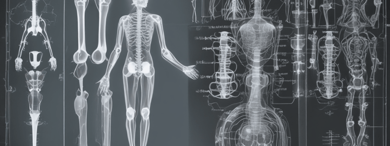
Focal Spot and Image Receptor
- The physical shape of the object may prevent its optimal orientation, resulting in shape distortion.
- To minimize shape distortion, the practitioner should align the tube, object, and image receptor carefully.
- The image receptor should be parallel to the long axis of the object.
- The central ray of the x-ray beam should be perpendicular to the object and image receptor.
Image Shape Distortion
- Image shape distortion occurs when the object and image receptor are not parallel, or the central ray is not directed at right angles to each.
- Distortion is most evident on maxillary molar views, where the palatal roots appear disproportionately longer than the buccal roots.
Paralleling Technique
- This technique is the preferred method for making intraoral radiographs.
- The image receptor is placed parallel to the long axis of the tooth, minimizing image distortion.
- Image receptor holders should be used to support the image receptor in the patient’s mouth.
Factors Affecting Image Sharpness and Magnification
- Increasing the distance between the focal spot and the object results in an image with increased sharpness and less magnification.
- Decreasing the distance between the object and the image receptor increases the sharpness and results in less magnification.
Object Localization
- Three methods are used to obtain three-dimensional information from radiographs:
- The SLOB rule (same lingual, opposite buccal) helps identify the location of an object.
- Comparing the anatomy displayed on the images helps distinguish changes in horizontal or vertical angulation.
- The relative positions of osseous landmarks with respect to the teeth help identify changes in horizontal or vertical angulation.
Eggshell Effect
- Plain images may produce an eggshell effect, where the periphery of an expanded cortex appears more opaque than the region inside.
- The cortical bone is not thicker on the cortex, but rather the x-ray beam is more attenuated in this region due to the longer path length of photons through the bony cortex on the periphery.
Studying That Suits You
Use AI to generate personalized quizzes and flashcards to suit your learning preferences.


