Podcast
Questions and Answers
What happens to the Z bands during muscle contraction?
What happens to the Z bands during muscle contraction?
- They remain the same
- They become farther apart
- They become closer together (correct)
- They disappear
What is the primary source of ATP for muscle contraction during anaerobic respiration?
What is the primary source of ATP for muscle contraction during anaerobic respiration?
- Aerobic respiration in mitochondria
- Oxidation of lactic acid
- Phosphorylation by myokinase and creatin phosphate
- Glycolysis from stored glycogen (correct)
What is the byproduct of glycolysis in muscle cells?
What is the byproduct of glycolysis in muscle cells?
- ATP
- Glucose
- Pyruvate
- Lactic acid (correct)
What is the mechanism of smooth muscle contraction?
What is the mechanism of smooth muscle contraction?
What is the role of calmodulin in smooth muscle contraction?
What is the role of calmodulin in smooth muscle contraction?
What is the ratio of actin to myosin filaments in smooth muscle cells?
What is the ratio of actin to myosin filaments in smooth muscle cells?
What is the result of muscle fatigue?
What is the result of muscle fatigue?
What is the purpose of oxygen debt?
What is the purpose of oxygen debt?
What is the characteristic of smooth muscle fibers?
What is the characteristic of smooth muscle fibers?
What is the function of myosin light chain kinase?
What is the function of myosin light chain kinase?
Flashcards are hidden until you start studying
Study Notes
Universal Characteristics of Muscle
- Excitability: responds to stimuli (e.g., nervous impulses)
- Conductivity: ability to produce a local effect
- Contractility: able to shorten in length
- Extensibility: stretches when pulled
- Elasticity: tends to return to original shape & length after contraction or extension
Classification of Muscle Tissue
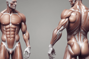
Skeletal Muscle
- Voluntary striated muscle attached to one or more bones
- Diameter: 100 µm in diameter and 3 cm long (500um/30 cm)
- Length!!!Sk Muscle cells = muscle/myofibers
- Exhibits alternating light and dark transverse bands, or striations
- Composed of:
- Endomysium: surrounds each muscle fiber
- Perimysium: bundles muscle fibers together into fascicles
- Epimysium: encloses the entire muscle
- Long, multinucleated, formed by fusion of myoblasts
- Contains satellite cells, Sarcolemma, T-tubules, Glycogen, Myoglobin, Mitochondria, and other organelles
- Sarcoplasmic reticulum surrounds myofibrils longitudinally, acts as a Ca2+ reservoir
Muscle Fibers
Myofibrils
- Consist of three types of myofilaments:
- Thick filaments: 300 myosin molecules with globular heads that form cross-bridges between thick and thin filaments
- Thin filaments: actin strands contain binding sites for myosin heads
- Elastic filaments (titin): attaches thick filaments to Z disc, holds thick filaments in place, and prevents muscle cell overstretching
- Dystrophin: an accessory protein that links thin filaments to sarcolemma and endomysium via integral proteins
Striations and Sarcomeres
- A band: Dark band formed by parallel thick filaments that partly overlap with thin filaments
- H band: Lighter region in the middle of an A band that contains thick filaments only
- M line: Dark line in the middle of an H band, a meshwork of proteins that anchor thick filaments
- I band: Light band composed of thin filaments only, also contains elastic filaments
- Z disc: Protein disc that anchors thin and elastic filaments at each end of a sarcomere
Motor Unit
- Axons (fibers) of a somatic motor neuron divide as they enter the muscle
- Motor unit: one nerve fiber and all muscle fibers innervated by it
- Each muscle gets at least one motor nerve that contains hundreds of motor neurons with their axons
- One axon branches to many terminals, each forming a junction with one muscle fiber
- Muscle fibers in a motor unit spread throughout the muscle, not clustered together
Neuromuscular Junction
- Each fiber ending forms a neuromuscular junction (synapse) with a muscle fiber at the motor end plate
- Synaptic cleft: gap between the axonal ending and the motor end plate
- Axonal ending has synaptic vesicles containing ACh
- Sarcolemma of the motor end plate has receptors for ACh and ion channels
Sequence of Events during Transmission
- Nerve impulse reaches the end of the axon, opening voltage-gated Ca2+ channels
- Ca2+ influx causes vesicles to fuse with the axon membrane, releasing ACh
- ACh binds to receptors on the sarcolemma, triggering depolarization and an action potential
- Acetylcholinesterase on the sarcolemma breaks down ACh, preventing continued muscle contraction
Generation of Action Potential
- Recall resting membrane potential
- Binding of ACh opens chemically gated ion channels, causing depolarization at the motor end plate
- Voltage-gated channels on the sarcolemma open, creating an action potential that propagates along the membrane
- Na+ gates close and K+ gates open, causing repolarization
Excitation-Contraction Coupling
- Action potential moves down the T-tubule in the triad
- Terminal cisternae of the SR release Ca2+ into the sarcoplasm via voltage-gated channels
- Ca2+ binds to troponin, shifting tropomyosin, and exposing binding sites on actin
- Myosin heads attach and pull thin filaments toward the center of the sarcomere
- ATP hydrolysis activates myosin heads, and ATP binding releases them for another stroke
- Ca2+ reuptake by the SR causes relaxation
Sliding Filament Contraction Model
- Thin filaments slide past thick filaments to overlap to a greater extent than when relaxed
- Nerve impulse → myosin heads attach to actin in thin filaments → thin filaments move to the center of the sarcomere
- Z bands become closer together, I bands shorten, H zone disappears, and the muscle fiber shortens
Relaxation
- Ca2+ reuptake by the SR causes relaxation
- Muscle fatigue causes: consumption of glycogen and ACh, slowing of the Na+/K+ pump, lactic acid accumulation, and K+ accumulation in extracellular fluid
Muscle Metabolism
- ATP sources:
- Phosphorylation by myokinase and creatin phosphate (phosphagen system)
- Glycolysis from stored glycogen (anaerobically)
- Aerobic respiration in mitochondria
- Oxygen debt: O2 reserves replacement, phosphagen system replenishment, oxidation of lactic acid, and elevated metabolic rate requirements
Smooth Muscle
- Each smooth muscle fiber is a spindle-shaped cell with a diameter ranging from 2 to 10 µm
- Smooth muscle fibers have a single nucleus
- Two types of filaments: thick myosin-containing filaments and thin actin-containing filaments
- Contraction depends on a rise in free intracellular Ca2+, which binds with calmodulin to activate myosin light chain kinase, leading to myosin head phosphorylation and contraction.
Studying That Suits You
Use AI to generate personalized quizzes and flashcards to suit your learning preferences.