Podcast
Questions and Answers
What is the primary function of the periodontal fibers?
What is the primary function of the periodontal fibers?
- To supply blood to the tooth
- To provide sensation to the tooth
- To form the root of the tooth
- To prevent tooth tipping and resists luxation (correct)
What is the origin of the small, fine brush-like fibrils detected in the development of principal fibers of PDL?
What is the origin of the small, fine brush-like fibrils detected in the development of principal fibers of PDL?
- From the alveolar bone
- From the root cementum (correct)
- From the dental pulp
- From the periodontal ligament space
What is the main function of the lymphatic vessels in the periodontal ligament?
What is the main function of the lymphatic vessels in the periodontal ligament?
- To supply blood to the tooth
- To drain lymph from the periodontal ligament (correct)
- To form the root of the tooth
- To provide sensation to the tooth
What is the primary source of nerve supply to the periodontal ligament?
What is the primary source of nerve supply to the periodontal ligament?
What are cementicles?
What are cementicles?
What is the primary function of the periodontal ligament in relation to occlusal forces?
What is the primary function of the periodontal ligament in relation to occlusal forces?
What is the role of the periodontal ligament in relation to the synthesis and resorption of cementum, ligament, and alveolar bone?
What is the role of the periodontal ligament in relation to the synthesis and resorption of cementum, ligament, and alveolar bone?
What is the significance of the rich vascular supply of the periodontal ligament?
What is the significance of the rich vascular supply of the periodontal ligament?
What is the typical width of the periodontal ligament space in normal functions?
What is the typical width of the periodontal ligament space in normal functions?
What is the relationship between the width of the periodontal ligament space and the degree of stress to which the tooth was subjected?
What is the relationship between the width of the periodontal ligament space and the degree of stress to which the tooth was subjected?
Flashcards are hidden until you start studying
Study Notes
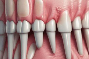
Periodontal Fibers
- There are five groups of principal fibers of the PDL.
- Inter-radicular fibers prevent tooth tipping, resist luxation, and protect the blood, lymph, and nerve supply of the tooth.
Development of Principal Fibers of PDL
- Small, fine brush-like fibrils are detected arising from the root cementum and projecting into the PDL space.
- Small fibers are seen on the surface of the bone but only in thin, small numbers.
- The number and thickness of fibers originating from the bone increase and elongate, radiating towards the loose connective tissue in the mid portion of the periodontal ligament.
- The fibers originating from the cementum also increase in length and thickness and fuse with the fibers originating from the alveolar bone in the periodontal ligament space.
- Following tooth eruption, the principal fibers become organized in bundles and run continuously from bone to cementum.
Connective Tissue
- The periodontal ligament is supplied by branches derived from three sources: dental, inter-radicular, and interdental arteries.
- Lymphatic vessels follow the path of blood vessels in the periodontal ligament.
- The periodontal ligament is mainly supplied by dental branches of the alveolar nerve.
- The periodontal ligament has mechanoreceptors providing sense of touch, pressure, pain, and proprioception during mastication.
Functions of the PDL
- Physical functions:
- Provide soft tissue “casing” to protect the vessels and nerves from injury by mechanical forces.
- Transmit occlusal forces to the bone.
- Attach the teeth to the bone.
- Maintain the gingival tissues in their proper relationship to the teeth.
- Resist the impact of occlusal forces (shock absorption).
- Formative and remodeling functions:
- Cells of the periodontal ligament have the capacity to control the synthesis and resorption of the cementum, ligament, and alveolar bone.
- Periodontal ligament undergoes constant remodeling; old cells and fibers are broken down and replaced by new ones.
- Nutritive and sensory functions:
- Since PDL has a rich vascular supply, it provides nutrition to the cementum, bone, and gingiva.
- The PDL is supplied with sensory nerve fibers which transmit the sensation of touch, pressure, and pain to higher centers.
Clinical Consideration
- The width of PDL space varies with age, location of the tooth, and degree of stress to which the tooth was subjected.
- The width of PDL space is about 0.25mm in normal functions.
- It is widest at the cervical and apical portions of the root and narrowest at the middle.
- The PDL has adaptability to rapidly changing applied force and its capacity to maintain its width at constant dimensions throughout its lifetime.
Studying That Suits You
Use AI to generate personalized quizzes and flashcards to suit your learning preferences.