Podcast
Questions and Answers
What does a protuberant abdomen typically indicate?
What does a protuberant abdomen typically indicate?
- Abdominal distension (correct)
- Deeply sunken featuring obesity
- Normal rounded shape
- Everted umbilicus
Which observation is an indication of pain during abdominal assessment?
Which observation is an indication of pain during abdominal assessment?
- Restlessness and constant turning (correct)
- Relaxed and even respirations
- Symmetric abdominal shape
- Inverted umbilicus
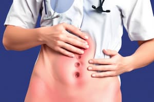
What is indicated by a bluish periumbilical color?
What is indicated by a bluish periumbilical color?
- Obesity
- Normal finding
- Intraperitoneal bleeding (correct)
- Ascites
What could a scaphoid abdomen suggest during an inspection?
What could a scaphoid abdomen suggest during an inspection?
Which of the following findings is NOT considered normal during abdominal inspection?
Which of the following findings is NOT considered normal during abdominal inspection?
What is the primary purpose of using the abdominal quadrant approach during an abdominal assessment?
What is the primary purpose of using the abdominal quadrant approach during an abdominal assessment?
Which technique is NOT a component of abdominal assessment?
Which technique is NOT a component of abdominal assessment?
Which of the following complaints is associated with dysphagia?
Which of the following complaints is associated with dysphagia?
When documenting observations made during an abdominal assessment, which of the following should be prioritized?
When documenting observations made during an abdominal assessment, which of the following should be prioritized?
Which of the following best describes the role of auscultation in abdominal assessment?
Which of the following best describes the role of auscultation in abdominal assessment?
Flashcards
Abdominal Assessment
Abdominal Assessment
A systematic examination of the abdomen using visual, listening, tapping, and feeling techniques.
Abdominal Quadrants
Abdominal Quadrants
Dividing the abdomen into four sections (RUQ, RLQ, LUQ, LLQ) for focused examination.
Assessment Techniques
Assessment Techniques
Methods used to examine the abdomen, including inspection, auscultation, percussion, and palpation.
Client Complaints
Client Complaints
Signup and view all the flashcards
Digestive System Anatomy/Physiology
Digestive System Anatomy/Physiology
Signup and view all the flashcards
Abdominal Inspection
Abdominal Inspection
Signup and view all the flashcards
Umbilical Location
Umbilical Location
Signup and view all the flashcards
Abdominal Distension
Abdominal Distension
Signup and view all the flashcards
Colicky Pain
Colicky Pain
Signup and view all the flashcards
Study Notes
Assessment of Abdomen
- Presenter: Mr. Hamza Chehade
- College: College of Health Sciences
- Programme: Nursing
- Semester: Fall 2024/25
Course Outcomes
- Discuss the concept of health assessment and its types
- Identify the steps in performing selected examination procedures
- Perform comprehensive health assessment correctly and confidently
- Apply appropriate sequencing in conducting a physical health examination systematically
- Analyze the relationship of nursing process health assessment and its implications in comprehensive health assessment
- Select examination techniques appropriate for clients of different ages
- Reflect on social and ethical responsibilities required to function as a professional
- Demonstrate good understanding of the values and ethics of the profession
Learning Objectives
- Review the anatomy and physiology of the digestive system
- Identify equipment needed to assess the abdomen
- Apply techniques used to assess the abdomen
- Identify normal observations during abdominal assessment
- Distinguish abnormal observations in abdominal assessment
- Relate abnormal observations to related disorders
- Document observations made during abdominal assessment
Anatomy and Physiology- Digestive System
- Link to online video: https://www.youtube.com/watch?v=i5MH6ddyi74
Common Client Complaints
- Appetite
- Dysphagia
- Food intolerance
- Abdominal pain
- Nausea
- Vomiting
- Bowel habits (diarrhea/constipation)
- Past history of abdominal diseases
Components and Techniques of Abdominal Assessment
- General appearance
- Inspection
- Auscultation
- Percussion
- Palpation
Exam of the Abdomen
- Think Anatomically: Examine abdomen using four-quadrant system (RUQ, RLQ, LUQ, LLQ)
- Anatomical Awareness: Visualize organs in each quadrant during assessment (inspection, auscultation, palpation, percussion)
- Differential Diagnosis: Understanding organ locations helps in identifying normal/abnormal findings and potential pathologies
Topical Anatomy of the Abdomen
- Diagram showing liver, stomach, spleen, transverse colon, aorta, and descending colon
General Appearance- Abnormal Observations
- Restlessness and constant turning with colicky pain
- Absolute stillness
- Knees flexed up, facial grimacing, and rapid, uneven respirations
Abdomen- Inspection- Normal Findings
- Normal contour/shape (rounded or flat)
- Symmetry
Abdomen- Inspection- Abnormal Observations
- Scaphoid abdomen (caves in)
- Protuberant abdomen (indicates abdominal distension)
- Hernia (protrusion of abdominal viscera through abnormal opening in muscle)
- Bulging
- Visible mass
- Asymmetric shape
Abdomen- Inspection- Normal Findings
- Umbilicus is midline, inverted, without discolouration, inflammation, or hernia
- Umbilicus may become everted and pushed upward with pregnancy
Abdomen- Inspection- Abnormal Observations
- Everted umbilicus with ascites or underlying mass
- Deeply sunken umbilicus with obesity
- Enlarged, everted umbilicus with umbilical hernia
- Bluish periumbilical color with intraperitoneal bleeding (Cullen Sign)
Abdomen- Inspection- Normal Findings
- Smooth and even skin surface
- Absence of surgical scars
- Normal skin turgor
- Respiratory movements
Abdomen- Inspection- Abnormal Observations
- Redness indicates inflammation
- Striae (glistening, taut skin) may indicate ascites
- Surgical scars (possible adhesions, excess fibrous tissue)
- Spider angiomas (liver disease)
- Decreased skin turgor
Abnormalities on Inspection (Causes)
- Scars (past surgery/trauma)
- Striae (obesity, ascites, pregnancy, tumors, steroid use)
- Venous pattern (prominent in fair-skinned people, portal congestion)
- Discoloration (jaundice, Addison's disease, trauma)
- Visible peristalsis (bowel obstruction in older adults, pyloric stenosis in newborns)
- Pulsations (aortic aneurysm)
- Distension (fat, fluid, feces, fetus, flatus, fibroid, full bladder, false pregnancy)
Examples of abnormalities (Images)
- Obese abdomen
- Hepatomegaly
- Markedly enlarged gall bladder
- Ascites
- Umbilical hernia
Abdomen- Inspection- Normal Findings
- Peristaltic waves
- Aortic pulsation
Abdomen- Inspection- Abnormal Observations
- Marked aorta pulsation with widened pulse pressure (hypertension, aortic aneurysm)
- Marked visible peristalsis with a distended abdomen (intestinal obstruction)
Abdomen- Auscultation (Bowel sounds)- Normal Findings
- Diaphragm of stethoscope- Start at RLQ
- High-pitched, gurgling, cascading sounds irregularly from 5 to 30 times/minute
- Hyperperistalsis (borborygmus)
Abdomen- Auscultation (Bowel sounds)- Abnormal Observations
- Hyperactive sounds (loud, high-pitched, rushing, tinkling)
- Hypoactive or absent bowel sounds (after abdominal surgery or peritonitis)
Vascular Sound Locations
- Stethoscope bell, firm pressure over aorta, renal, iliac, femoral arteries
Abdomen- Auscultation (Vascular sounds)- Normal Findings
- Absence of vascular sounds or bruits over aorta, renal, iliac & femoral arteries
- Possible normal bruit from celiac artery (systolic, medium/low pitch) between xiphoid & umbilicus (4-20% of healthy ppl)
Abdomen- Auscultation (Vascular sounds)- Abnormal Observations
- Systolic bruit (pulsatile, blowing sound) - stenosis, partial occlusion, or aneurysm of an artery.
Abdominal Quadrants and Their Structures
- Diagram showing organs in each quadrant (RUQ, RLQ, LUQ, LLQ)
Abdomen- Percussion- Normal Findings
- Move clockwise, determine existing tympany and dullness
- Tympany should predominate
- Shifting dullness/fluid wave
Abdomen- Percussion- Abnormal Observations
- Dullness (distended bladder, adipose tissue, fluid, mass)
- Hyperresonance (gaseous distension)
- Ascites (heart failure, hypertension, cirrhosis, hepatitis, pancreatitis, or cancer)
- Test for fluid wave (generated for ascites presence)
- Distinct tap in left hand
Drum and Humdrum!
- Tympany (hollow sound over hollow organs)
- Dullness (solid organs)
Abdomen- Percussion (Kidneys)- Normal Findings
- Assess kidneys by placing one hand over 12th rib (costovertebral angle) and thumping with ulnar edge of the other fist; a thud confirms normal findings.
Abdomen- Percussion (Kidneys)- Abnormal Observations
- Costovertebral angle tenderness
- Sharp pain due to kidney or paranephric area inflammation
STOP!
- Don't percuss if the patient has abdominal aortic aneurysm or transplanted abdominal organs.
Percussing The Kidneys- Techniques
- Patient sits up -Place nondominant hand on patient's back at the costovertebral angle
- Strike hand with other hand's ulnar surface
- Causing a perceptible thud.
Normal Versus Abnormal Abdominal Percussion findings
- Liver: Usually mostly covered by ribs, occasionally a small edge below the costal margin; generally dull sound
- Spleen: Usually resonant but dull only with splenomegaly (enlarged spleen)
- Intestines: Tympanic (drum-like) sound due to gas, possible dullness if fluid-filled
- Stomach: Tympanic sound in the left upper quadrant, near the sternum
- Overall: Percussion helps determine the cause of abdominal distension.
Abdomen- Palpation- Normal Findings
- Palpate the liver in RUQ; edge should feel firm, regular ridge, normally not palpable
- Palpate spleen, typically not palpable
Abdomen- Palpation- Abnormal Observations
- Liver displaced downwards from hyper-inflated lung
- Hepatomegaly (liver palpated more than 1-2 cm below right costal margin)
- Pain during inspiration (inflamed gallbladder)- Note for McBurney point tenderness.
- Enlarged spleen bumping fingertips leads to rupture; stop palpating.
- Muscle guarding, rigidity, large masses or tenderness
Abdomen- Palpation- Normal Findings
- Palpate the kidneys:
- Lower pole of the right kidney is felt as a round, smooth mass; the left kidney is higher
- Normally not palpable
Abdomen- Palpation- Abnormal Observations
- Enlarged kidneys with kidney masses
Abdomen- Palpation- Normal Findings (Rebound tenderness)
- Palpate for rebound tenderness. Normally, pain is not felt.
Abdomen- Palpation- Abnormal Observations (Rebound tenderness, Rovsing's sign)
Pain on release of pressure. Rebound tenderness indicates peritoneal inflammation, usually accompanying appendicitis. Rovsing's sign pain in the RLQ with pressure in the LLQ.
Rebound Tenderness (Blumberg's sign): Instructions
- Patient in supine position, knees flexed
- Place hands at right lower quadrant and press deeply, then release quickly
- Pain on release is a positive sign.
Eliciting rebound tenderness in children: Techniques
- Elicit minimal tenderness
- Have the child jump/hop
- Monitor for signs of pain
- Gain child's cooperation
Rovsing's sign - explanation
- Press gently on the left lower quadrant then release pressure.
- If pain moves to the right lower quadrant (where appendix is), then it's a positive Rovsing's sign.
Is Rovsing's sign the same as rebound tenderness?
- Rebound tenderness: pain felt when pressure is released, a general indicator for peritonitis.
- Rovsing's sign: pressure on the left lower quadrant causing pain in the right lower quadrant, more specific for appendicitis due to gas distention pressure
Documentation-Sample
- Abdomen's flat/scaphoid/protuberant, and symmetric
- No masses or scars, or striae
- Bowel sounds are present (hyper/hypo/absent)
- No bruits
- Tympany is predominant, with dullness noted over RLQ
- Abdomen is soft, with no masses or tenderness around the umbilicus.
References
- Taylor et al (2008): Fundamentals of Nursing
- Jarvis: Physical Examination & Health Assessment (2020)
- Wilson, Giddens: Health Assessment for Nursing Practice (2013)
- Rastogi et al (2018): Abdominal Physical Signs and Medical Eponyms
- Google photos
Studying That Suits You
Use AI to generate personalized quizzes and flashcards to suit your learning preferences.