Podcast
Questions and Answers
Which of the following correctly describes the function of glial cells in the CNS?
Which of the following correctly describes the function of glial cells in the CNS?
What distinguishes white matter from grey matter in the nervous system?
What distinguishes white matter from grey matter in the nervous system?
Which component of the nervous system is correctly matched with its function?
Which component of the nervous system is correctly matched with its function?
Which of the following accurately describes the structure of the meninges?
Which of the following accurately describes the structure of the meninges?
Signup and view all the answers
What role do ependymal cells play in the central nervous system?
What role do ependymal cells play in the central nervous system?
Signup and view all the answers
What characteristic appearance do oligodendrocytes exhibit under H&E stain?
What characteristic appearance do oligodendrocytes exhibit under H&E stain?
Signup and view all the answers
Which statement accurately differentiates the function of Schwann cells and oligodendrocytes?
Which statement accurately differentiates the function of Schwann cells and oligodendrocytes?
Signup and view all the answers
Which structural feature characterizes both oligodendrocytes and astrocytes as observed under H&E stain?
Which structural feature characterizes both oligodendrocytes and astrocytes as observed under H&E stain?
Signup and view all the answers
What is the primary role of oligodendrocytes in the central nervous system?
What is the primary role of oligodendrocytes in the central nervous system?
Signup and view all the answers
Which of the following statements about the cytoplasmic extensions of oligodendrocytes is true?
Which of the following statements about the cytoplasmic extensions of oligodendrocytes is true?
Signup and view all the answers
What characterizes Betz cells in the motor cortex?
What characterizes Betz cells in the motor cortex?
Signup and view all the answers
In which layers of the cerebral cortex are pyramidal cells predominantly found?
In which layers of the cerebral cortex are pyramidal cells predominantly found?
Signup and view all the answers
Which of the following describes the molecular layer of the cerebellar cortex?
Which of the following describes the molecular layer of the cerebellar cortex?
Signup and view all the answers
What is the primary function of Purkinje cells in the cerebellum?
What is the primary function of Purkinje cells in the cerebellum?
Signup and view all the answers
How many principal layers make up the cerebellar cortex?
How many principal layers make up the cerebellar cortex?
Signup and view all the answers
Which of the following statements is NOT true about pyramidal cells?
Which of the following statements is NOT true about pyramidal cells?
Signup and view all the answers
Which type of neurons is classified as smaller in the cerebellar cortex?
Which type of neurons is classified as smaller in the cerebellar cortex?
Signup and view all the answers
What distinguishes the architecture of the six layers of the cerebral cortex?
What distinguishes the architecture of the six layers of the cerebral cortex?
Signup and view all the answers
What characterizes the appearance of grey matter?
What characterizes the appearance of grey matter?
Signup and view all the answers
Which type of neuron is most abundant in the cerebral cortex?
Which type of neuron is most abundant in the cerebral cortex?
Signup and view all the answers
What distinguishes white matter from grey matter?
What distinguishes white matter from grey matter?
Signup and view all the answers
What is the primary function of pyramidal neurons?
What is the primary function of pyramidal neurons?
Signup and view all the answers
Which feature is typical of the neuropil found in grey matter?
Which feature is typical of the neuropil found in grey matter?
Signup and view all the answers
What aspect of pyramidal neurons is often poorly represented in silver stains?
What aspect of pyramidal neurons is often poorly represented in silver stains?
Signup and view all the answers
What is the role of oligodendrocytes primarily found in white matter?
What is the role of oligodendrocytes primarily found in white matter?
Signup and view all the answers
Which of the following statements is incorrect regarding the composition of grey matter?
Which of the following statements is incorrect regarding the composition of grey matter?
Signup and view all the answers
Which structure primarily connects the pia mater and arachnoid?
Which structure primarily connects the pia mater and arachnoid?
Signup and view all the answers
What type of appearance do ependymal cells have?
What type of appearance do ependymal cells have?
Signup and view all the answers
What is the main anatomical feature of the choroid plexus?
What is the main anatomical feature of the choroid plexus?
Signup and view all the answers
What role do ependymal cells play in relation to cerebrospinal fluid?
What role do ependymal cells play in relation to cerebrospinal fluid?
Signup and view all the answers
Which maters are directly related to the structural support of the CNS?
Which maters are directly related to the structural support of the CNS?
Signup and view all the answers
Which statement about the subarachnoid space is correct?
Which statement about the subarachnoid space is correct?
Signup and view all the answers
What characterizes the ventricular lining cells?
What characterizes the ventricular lining cells?
Signup and view all the answers
What does the choroid plexus utilize to produce CSF?
What does the choroid plexus utilize to produce CSF?
Signup and view all the answers
What is the primary function of large somatic motor neurons located in the ventral horn of the spinal cord?
What is the primary function of large somatic motor neurons located in the ventral horn of the spinal cord?
Signup and view all the answers
Which of the following statements accurately describes the neurons in the dorsal horn of the spinal cord?
Which of the following statements accurately describes the neurons in the dorsal horn of the spinal cord?
Signup and view all the answers
Which of the following accurately describes one of the layers of the meninges?
Which of the following accurately describes one of the layers of the meninges?
Signup and view all the answers
What role does the pia mater play in relation to the central nervous system?
What role does the pia mater play in relation to the central nervous system?
Signup and view all the answers
What characterizes the metabolic demands of neurons in the dorsal horn compared to those in the ventral horn?
What characterizes the metabolic demands of neurons in the dorsal horn compared to those in the ventral horn?
Signup and view all the answers
Which of the following best describes the composition of the meninges?
Which of the following best describes the composition of the meninges?
Signup and view all the answers
What is the structural feature of the arachnoid mater?
What is the structural feature of the arachnoid mater?
Signup and view all the answers
Where are large motor neurons primarily located within the spinal cord?
Where are large motor neurons primarily located within the spinal cord?
Signup and view all the answers
What are the prominent organelles typically found in neuronal cytoplasm?
What are the prominent organelles typically found in neuronal cytoplasm?
Signup and view all the answers
Which structure is specifically involved in the production of cerebrospinal fluid (CSF)?
Which structure is specifically involved in the production of cerebrospinal fluid (CSF)?
Signup and view all the answers
Which characteristic helps distinguish between grey matter and white matter in the nervous system?
Which characteristic helps distinguish between grey matter and white matter in the nervous system?
Signup and view all the answers
What is the general structural organization of the meninges?
What is the general structural organization of the meninges?
Signup and view all the answers
Which cellular feature is typically associated with aging neurons?
Which cellular feature is typically associated with aging neurons?
Signup and view all the answers
What unique appearance do oligodendrocytes exhibit when stained with H&E?
What unique appearance do oligodendrocytes exhibit when stained with H&E?
Signup and view all the answers
Which statement correctly differentiates the myelination process in the PNS and CNS?
Which statement correctly differentiates the myelination process in the PNS and CNS?
Signup and view all the answers
What structural feature is primarily observed in oligodendrocytes compared to astrocytes under H&E stain?
What structural feature is primarily observed in oligodendrocytes compared to astrocytes under H&E stain?
Signup and view all the answers
Which of the following statements about the cytoplasmic extensions of oligodendrocytes is accurate?
Which of the following statements about the cytoplasmic extensions of oligodendrocytes is accurate?
Signup and view all the answers
What is a distinguishing feature of microglia when observed under H&E stain?
What is a distinguishing feature of microglia when observed under H&E stain?
Signup and view all the answers
What is the primary implication of the 'fried egg' appearance observed in oligodendrocytes when stained?
What is the primary implication of the 'fried egg' appearance observed in oligodendrocytes when stained?
Signup and view all the answers
What unique appearance do astrocytes exhibit in histological preparations?
What unique appearance do astrocytes exhibit in histological preparations?
Signup and view all the answers
In which type of nervous tissue are microglia primarily found?
In which type of nervous tissue are microglia primarily found?
Signup and view all the answers
During CNS injury, how do microglia respond?
During CNS injury, how do microglia respond?
Signup and view all the answers
What is the primary composition of white matter in the CNS?
What is the primary composition of white matter in the CNS?
Signup and view all the answers
What type of particles predominantly characterize gray matter?
What type of particles predominantly characterize gray matter?
Signup and view all the answers
Which cells act as the resident macrophages in the CNS?
Which cells act as the resident macrophages in the CNS?
Signup and view all the answers
What structural feature is characteristic of oligodendrocytes compared to astrocytes?
What structural feature is characteristic of oligodendrocytes compared to astrocytes?
Signup and view all the answers
What is a characteristic feature of grey matter?
What is a characteristic feature of grey matter?
Signup and view all the answers
Which statement is true regarding the appearance of white matter?
Which statement is true regarding the appearance of white matter?
Signup and view all the answers
Which component is predominantly found in the neuropil of grey matter?
Which component is predominantly found in the neuropil of grey matter?
Signup and view all the answers
What structural feature often characterizes pyramidal neurons in staining procedures?
What structural feature often characterizes pyramidal neurons in staining procedures?
Signup and view all the answers
What role do oligodendrocytes play in the CNS?
What role do oligodendrocytes play in the CNS?
Signup and view all the answers
How are pyramidal neurons functionally characterized?
How are pyramidal neurons functionally characterized?
Signup and view all the answers
What is a defining feature of axons within white matter?
What is a defining feature of axons within white matter?
Signup and view all the answers
What is the primary role of astrocytes in the central nervous system?
What is the primary role of astrocytes in the central nervous system?
Signup and view all the answers
Which glial cell type is primarily responsible for myelin production in the CNS?
Which glial cell type is primarily responsible for myelin production in the CNS?
Signup and view all the answers
What unique marker is used to identify astrocytes in diagnostic studies?
What unique marker is used to identify astrocytes in diagnostic studies?
Signup and view all the answers
Which function is associated with microglial cells?
Which function is associated with microglial cells?
Signup and view all the answers
What is the significance of astrocytic processes in the central nervous system?
What is the significance of astrocytic processes in the central nervous system?
Signup and view all the answers
Which of the following cells line the cavities of the CNS, including the ventricles?
Which of the following cells line the cavities of the CNS, including the ventricles?
Signup and view all the answers
What feature distinguishes astrocytes from other glial cells?
What feature distinguishes astrocytes from other glial cells?
Signup and view all the answers
Which glial cells are considered the most numerous in the central nervous system?
Which glial cells are considered the most numerous in the central nervous system?
Signup and view all the answers
What is the primary function of large somatic motor neurons in the ventral horn of the spinal cord?
What is the primary function of large somatic motor neurons in the ventral horn of the spinal cord?
Signup and view all the answers
Which layer of the meninges is the thickest and most fibrous?
Which layer of the meninges is the thickest and most fibrous?
Signup and view all the answers
What characteristic differentiates the neurons found in the dorsal horn from those in the ventral horn?
What characteristic differentiates the neurons found in the dorsal horn from those in the ventral horn?
Signup and view all the answers
What is the primary reason for the smaller cell bodies of neurons in the dorsal horn compared to those in the ventral horn?
What is the primary reason for the smaller cell bodies of neurons in the dorsal horn compared to those in the ventral horn?
Signup and view all the answers
Which structure provides a barrier between the skull and the central nervous system tissues?
Which structure provides a barrier between the skull and the central nervous system tissues?
Signup and view all the answers
Which statement correctly describes the layers of the meninges?
Which statement correctly describes the layers of the meninges?
Signup and view all the answers
What purpose do the villi and trabecular extensions of the arachnoid mater serve?
What purpose do the villi and trabecular extensions of the arachnoid mater serve?
Signup and view all the answers
Which component of the nervous system is primarily supported by the pia mater?
Which component of the nervous system is primarily supported by the pia mater?
Signup and view all the answers
Which component of a neuron is primarily responsible for synthesizing proteins?
Which component of a neuron is primarily responsible for synthesizing proteins?
Signup and view all the answers
What role does the choroid plexus play within the central nervous system?
What role does the choroid plexus play within the central nervous system?
Signup and view all the answers
Which layer of the meninges provides the most direct structural support to the brain?
Which layer of the meninges provides the most direct structural support to the brain?
Signup and view all the answers
What type of cytoplasmic inclusions are considered common in neurons as they age?
What type of cytoplasmic inclusions are considered common in neurons as they age?
Signup and view all the answers
In what structure do ependymal cells primarily function within the central nervous system?
In what structure do ependymal cells primarily function within the central nervous system?
Signup and view all the answers
What is the primary distinctive feature of oligodendrocytes when observed under high magnification with special stains?
What is the primary distinctive feature of oligodendrocytes when observed under high magnification with special stains?
Signup and view all the answers
How do oligodendrocytes contribute to the myelination of axons in the central nervous system?
How do oligodendrocytes contribute to the myelination of axons in the central nervous system?
Signup and view all the answers
What accurately describes the nuclei of oligodendrocytes compared to other glial cells?
What accurately describes the nuclei of oligodendrocytes compared to other glial cells?
Signup and view all the answers
Which statement correctly differentiates oligodendrocytes from Schwann cells in terms of myelination?
Which statement correctly differentiates oligodendrocytes from Schwann cells in terms of myelination?
Signup and view all the answers
What protein is used as a unique marker for astrocytes during immunostaining?
What protein is used as a unique marker for astrocytes during immunostaining?
Signup and view all the answers
What appearance do the cell bodies of oligodendrocytes and astrocytes typically exhibit under H&E stain?
What appearance do the cell bodies of oligodendrocytes and astrocytes typically exhibit under H&E stain?
Signup and view all the answers
Which glial cell type is primarily responsible for the production of myelin in the CNS?
Which glial cell type is primarily responsible for the production of myelin in the CNS?
Signup and view all the answers
Which characteristic function of astrocytes supports their role in maintaining the blood-brain barrier?
Which characteristic function of astrocytes supports their role in maintaining the blood-brain barrier?
Signup and view all the answers
What type of glial cell serves as the resident macrophages of the CNS?
What type of glial cell serves as the resident macrophages of the CNS?
Signup and view all the answers
Ependymal cells are primarily associated with which of the following functions in the CNS?
Ependymal cells are primarily associated with which of the following functions in the CNS?
Signup and view all the answers
Which of the following glial cells in the CNS has the most abundant presence?
Which of the following glial cells in the CNS has the most abundant presence?
Signup and view all the answers
The primary role of astrocytes in response to CNS damage is to:
The primary role of astrocytes in response to CNS damage is to:
Signup and view all the answers
What is one of the key roles of oligodendrocytes apart from myelin production?
What is one of the key roles of oligodendrocytes apart from myelin production?
Signup and view all the answers
What is the primary structure that connects the pia mater and arachnoid?
What is the primary structure that connects the pia mater and arachnoid?
Signup and view all the answers
What type of cells comprise the choroid plexus?
What type of cells comprise the choroid plexus?
Signup and view all the answers
What characteristic distinguishes ependymal cells from other glial cells?
What characteristic distinguishes ependymal cells from other glial cells?
Signup and view all the answers
Which structure does NOT line the ventricles of the brain?
Which structure does NOT line the ventricles of the brain?
Signup and view all the answers
Which of the following is a key function of cerebrospinal fluid (CSF)?
Which of the following is a key function of cerebrospinal fluid (CSF)?
Signup and view all the answers
What type of appearance do ependymal cells exhibit?
What type of appearance do ependymal cells exhibit?
Signup and view all the answers
Which of the following structures is most closely associated with CSF production?
Which of the following structures is most closely associated with CSF production?
Signup and view all the answers
What role do tight junctions play in the choroid plexus?
What role do tight junctions play in the choroid plexus?
Signup and view all the answers
What is the appearance of microglial nuclei when stained?
What is the appearance of microglial nuclei when stained?
Signup and view all the answers
Which statement accurately describes astrocytes?
Which statement accurately describes astrocytes?
Signup and view all the answers
In which type of brain matter would you find most of the myelinated axons?
In which type of brain matter would you find most of the myelinated axons?
Signup and view all the answers
What occurs to microglia in response to areas of injury and cell death?
What occurs to microglia in response to areas of injury and cell death?
Signup and view all the answers
How are microglia derived in the central nervous system?
How are microglia derived in the central nervous system?
Signup and view all the answers
What is a common structural distinction between glial nuclei and neuronal nuclei?
What is a common structural distinction between glial nuclei and neuronal nuclei?
Signup and view all the answers
Which of the following accurately describes the cellular makeup of gray matter?
Which of the following accurately describes the cellular makeup of gray matter?
Signup and view all the answers
What appearance distinguishes astrocytes under microscopic examination?
What appearance distinguishes astrocytes under microscopic examination?
Signup and view all the answers
What is primarily abundant in the neuropil found in grey matter?
What is primarily abundant in the neuropil found in grey matter?
Signup and view all the answers
Which neuron type is mainly involved in the integration of sensory information in the cerebral cortex?
Which neuron type is mainly involved in the integration of sensory information in the cerebral cortex?
Signup and view all the answers
What is the primary structural feature of the grey matter compared to white matter?
What is the primary structural feature of the grey matter compared to white matter?
Signup and view all the answers
Which staining method is most likely to depict details of the neuropil?
Which staining method is most likely to depict details of the neuropil?
Signup and view all the answers
What is the likely reason why the axon of a pyramidal neuron does not stain well in most cases?
What is the likely reason why the axon of a pyramidal neuron does not stain well in most cases?
Signup and view all the answers
Which of the following correctly describes the appearance of white matter?
Which of the following correctly describes the appearance of white matter?
Signup and view all the answers
Which glial cells are primarily supporting the conduction in white matter?
Which glial cells are primarily supporting the conduction in white matter?
Signup and view all the answers
What is one important characteristic of the layers in the cerebral cortex?
What is one important characteristic of the layers in the cerebral cortex?
Signup and view all the answers
Study Notes
Session 1: Brain Tissue CNS Histology
- The course director is Brent Thompson, PhD
- His office is 109 DCOMK
- His phone number is 865-338-5682
- His email address is [email protected]
Lecture Objectives
- Review the organization of the nervous system
- Identify and review the major structural components of a neuron and their functions
- Describe the structure and function of glial cells in the CNS and PNS
- Identify the characteristic organization of tissue components, cells, and layers of the cerebrum, cerebellum, and spinal cord
- Differentiate between white and gray matter
- Identify regions of the brain and spinal cord in a micrograph
- Describe the structure and function of the meninges
- Describe the structure and function of the choroid plexus and ependymal cells
Neuronal Cytoplasm
- Typical cellular organelles are present, including prominent Golgi, abundant mitochondria, RER, and neurofilaments
- Intracytoplasmic inclusions are normal and common, especially with increasing age
- Review of the structure and function of neurons
Supporting Cells of Nervous Tissue: Glia
- CNS Glial Cells: Oligodendrocytes, Astrocytes, Ependymal cells, Microglia
- PNS Glial Cells: Schwann cells, Capsular/satellite cells
Functions of CNS Glial Cells
- Astrocytes: structural support, maintenance, and repair; forming the blood-brain barrier
- Oligodendrocytes: CNS myelin production
- Microglia: resident macrophages
- Ependymal cells: line the cavities of the CNS (ventricles and central canal of spinal cord)
CNS Glial Cells
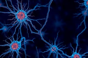
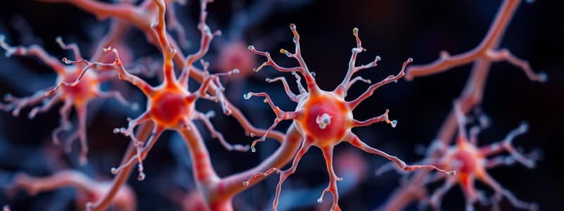
- Images depicting each type of cell and their locations.
Astrocytes
- Most numerous glial cells in the CNS
- Numerous cytoplasmic processes extend from the cell body (processes not visible with H&E but readily seen with gold staining)
- Easily characterized by their expression of the intermediate filament called Glial Fibrillary Acidic Protein (GFAP)
- Immunostaining of GFAP is visible using immunostaining (unique marker for astrocytes, helpful in diagnosis of astrocytomas)
Astrocytes
- Support neurons (regulate extracellular environment, secrete metabolic factors)
- Play a role in CNS development
- Form scar tissue following CNS damage
- Help form the blood-brain barrier
Astrocytes and the Blood-Brain Barrier
- Images depicting astrocyte interactions with capillaries to create blood-brain barrier. In these images, the role of astrocytes in forming the blood-brain barrier is visible.
Oligodendrocytes
- In H&E stained sections they may have a fried egg appearance, with dense nuclei
- They have multiple cytoplasmic extensions
Myelination of PNS and CNS Axons
- PNS: Schwann cells extend one cytoplasmic process that wraps around a single section of one axon in the peripheral nervous system
- CNS: Oligodendrocytes extend multiple cytoplasmic processes that wrap around multiple sections of axons, producing myelin sheaths within the white matter of the CNS.
Astrocytes vs. Oligodendrocytes
- Cell bodies are usually not visible for either cell type on H&E stain (look for nuclei)
- Oligodendrocytes are smaller, with condensed round nuclei
- Astrocytes are larger, with less dense oval nuclei
Microglia
- Small cells with short, irregular branched processes
- Resident macrophages of the CNS, derived from monocytes
- Dense elongated nuclei, frequently seen with H&E stain
- Other glial nuclei are typically spherical and more lightly stained
Microglia
- Microglia can become reactive and aggregate around areas of injury and cell death, causing formation of nodules.
- Microglial nuclei may take on a shape resembling rods or cigars
Cerebellum, Summary of Glia, White vs. Grey matter
- Summary of cerebellum glial cells, with examples of white and grey matter. Images show sections of cerebrum, cerebellum, and spinal cord, highlighting grey and white matter.
Cerebellar Cortex
- Describe histological sections of the cerebellum, showing layers
- Identify the main layers (molecular, Purkinje, granular)
- Characterize the cells in each layer, including Purkinje cells.
- Diagram of the cerebellar cortex, with detailed descriptions of granule and Purkinje neurons
Purkinje Cells
- Large, flask-shaped neurons with enormous dendritic trees (arborization) that extend up into the molecular layer
- Myelinated axons project through the underlying granular layer and into the white matter
- Only neurons of the cerebellar cortex that leave the CNS acting as efferent neurons
Granule Cells of the Cerebellum
- Small abundant neurons in the cerebellar cortex
- Axons project into the upper molecular layer
- Involve synapses with Purkinje cell dendrites
Cerebellar Cortex
- Images showing sections of cerebellar cortex indicating positions of the main cell types (molecular layer, Purkinje cell layer, granular layer). Specific cell types are identified in images.
Spinal Cord
- Images describing location of white matter, grey matter, spinal ganglion, and central canal. Images show the different positions of white and gray matter.
Spinal Cord Contains Large Motor Neurons
- Images showing spinal cord and its sections demonstrating large motor neurons in the grey matter. Details of large motor neuron location is highlighted.
White and Gray Matter of the Ventral Horn of the Spinal Cord
- Large somatic motor neurons are located in the ventral horn of the cord
- These neurons innervate the skeletal muscles of the limbs and trunk
- Neurons in the dorsal horn are essentially interneurons
Ventral Spinal Cord
- Images are provided indicating large motor neurons are found in the ventral spinal cord. Location of large motor neurons is highlighted.
Meninges
- Connective tissue membranes located between bone (skull and vertebral column) and CNS tissue
- Consists of three layers: dura mater, arachnoid, pia mater
- Dura mater: Dense connective tissue continuous with the periosteum
- Arachnoid: Two layers of connective tissue (flat layer adjacent to the dura and trabecular layer continuous with the underlying pia mater)
- Pia mater: Thin layer of flattened cells that covers the entire surface of CNS tissue, including blood vessels
Histological Section Through Spinal Meninges
- Images detailing the structure of the spinal meninges, showing various layers (dura, arachnoid, pia). The various layers are highlighted.
Ependymal cells
- Ependymal cells are a type of glial cell
- Line the ventricles and central canal of the spinal cord
- Help produce, circulate, and reabsorb cerebrospinal fluid (CSF)
- Cuboidal or low columnar cells with an "epithelial-like" appearance, lacking a basal lamina
Choroid Plexus
- Area within the ventricles that produces CSF through selective transport
- Consists of ependymal cells joined by tight junctions. Images show the structure of the Choroid Plexus
Ependymal cells basal projections
- Ependymal cells basal projections connect to underlying tissue. Detail images of these structures
Summary of CNS Cells
- Diagram illustrating the various cells of the CNS, including neurons, astrocytes, oligodendrocytes, microglia, and ependymal cells. The diagram shows the different cells and their locations within the CNS.
Studying That Suits You
Use AI to generate personalized quizzes and flashcards to suit your learning preferences.
Related Documents
Description
Test your knowledge on the functions and structures of glial cells in the central nervous system, as well as the distinctions between white and grey matter. This quiz covers critical concepts related to the organization and roles of various cell types in neuroanatomy. Assess your understanding of key histological features and cellular functions.