Podcast Beta
Questions and Answers
What effect does tetrodotoxin have on sensory neurons?
What characterizes the absolute refractory period?
During the relative refractory period, what must happen for another action potential to occur?
What role does myelin play in action potential propagation?
Signup and view all the answers
Why can action potentials only propagate in one direction?
Signup and view all the answers
What happens to the voltage-gated K+ channels during the relative refractory period?
Signup and view all the answers
What results from the presence of nodes of Ranvier along myelinated axons?
Signup and view all the answers
How does the refractory period influence the frequency of action potentials?
Signup and view all the answers
What is the main consequence of Na+ ions diffusing inside neighboring parts of the membrane?
Signup and view all the answers
What occurs once the cell reaches resting potential after an action potential?
Signup and view all the answers
What defines saltatory conduction in myelinated neurons?
Signup and view all the answers
Which mechanism allows the presynaptic neuron to release neurotransmitters?
Signup and view all the answers
What is the primary role of Ca2+ ions at the axon terminal?
Signup and view all the answers
What is a key characteristic of chemical synapses?
Signup and view all the answers
Which of the following best describes electrical synapses?
Signup and view all the answers
What happens when a neurotransmitter binds to ligand-gated ion channels on the postsynaptic neuron?
Signup and view all the answers
Why is the ionic concentration of calcium essential in the axon terminal?
Signup and view all the answers
What effect does myelination have on action potential propagation?
Signup and view all the answers
What characterizes grey matter in the nervous system?
Signup and view all the answers
Which of the following accurately describes a presynaptic neuron?
Signup and view all the answers
How do interneurons primarily function in the nervous system?
Signup and view all the answers
What is the resting membrane potential of most neurons typically around?
Signup and view all the answers
What causes depolarization of the membrane potential?
Signup and view all the answers
Which ion primarily contributes to hyperpolarization of the membrane potential?
Signup and view all the answers
What is the primary mechanism through which the resting membrane potential is established?
Signup and view all the answers
What distinguishes action potentials from graded potentials?
Signup and view all the answers
What role do voltage-gated K+ channels play during an action potential?
Signup and view all the answers
Which factor is responsible for maintaining the concentration gradient of Na+ and K+ ions during resting membrane potential?
Signup and view all the answers
What type of feedback mechanism is involved when Na+ channels open during an action potential?
Signup and view all the answers
What is the role of SNARE proteins in synaptic transmission?
Signup and view all the answers
What is a principal function of the Na+/K+ pump in neurons?
Signup and view all the answers
Which mechanism is NOT a way neurotransmitters are removed from the synaptic cleft?
Signup and view all the answers
How do leakage ion channels affect membrane potential over time?
Signup and view all the answers
What effect does a drug that blocks voltage-gated Ca2+ channels in presynaptic neurons have?
Signup and view all the answers
What phenomenon describes the process whereby the membrane potential returns to resting levels after an action potential?
Signup and view all the answers
What is the primary action of excitatory neurotransmitters on postsynaptic cells?
Signup and view all the answers
What type of summation involves multiple presynaptic neurons sending signals simultaneously?
Signup and view all the answers
Which neurotransmitter is commonly associated with the 'rest and digest' functions?
Signup and view all the answers
What characterizes an inhibitory postsynaptic potential (IPSP)?
Signup and view all the answers
What effect do agonists have on neurotransmitter action?
Signup and view all the answers
What triggers the conformational change in SNAREs necessary for neurotransmitter release?
Signup and view all the answers
What is the primary function of the enteric nervous system?
Signup and view all the answers
What is the function of cholinergic neurons?
Signup and view all the answers
Which class of neurotransmitters includes serotonin and dopamine?
Signup and view all the answers
What neurotransmitter is commonly released in response to stress, often associated with the fight or flight response?
Signup and view all the answers
What is the primary role of neurotransmitter reuptake in synaptic transmission?
Signup and view all the answers
Which type of summation occurs when a single presynaptic neuron fires multiple times in quick succession?
Signup and view all the answers
What effect does inhibitory neurotransmitter binding have on a cell?
Signup and view all the answers
What is the primary role of the axons of ganglion cells in vision?
Signup and view all the answers
How does binocular vision aid in perception?
Signup and view all the answers
What primarily influences the loudness of sound?
Signup and view all the answers
Which structure is responsible for amplifying vibrations in the ear?
Signup and view all the answers
What function do hair cells in the cochlea serve?
Signup and view all the answers
What happens when stereocilia bend in the opposite direction?
Signup and view all the answers
What is the role of the tectorial membrane in the auditory system?
Signup and view all the answers
Which frequency of sound produces vibrations closer to the base of the basilar membrane?
Signup and view all the answers
How does the cochlea respond to increased sound amplitude?
Signup and view all the answers
What characteristic of sound is measured in hertz (Hz)?
Signup and view all the answers
What is a consequence of the sound transmission process involving the oval window?
Signup and view all the answers
What occurs during the first step of sound transmission?
Signup and view all the answers
What type of potential change signals the conversion of sound to neural impulses in hair cells?
Signup and view all the answers
Study Notes
Synaptic Transmission
- Vesicles containing neurotransmitters (NTs) are tethered to the presynaptic neuron's membrane by proteins called SNAREs.
- In the absence of calcium, the vesicle is loosely connected.
- When calcium concentration increases, it binds to SNAREs, causing a conformational change that tightens the connection between the vesicle and the membrane.
- This fusion releases the NT into the synapse.
Synaptic Cleft
- Neurotransmitters are removed from the synapse by:
- Reuptake into the presynaptic neuron (recycling).
- Degradation by enzymes produced by the postsynaptic neuron.
- Diffusion away from the synapse.
- Glial cells often contribute to removing excess neurotransmitters.
- NT removal is essential to ensure that neurotransmitter release is transient and does not overstimulate the postsynaptic neuron.
Effects of Drugs on Synapses
- Drugs can interfere with synaptic transmission in various ways:
- Increase leakage of NT from the vesicle to the cytoplasm, leading to enzymatic breakdown and decreased communication.
- Increase transmitter release into the cleft, boosting communication.
- Block transmitter release, reducing communication.
- Inhibit transmitter synthesis, decreasing communication because NT cannot be released.
- Block transmitter reuptake, enhancing communication by prolonging NT's presence in the synapse.
- Block cleft or intracellular enzymes that metabolize transmitter, increasing communication by extending the duration of NT binding to receptors.
- Bind to receptors on the postsynaptic membrane to block (antagonist) or mimic (agonist) transmitter action. Antagonists decrease communication, while agonists increase it.
- Inactivate or block voltage-gated Ca2+ channels in the presynaptic neuron, reducing communication by preventing Ca2+ influx.
Neurotransmitter Effects on Postsynaptic Cells
-
NT can be excitatory or inhibitory.
-
Excitatory postsynaptic potential (EPSP):
- Caused by excitatory neurotransmitters like glutamate and acetylcholine.
- Makes the membrane potential more positive, bringing it closer to the threshold for an action potential.
- Opens ligand-gated Na+ channels, allowing Na+ to flow into the cell due to its concentration gradient.
- This depolarizes the cell.
-
Inhibitory postsynaptic potential (IPSP):
- Causes the membrane potential to become more negative, making it harder to elicit future action potentials.
- Caused by inhibitory neurotransmitters, such as serotonin and GABA.
- Opens ligand-gated K+ channels or ligand-gated Cl- channels.
- K+ flows out, and Cl- flows in, making the membrane potential more negative, moving it further from reaching an action potential threshold.
Synaptic Summation
-
Most neurons have multiple synapses (7000 to 10,000+).
-
Postsynaptic neurons integrate information arriving from different synapses, which is called synaptic summation.
-
A single EPSP is insufficient to reach the threshold and trigger an action potential.
-
Multiple EPSPs occurring before the neuron has time to repolarize can open more Na+ channels, leading to greater depolarization and potentially reaching the threshold for an action potential.
-
EPSPs and IPSPs can occur in the same neuron and cancel each other out.
-
Temporal Summation:
- The same presynaptic neuron sends multiple signals in quick succession (close in time).
- NTs are released multiple times from the same synapse, close in time.
-
Spatial Summation:
- Several different presynaptic neurons send signals concurrently (at the same time).
- NTs are released from multiple synapses (different locations) at the same time.
-
Scenario 4: Both temporal and spatial summation occur when presynaptic neurons A and B each send two signals close in time. This creates both temporal (close in time) and spatial (from two different neurons) summation.
-
Scenario 5: Spatial summation takes place between excitatory and inhibitory synapses. When excitatory synapse A and inhibitory synapse C fire simultaneously, their effects cancel each other out. This mechanism might be used to block pain signals reaching the brain during times of extreme stress.
-
Achieving sufficient threshold for an action potential often requires both temporal and spatial summation.
Classes of Neurotransmitters
-
Acetylcholine:
- Used in both the PNS and CNS by cholinergic neurons.
- Synthesized by combining acetyl CoA and choline.
-
Types:
- Nicotinic: Ligand-gated ion channels used by some neurons, triggering skeletal muscle contraction.
- Muscarinic: G protein-coupled receptors used by the parasympathetic nervous system, brain, and smooth and cardiac muscle. They slow heart rate by affecting the heart's pacemaker.
- Degraded by the cholinesterase enzyme. Inhibiting cholinesterase, like in the case of Sarin gas or Novichok agents, leads to prolonged acetylcholine action, causing muscle contraction, slowed heart rate, and breathing difficulties.
-
Biogenic Amines:
- Small, charged molecules synthesized from amino acids.
-
Examples:
- Dopamine: Movement, learning, attention, reward, and addiction.
- Norepinephrine: Alertness and mood.
- Serotonin: Mood, sleep, hunger, and digestion.
- Histamine: Allergic reactions, stomach acid production, and brain activity.
Structure of the Nervous System
-
Peripheral Nervous System (PNS):
-
Afferent Division:
- Somatic sensory
- Visceral sensory
- Special sensory
-
Efferent Division:
- Somatic motor
- Autonomic motor
- Regulates involuntary functions.
- Sympathetic Nervous System (SNS): "Fight or flight," NT: norepinephrine, dilates pupils, bronchioles, vessels, increases heart rate, and inhibits digestion.
- Parasympathetic Nervous System (PSNS): "Rest and digest," NT: acetylcholine, constricts pupils, bronchioles, vessels, decreases heart rate, and stimulates digestion.
-
Enteric Nervous System (ENS): Controls motility, secretion, and sensation of the GI tract.
- Contains sensory, motor, and interneurons.
- Functions largely independently of the CNS.
- Uses the same neurotransmitters as the CNS (mostly serotonin).
-
Afferent Division:
Grey Matter and White Matter
- White Matter: Myelinated axons of neurons.
- Grey Matter: Cell bodies of neurons.
Synapses
- Synapses are the sites of communication between neurons.
- Presynaptic Neuron: Releases neurotransmitters (sending the signal).
- Postsynaptic Neuron: Responds to neurotransmitters (receiving the signal).
Functional Classes of Neurons
- Afferent Neurons: Transmit information from the PNS to the CNS.
- Interneurons: Transmit information within the CNS.
- Efferent Neurons: Transmit information from the CNS to the PNS (motor neurons).
- Mnemonic: SAME (sensory afferent motor efferent).
Membrane Potential
- The difference in electrical charge across a cell membrane, particularly in neurons.
- Caused by the difference in charged ion concentration inside and outside the cell.
-
Inside: More negative relative to the outside.
- Contains large negatively charged nonpenetrating ions (DNA, RNA, PO43-).
- Fewer positive ions compared to the outside.
-
Types:
- Resting potential
- Graded potential
- Action potential
Resting Membrane Potential
- At rest, the cell is negatively charged inside compared to the outside (extracellular fluid).
- The membrane is considered polarized.
- For most neurons, the resting potential is around -70mV.
-
Contributing Factors:
- Many large negatively charged molecules inside.
- Na+/K+ pump.
- Ungated (leak) K+ channels.
- Opening of gated channels causes changes in membrane potential.
Establishing Resting Membrane Potential
-
Na+/K+ Pump:
- Actively pumps 3 Na+ out of the cell and 2 K+ inside.
- This process moves more positive ions out than in, making the inside slightly more negative.
-
Leak Channels:
- Neurons have more K+ leak channels than Na+ leak channels.
- This allows more K+ to leave the cell than Na+ entering, leading to a net loss of positive ions.
Changes in Membrane Potential
- Caused by opening and closing of gated ion channels.
- Depolarization: Potential becomes less negative or more positive (e.g., opening of gated Na+ channels).
- Hyperpolarization: Potential becomes more negative (e.g., opening of gated K+ channels).
Graded Potential
- A transient change in membrane potential, confined to a small region of the plasma membrane.
- Usually occurs on dendrites.
- Involves opening of only a few ion channels.
- Typically involves ligand-gated or mechanically-gated ion channels.
- Can be either depolarizing or hyperpolarizing.
- Dissipates quickly due to leak channels and the Na+/K+ pump.
Action Potential
- A large, "all-or-none" change in membrane potential.
- Fast (milliseconds) and can be repeated.
- Triggered by depolarization to a threshold (typically -55mV), often caused by a graded potential.
- Involves opening of numerous voltage-gated channels:
- Voltage-gated Na+ Channels: Cause depolarization.
- Voltage-gated K+ Channels: Cause repolarization and hyperpolarization.
- Other channels, like voltage-gated Ca2+ channels, are essential for triggering neurotransmitter release.
Voltage-gated Channels and Action Potentials
-
Na+ Channels: Have three states:
- Closed: Before stimulation.
- Open: In response to depolarization to threshold.
- Inactivated: Once the membrane potential becomes positive; lasts until the membrane potential returns to resting, at which point the channel is closed but capable of opening again.
-
K+ Channels: Have two states:
- Closed: Before stimulation.
- Open: In response to depolarization to threshold.
- Close in response to the membrane potential returning to resting.
- Key difference from Na+ channels: K+ channels open and close more slowly.
Action Potential Summary
- Resting Potential: All gated channels are closed.
- Ligand-gated Na+ Channels Open: Na+ flows in; if enough Na+ enters, the cell may depolarize to threshold (-55mV).
- Voltage-gated Na+ Channels Open: Na+ floods into the cell, further depolarizing the cell, positive feedback effect, opens more Na+ channels, rapid rise in membrane potential to a positive value.
- Voltage-gated Na+ Channels Inactivate: Stops depolarization, no more Na+ can enter.
- Voltage-gated K+ Channels Open: K+ flows out of the cell, repolarizing the membrane back to resting potential.
- Voltage-gated K+ Channels Close Slowly: K+ continues to flow out, causing hyperpolarization.
- Return to Resting Potential: Na+/K+ pump restores the original ion concentration gradients.
Control Mechanisms
- Na+ Channel Opening: Positive feedback, increases depolarization..
- K+ Channel Opening: Negative feedback, causing repolarization and hyperpolarization.
Threshold Stimuli and Action Potentials
- Action potentials only occur when the stimulus is strong enough to trigger a depolarization to the threshold potential (typically -55mV).
- Stimuli below the threshold will not cause an action potential.
- The strength of the stimulus does not impact the size of the action potential, it's all or none, but it can impact the frequency of action potentials.
Tetrodotoxin
- Tetrodotoxin blocks voltage-gated Na+ channels
- It is produced by pufferfish
Refractory Period
- It is the time after an action potential in which it is impossible (absolute) or difficult (relative) to trigger another action potential.
-
Absolute refractory period:
- No stimulus can cause another action potential.
- All voltage-gated Na+ channels are open or inactivated.
-
Relative refractory period:
- Another action potential can be produced only if the stimulus is much stronger than usual.
- The cell is hyperpolarized because some K+ is still flowing out.
- Graded potential must be bigger to depolarize the cell to threshold.
- At relative refractory period, voltage-gated K+ channels are open (voltage-gated Na+ channels have reset from inactivated to closed).
- The refractory period limits how many action potentials a cell can do in a given time.
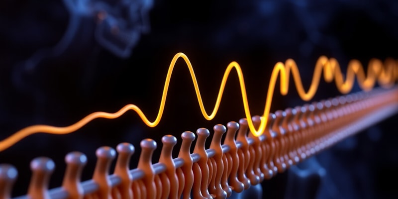
Action Potential Propagation
- Action potentials are unidirectional.
- They propagate from the cell body to the axon terminals.
- Na+ ions diffuse inside the neuron, depolarizing neighboring regions of membrane.
- The depolarization triggers an action potential in the neighboring area ahead of the action potential.
- The area behind the action potential is in the refractory period and cannot cause another action potential.
Action Potential Propagation & Myelin
- Myelin covers the membrane and prevents ions from entering or leaving the axon.
- Ions can only enter or leave at the nodes of Ranvier, which are unmyelinated gaps of exposed membrane along the axon.
- Myelin speeds up the propagation of the action potential along the axon because:
- There is less membrane to depolarize.
- Fewer places for ions to leak out and weaken the current.
- Fewer Na+/K+ pumps are needed to restore resting potential.
- This mechanism is called saltatory conduction: conduction through myelinated nerve fiber.
- Action potentials are faster through an axon with fewer nodes of Ranvier.
What Happens When an Action Potential Reaches the Axon Terminal?
-
Chemical synapses:
- The presynaptic neuron releases neurotransmitters from the axon terminal exocytosis.
- Neurotransmitters bind to ligand-gated ion channels on the postsynaptic neuron's membrane.
- This opens ion channels and leads to a graded potential (that may or may not lead to an action potential).
-
Electrical synapses:
- Membranes are connected by gap junctions, and ions flow directly between cells.
- Transmission is faster than in chemical synapses.
- These are used by some neurons and cardiac muscle.
Neurotransmitter Release
- An action potential arriving at the axon terminal depolarizes the membrane.
- This depolarization opens voltage-gated Ca2+ channels.
- Ca2+ moves from high to low concentration into the cell.
- Ca2+ triggers exocytosis of vesicles containing neurotransmitters.
- Neurotransmitters diffuse across the synapse and bind to ligand-gated ion channels on the postsynaptic neuron.
- Neurotransmitters are removed from the synapse.
Binocular Vision
- Axons of ganglion cells form the optic nerve.
- The two optic nerves meet at the optic chiasm in the brain, where some fibers cross.
- Some visual information from the left eye goes to the right side of the visual cortex and vice versa.
- The afferent pathway ends at the visual cortex.
- Binocular vision enables depth perception (the ability to gauge distances of objects from you).
Sound
- Sound is caused by energy transferred through a medium (gas, liquid, or solid), causing the molecules to vibrate.
- Sound cannot occur in a vacuum (there is no medium).
Sound Waves
-
Loudness: Determined by the wave's amplitude (height of the wave).
- Measured in decibels (log scale).
-
Pitch: Determined by the wave's frequency (width of the wave or waves/sec).
- Measured in hertz (Hz; cycles/sec).
- Deeper pitch has a wider width.
Sound Transmission
-
Step 1:
- Sound is collected and funneled into the ear canal by the pinna (external ear).
- Sound enters the external auditory canal.
- This causes the tympanic membrane (eardrum) to vibrate.
-
Step 2:
- The tympanic membrane is connected to the bones of the middle ear (ossicles).
- Bones amplify vibrations, causing the fluid in the cochlea to move.
- The vibration of the tympanic membrane causes the ossicles to vibrate.
- The ossicles are attached to the oval window, a membrane at the entrance to the cochlea.
- The basilar membrane contains sensory receptors.
- The cochlea is filled with fluid, which is compressed and vibrates when the stapes pushes against it, causing the membrane to move.
- The cochlea is a spiral-shaped, fluid-filled space within the temporal bone containing several membranes that divide it lengthwise into separate sections.
-
Step 3:
- Vibrations of the oval window cause fluid in the cochlea to vibrate.
- The basilar membrane vibrates, causing hair cells to move.
- This change in graded potential causes neurotransmitters to be released from sensory cells to afferent neurons.
- Hair cells have mechanically-gated ion channels (stimulus: mechanical movement of sensory cells against membranes).
Sensory Receptors of the Inner Ear: Hair Cells
- Located within the Organ of Corti, which sits on the basilar membrane.
- Hair cells have movable "hair" at one end called stereocilia (attached to the tectorial membrane).
- They are embedded in an overlaying membrane called the tectorial membrane.
- The basilar membrane vibrates in response to sound, and the tectorial membrane stays still. This causes hair cells to move against the tectorial membrane.
Auditory Signal Transduction
-
Step 4:
- The basilar membrane moves in response to sound.
- This pulls stereocilia against the tectorial membrane and causes them to bend.
- The stereocilia bend in a particular direction, and the proteins connecting each individual stereocilia open mechanically-gated ion channels, depolarizing the cell.
- This causes a graded potential, which releases neurotransmitters onto the afferent neurons, leading to action potentials in afferent neurons.
- Stereocilia always bend in the longer direction.
Audio Video Summary
- Sound enters the ear through the external auditory canal.
- The tympanic membrane vibrates in response to sound.
- Lower pitch sound = slower rate of vibration.
- Lower volume = less dramatic vibration.
- Higher frequency sound = faster vibrations.
- Tympanic membrane vibrations are transferred to the auditory ossicles (3 bones).
- Vibrations are then transferred to the footplate of the stapes.
- The stapes move with a piston-like action, sending vibrations to the bony labyrinth.
- Labyrinth is filled with perilymph (fluid).
- Vibrations produced by the stapes are drawn into the cochlea and return to meet the round window.
- The cochlear duct is filled with endolymph.
- Reissner's membrane and the basilar membrane move in response to vibrations traveling up the scala vestibuli.
- The movement of these membranes sends vibrations back down to the scala tympani.
- Hair cells in the Organ of Corti on the basilar membrane send nerve impulses to the brain via the cochlear nerve.
- Hair cells are covered by the tectorial membrane.
- As the basilar membrane vibrates, hair cells are bent against the tectorial membrane, triggering them to fire.
- Specific areas along the basilar membrane move variably in response to different frequencies of sounds.
- Lower frequency: vibrations closer to the apex.
- Higher frequency: vibrations closer to the base.
Auditory Signal Transduction: Distinguishing Amplitude
- Louder sounds cause stronger vibrations.
- Hair cells have larger or longer graded potentials.
- They release more neurotransmitters onto the afferent neuron.
- The frequency of action potentials in afferent neurons tells the brain whether the stimulus is mild or strong.
Auditory Signal Transduction: Distinguishing Pitch
- The basilar membrane is narrower and stiffer near the base.
- Higher frequency sounds cause smaller movements near the base.
- Lower frequency sounds travel to the wider and less stiff apex.
- The brain interprets the location of the activated neurons to determine pitch.
Studying That Suits You
Use AI to generate personalized quizzes and flashcards to suit your learning preferences.
Related Documents
Description
This quiz covers key concepts of neuroscience related to action potentials and synaptic transmission. Topics include the effects of tetrodotoxin, the role of myelin, refractory periods, and mechanisms of neurotransmitter release. Test your understanding of neuronal communication and physiology.