Podcast
Questions and Answers
Which type of cell is responsible for sending information away from the neuron?
Which type of cell is responsible for sending information away from the neuron?
What is the primary function of motor neurons?
What is the primary function of motor neurons?
Which structure is considered the ‘trigger zone’ for action potentials?
Which structure is considered the ‘trigger zone’ for action potentials?
Which type of supporting cell is mainly found in the central nervous system?
Which type of supporting cell is mainly found in the central nervous system?
Signup and view all the answers
What characteristic distinguishes oligodendrocytes from Schwann cells?
What characteristic distinguishes oligodendrocytes from Schwann cells?
Signup and view all the answers
Which of the following features is NOT a characteristic of neurons?
Which of the following features is NOT a characteristic of neurons?
Signup and view all the answers
What role do dendrites play in neuronal function?
What role do dendrites play in neuronal function?
Signup and view all the answers
Which cytoskeletal protein is specifically found in dendrites?
Which cytoskeletal protein is specifically found in dendrites?
Signup and view all the answers
What is the primary function of Nissl substance in neurons?
What is the primary function of Nissl substance in neurons?
Signup and view all the answers
Where are the cell bodies of multipolar motor neurons located?
Where are the cell bodies of multipolar motor neurons located?
Signup and view all the answers
Which staining method is used for visualizing neurons in nervous tissue?
Which staining method is used for visualizing neurons in nervous tissue?
Signup and view all the answers
What surrounds individual ganglion cells in dorsal root ganglia?
What surrounds individual ganglion cells in dorsal root ganglia?
Signup and view all the answers
Interneurons play a crucial role in which of the following processes?
Interneurons play a crucial role in which of the following processes?
Signup and view all the answers
What characteristic distinguishes the six layers of the cerebral cortex?
What characteristic distinguishes the six layers of the cerebral cortex?
Signup and view all the answers
Which type of neuron is primarily responsible for carrying information to the central nervous system?
Which type of neuron is primarily responsible for carrying information to the central nervous system?
Signup and view all the answers
What is the primary purpose of a connective tissue capsule surrounding dorsal root ganglia?
What is the primary purpose of a connective tissue capsule surrounding dorsal root ganglia?
Signup and view all the answers
What is the primary function of oligodendrocytes in the central nervous system (CNS)?
What is the primary function of oligodendrocytes in the central nervous system (CNS)?
Signup and view all the answers
Which type of neuron is considered the most abundant in the brain?
Which type of neuron is considered the most abundant in the brain?
Signup and view all the answers
What role do astrocytes play in the brain?
What role do astrocytes play in the brain?
Signup and view all the answers
Which statement is true regarding the blood-brain barrier?
Which statement is true regarding the blood-brain barrier?
Signup and view all the answers
What is the structure specialized for receiving input in Purkinje neurons?
What is the structure specialized for receiving input in Purkinje neurons?
Signup and view all the answers
What distinguishes Schwann cells from oligodendrocytes?
What distinguishes Schwann cells from oligodendrocytes?
Signup and view all the answers
Which of the following cell types serves an immune function in the CNS?
Which of the following cell types serves an immune function in the CNS?
Signup and view all the answers
Which layer of the cerebellar cortex contains Purkinje cells?
Which layer of the cerebellar cortex contains Purkinje cells?
Signup and view all the answers
How is the myelin in the CNS formed compared to that in the PNS?
How is the myelin in the CNS formed compared to that in the PNS?
Signup and view all the answers
What is one key component of myelin?
What is one key component of myelin?
Signup and view all the answers
Study Notes
Neurohistology - BIOL 2051/2052
- Course taught by Dr. Melissa Andrews
- Lecture date: October 15, 2024
Lecture Objectives
- Identify and describe different cell types in the central and peripheral nervous systems (neurons, glia, and other support cells)
- Describe the histology and anatomy of:
- Cytoarchitecture of cerebral cortex, cerebellar cortex, and ganglia
- Supporting cell types (astrocytes, microglia)
- Distinguish myelination differences (oligodendrocytes and Schwann cells) in the central and peripheral nervous systems
- Recommended reading: Chapter 1 (Studying the Nervous System in Humans and Other Animals) in Neuroscience (3rd ed by Purves et al)
Nervous Tissue and Neurons
- Ectodermally derived
- One of four basic tissue types; consists of two principal cell types: neurons and supporting cells (neuroglia)
- Neurons:
- Excitable cells with long cytoplasmic extensions for stimulus reception and conduction of nerve impulses (action potentials)
- Do not undergo cell division and replication
- Supporting cells (neuroglia):
- Function in metabolism and support of neurons
- Different types in the central nervous system (CNS) and peripheral nervous system (PNS)
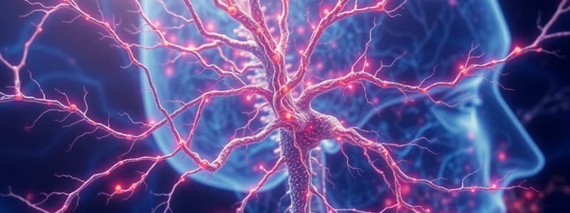
Neuronal Structure
- Dendrites receive information from adjacent axons
- Axons send information from one end of a neuron to another (faster if myelinated)
- Signals from the cell soma are summated at the axon hillock; the hillock is the trigger zone that must reach a threshold potential to achieve an action potential
- Both the axon hillock and dendrites participate in this process
Axon Hillock and Axon Initial Segment
- MAP2 (neuron-specific cytoskeletal protein) and BIV spectrin are proteins found in dendrites (specifically, microtubule-associated protein in dendrites)
Neuronal Subtypes
- 3 types of neurons:
- Motor neurons relay commands from the brain and spinal cord to muscles and glands
- Sensory neurons are excited by specific stimuli
- Interneurons integrate information from sensory to motor neurons
- Cell soma diameter ranges vary
Multipolar Neurons (Golgi Stain)
- Golgi stain is an indicator of nervous tissue treated with Potassium dichromate and Silver nitrate. The silver precipitation appears inside the neurons.
Neurons in the Spinal Cord
- Cell bodies of multipolar motor neurons are large and located in the ventral horn of the spinal cord
- They have a large, pale-staining nucleus
- These cells produce Nissl substance, which stains rough endoplasmic reticulum and polyribosomes. This substance is important in protein synthesis in neurons and largely absent in the axon.
Spinal Ganglia
- Aggregations of nerve cells (ganglion cells) outside the CNS
- Dorsal root ganglia are surrounded by a connective tissue capsule that connects to the peripheral nerve
- Individual ganglion cells are surrounded by flattened satellite (fibroblast) cells
Cerebal Cortex (Neocortex)
- The cerebral cortex is divided into six layers, each housing neurons with morphology that is characteristic of that layer
- Deep to the grey matter of the cortex is the white matter, composed of myelinated fibres
- Superficially, the meninges can be observed
- Note: This structure can also be observed in columns, shown by distinct neuron arrangements.*
Cerebellar Cortex
- Cytoarchitecture of the cerebellar cortex: Three layers, with three layers of cells in the grey matter (molecular layer, Purkinje cell layer, and granular layer).
- Molecular layer: basket cells and stellate cells
- Purkinje cell layer: Purkinje cells
- Granular layer: Granule cells (neurons most abundant in the brain)
- Three layers: outer molecular layer, single layer of Purkinje cells, and granular layer.
Purkinje Neurons (Cerebellum)
- Purkinje neurons are the largest cell in the cerebellum
- They have pear-shaped cell bodies and a distinctive dendritic tree in the molecular layer
Supporting Cells (Neuroglia) - Neuroglia
- Neuroglial cells function in the metabolism and support of neurons
- In the CNS, there are astrocytes, oligodendroglia, ependymal cells, and microglia
- In the PNS, there are Schwann cells and satellite cells
- Oligodendroglia and Schwann cells form myelin
Astrocytes
- Provide structural and metabolic support for neurons
- Types:
- Fibrous (white matter)
- Protoplasmic (gray matter)
- Müller glia (retina)
Blood-Brain Barrier
- Form glial-limiting membrane around blood vessels and along CNS surface (part of the blood-brain barrier)
- Barrier composed of endothelial cells joined by tight junctions
- Prevents diffusion of solutes and fluid into the brain and spinal cord (e.g., O2, CO2, lipid-soluble molecules, hormones)
- Molecules >500 daltons are not permissible
Microglia
- Serve an immune function within the CNS, able to phagocytose cell debris in response to injury
- Normally exist as resident microglia but become 'activated' upon CNS damage, moving towards sites of injury
- Release cytokines which can both help and hinder recovery
Oligodendrocytes
- Form myelin sheath around CNS axons; one oligodendrocyte can myelinate several axons
- Diseases that affect oligodendrocytes include multiple sclerosis and leukodystrophies
Schwann Cells
- Form myelin sheath around PNS axons; one Schwann cell can myelinate one axon
- Plays key role in the organization of connective tissue sheaths surrounding peripheral nerves during development
Myelination
- Myelin consists of ~80% lipid; provides insulation and enhances conduction velocity for action potentials
- Formed by oligodendrocytes and Schwann cells
Unmyelinated Axons
- Schwann cells envelope some unmyelinated axons
- Unmyelinated axons are not associated with glial cells; they have continuous conduction (as opposed to the “saltatory conduction” of myelinated neurons)
- Unmyelinated axons are often sensory fibers
Studying That Suits You
Use AI to generate personalized quizzes and flashcards to suit your learning preferences.
Related Documents
Description
Test your knowledge on the structure and function of nervous tissue and cell types within the central and peripheral nervous systems. This quiz will cover the histology and anatomy of various neuronal support cells and the key differences in myelination. Dive into Chapter 1 of Neuroscience to enhance your understanding.


