Podcast
Questions and Answers
Where can we find white matter in the central nervous system?
Where can we find white matter in the central nervous system?
- Cerebral cortex
- Grey horns of spinal cord
- Bundles of axons forming white matter pathways (correct)
- Cerebral nuclei
Which of the following best describes the cerebrum?
Which of the following best describes the cerebrum?
- It is responsible for receiving auditory impulses.
- It is located in the posterior cranial fossa.
- It is a single, undivided structure.
- It consists of an outer cerebral cortex and internal cerebral white matter. (correct)
What is the main function of the frontal lobe of the cerebral cortex?
What is the main function of the frontal lobe of the cerebral cortex?
- To receive taste impulses
- To receive visual impulses
- To receive general sensory stimuli
- To initiate motor impulses (correct)
Which of the following is not one of the four main lobes of the cerebral hemisphere?
Which of the following is not one of the four main lobes of the cerebral hemisphere?
What is the function of the parietal lobe of the cerebral cortex?
What is the function of the parietal lobe of the cerebral cortex?
What separates the two cerebral hemispheres?
What separates the two cerebral hemispheres?
Which area of the precentral gyrus controls the muscles for fine movements?
Which area of the precentral gyrus controls the muscles for fine movements?
What is the consequence of an injury to the precentral gyrus?
What is the consequence of an injury to the precentral gyrus?
Which part of the precentral gyrus is supplied by branches of the anterior cerebral artery?
Which part of the precentral gyrus is supplied by branches of the anterior cerebral artery?
What is the function of the homunculus in relation to the precentral gyrus?
What is the function of the homunculus in relation to the precentral gyrus?
Which of the following statements about the precentral gyrus is true?
Which of the following statements about the precentral gyrus is true?
Where is the Periaqueductal Gray Matter (PAG) located?
Where is the Periaqueductal Gray Matter (PAG) located?
Which cranial nerves emerge from the ponto-medullary sulcus in the pons?
Which cranial nerves emerge from the ponto-medullary sulcus in the pons?
What is one function of the pontine nuclei in the pons?
What is one function of the pontine nuclei in the pons?
Which area found within the pons adds more control to respiration?
Which area found within the pons adds more control to respiration?
What occupies the basilar sulcus on the anterior surface of the pons?
What occupies the basilar sulcus on the anterior surface of the pons?
How is the gray matter organized in the pons?
How is the gray matter organized in the pons?
What are the major functional areas in the medulla responsible for?
What are the major functional areas in the medulla responsible for?
Which of the following is NOT a function of the reticular formation?
Which of the following is NOT a function of the reticular formation?
What is the role of the reticular formation in sensory signals processing?
What is the role of the reticular formation in sensory signals processing?
Which system responds to stimuli, potentially disrupting the constant flow of signals?
Which system responds to stimuli, potentially disrupting the constant flow of signals?
What is the major role of the Reticular Activating System (RAS)?
What is the major role of the Reticular Activating System (RAS)?
What is an essential characteristic of the reticular formation's structure?
What is an essential characteristic of the reticular formation's structure?
What is the name given to the space between the dura mater and the arachnoid mater?
What is the name given to the space between the dura mater and the arachnoid mater?
What is the primary function of the pia mater?
What is the primary function of the pia mater?
Which of the following best describes the arachnoid mater?
Which of the following best describes the arachnoid mater?
What is the purpose of the denticulate ligaments within the spinal cord meninges?
What is the purpose of the denticulate ligaments within the spinal cord meninges?
What is the primary function of the epidural space surrounding the spinal cord?
What is the primary function of the epidural space surrounding the spinal cord?
What is the primary function of the somatosensory association area located in the parietal lobe?
What is the primary function of the somatosensory association area located in the parietal lobe?
What is the consequence of a lesion in the somatosensory association area?
What is the consequence of a lesion in the somatosensory association area?
What is the primary function of the visual association area located in the occipital lobe?
What is the primary function of the visual association area located in the occipital lobe?
What is the primary function of the auditory association area located on the lateral surface of the superior temporal gyrus?
What is the primary function of the auditory association area located on the lateral surface of the superior temporal gyrus?
What is the primary function of the common integrative area located among the sensory, visual, and auditory association areas?
What is the primary function of the common integrative area located among the sensory, visual, and auditory association areas?
How does the text describe the function of sharing different signals by an entire cerebral hemisphere?
How does the text describe the function of sharing different signals by an entire cerebral hemisphere?
What is the function of the arachnoid villus or arachnoid granulations?
What is the function of the arachnoid villus or arachnoid granulations?
What is the primary function of the dural folds or reflections?
What is the primary function of the dural folds or reflections?
Which layer of the meninges is described as having a 'web-like' appearance?
Which layer of the meninges is described as having a 'web-like' appearance?
What is the primary function of the pia mater?
What is the primary function of the pia mater?
What is the purpose of the epidural space surrounding the spinal cord?
What is the purpose of the epidural space surrounding the spinal cord?
What is the primary function of the basal ganglia nuclei, such as the caudate and lenticular nuclei?
What is the primary function of the basal ganglia nuclei, such as the caudate and lenticular nuclei?
What type of nerve fibers connect different gyri of one cerebral hemisphere to the corresponding gyri of the other hemisphere?
What type of nerve fibers connect different gyri of one cerebral hemisphere to the corresponding gyri of the other hemisphere?
Which of the following is an example of projectional fibers in the brain?
Which of the following is an example of projectional fibers in the brain?
Which of the following statements about the functional areas of the cerebral cortex is correct?
Which of the following statements about the functional areas of the cerebral cortex is correct?
What is the primary function of the association areas in the cerebral cortex?
What is the primary function of the association areas in the cerebral cortex?
What is the general idea about the primary sensory areas?
What is the general idea about the primary sensory areas?
Which part of the precentral gyrus is responsible for producing motor signals for the muscles of the leg and foot?
Which part of the precentral gyrus is responsible for producing motor signals for the muscles of the leg and foot?
What is the primary consequence of an injury to the precentral gyrus?
What is the primary consequence of an injury to the precentral gyrus?
Which area of the precentral gyrus is responsible for producing motor signals for the muscles of the hands, facial expression, and vocal apparatus?
Which area of the precentral gyrus is responsible for producing motor signals for the muscles of the hands, facial expression, and vocal apparatus?
What is the significance of the homunculus in relation to the precentral gyrus?
What is the significance of the homunculus in relation to the precentral gyrus?
How does the precentral gyrus extend itself across the cerebral surfaces?
How does the precentral gyrus extend itself across the cerebral surfaces?
What is the primary role of the reticular formation in relation to sensory stimuli?
What is the primary role of the reticular formation in relation to sensory stimuli?
Which of the following best describes the role of the reticular formation in regulating the sleep-wake cycle?
Which of the following best describes the role of the reticular formation in regulating the sleep-wake cycle?
What is the primary function of the reticulospinal pathway originating from the reticular formation?
What is the primary function of the reticulospinal pathway originating from the reticular formation?
How do general anesthetics exert their effects on the reticular formation?
How do general anesthetics exert their effects on the reticular formation?
Which of the following statements about the connections of the reticular formation is true?
Which of the following statements about the connections of the reticular formation is true?
Which of the following best describes the functional organization of the reticular formation?
Which of the following best describes the functional organization of the reticular formation?
What is the main function of the midbrain in the central nervous system?
What is the main function of the midbrain in the central nervous system?
Which of the following structures is located on the posterior aspect of the midbrain?
Which of the following structures is located on the posterior aspect of the midbrain?
Which of the following tracts is contained within the white matter of the midbrain, behind the crus cerebri?
Which of the following tracts is contained within the white matter of the midbrain, behind the crus cerebri?
What is the function of the substantia nigra within the midbrain?
What is the function of the substantia nigra within the midbrain?
Which of the following structures is described as connecting the midbrain to the pons inferiorly and the diencephalon superiorly?
Which of the following structures is described as connecting the midbrain to the pons inferiorly and the diencephalon superiorly?
What is the function of the nuclei deep within the superior and inferior colliculi of the midbrain?
What is the function of the nuclei deep within the superior and inferior colliculi of the midbrain?
Which layer of the meninges is described as the 'tough mother' due to its thickness?
Which layer of the meninges is described as the 'tough mother' due to its thickness?
Where in the spinal cord is the subdural space mainly found?
Where in the spinal cord is the subdural space mainly found?
What is the function of the epidural space surrounding the spinal cord?
What is the function of the epidural space surrounding the spinal cord?
What are denticulate ligaments thickening of, suspending the spinal cord in its dural sheath?
What are denticulate ligaments thickening of, suspending the spinal cord in its dural sheath?
Which layer of the meninges is tightly adhered to the neural tissue itself?
Which layer of the meninges is tightly adhered to the neural tissue itself?
Which of the following is NOT a basic function of the nervous system?
Which of the following is NOT a basic function of the nervous system?
What is the primary distinction between the central nervous system (CNS) and the peripheral nervous system (PNS)?
What is the primary distinction between the central nervous system (CNS) and the peripheral nervous system (PNS)?
What is the primary function of the afferent (sensory) division of the peripheral nervous system?
What is the primary function of the afferent (sensory) division of the peripheral nervous system?
Which of the following statements about the efferent (motor) division of the peripheral nervous system is correct?
Which of the following statements about the efferent (motor) division of the peripheral nervous system is correct?
Which of the following structures is NOT a part of the central nervous system?
Which of the following structures is NOT a part of the central nervous system?
What is the primary function of the endocrine system in relation to the nervous system?
What is the primary function of the endocrine system in relation to the nervous system?
Which of the following statements accurately describes the primary somatosensory area (cortex)?
Which of the following statements accurately describes the primary somatosensory area (cortex)?
What is the primary function of the primary motor area (cortex)?
What is the primary function of the primary motor area (cortex)?
Which of the following best describes the organization of the primary sensory areas in the cerebral cortex?
Which of the following best describes the organization of the primary sensory areas in the cerebral cortex?
What is the primary function of the association areas in the cerebral cortex?
What is the primary function of the association areas in the cerebral cortex?
Which of the following statements accurately describes the organization of the motor cortex?
Which of the following statements accurately describes the organization of the motor cortex?
Which of the following best describes the functional organization of the primary sensory areas in the cerebral cortex?
Which of the following best describes the functional organization of the primary sensory areas in the cerebral cortex?
What is the primary function of the inferior olivary nucleus in the medulla oblongata?
What is the primary function of the inferior olivary nucleus in the medulla oblongata?
What is the primary function of the nucleus gracilis and nucleus cuneatus in the medulla oblongata?
What is the primary function of the nucleus gracilis and nucleus cuneatus in the medulla oblongata?
What is the primary function of the decussation of pyramids at the junction of the medulla oblongata and spinal cord?
What is the primary function of the decussation of pyramids at the junction of the medulla oblongata and spinal cord?
Which of the following cranial nerves does NOT emerge from the anterolateral surface of the medulla oblongata?
Which of the following cranial nerves does NOT emerge from the anterolateral surface of the medulla oblongata?
What is the primary function of the pyramid, an elevation on the anterior surface of the medulla oblongata?
What is the primary function of the pyramid, an elevation on the anterior surface of the medulla oblongata?
What is the primary function of the olive, an elevation lateral to the pyramid on the anterior surface of the medulla oblongata?
What is the primary function of the olive, an elevation lateral to the pyramid on the anterior surface of the medulla oblongata?
Study Notes
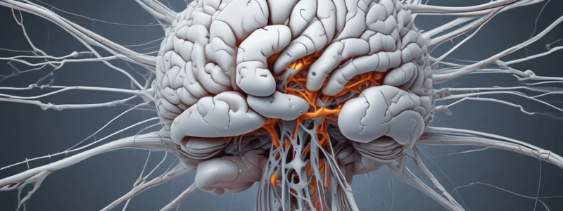
Central Nervous System (CNS)
- The CNS consists of the brain and spinal cord, located in bony cavities.
- The brain is divided into two subdivisions: cerebrum, cerebellum, brainstem, and diencephalon.
- The brainstem connects the cerebrum to the spinal cord and regulates automatic behaviors required for survival.
Brainstem
- Acts as a passageway for ascending and descending tracts between the cerebrum and spinal cord.
- Contains the nuclei of cranial nerves III to XII.
- Regulates automatic behaviors required for survival, such as respiration.
Midbrain
- Connects the pons inferiorly and the diencephalon superiorly.
- Anterior aspect: presents two columns, the cerebral peduncles, between which the CN III emerges.
- Posterior aspect: four elevations, superior and inferior colliculi, which are relay centers for visual and auditory reflexes.
Pons
- Connects the medulla oblongata inferiorly and the midbrain superiorly.
- Anterior surface: convex with a longitudinal sulcus, the basilar sulcus, occupied by the basilar artery.
- Cranial nerves VI, VII, and VIII emerge from the ponto-medullary sulcus.
- White matter: formed by ascending and descending tracts.
Medulla Oblongata
- Pyramidal in shape, separated from the pons by a transverse sulcus.
- Composed of gray and white matter, with arrangement similar to the spinal cord in the inferior half.
- Nuclei associated with cranial nerves V, VIII, IX, X, XI, and XII.
- Functional areas: cardiovascular center, respiratory center, and centers controlling vomiting, coughing, and sneezing.
Reticular Formation
- A collection of nuclei running vertically throughout the brainstem.
- Functions: control of skeletal muscle, pain modulation, control of autonomic and endocrine systems, circadian rhythms, and consciousness.
- Receives an enormous number of sensory signals from different parts of the body, filtering out up to 99% of them.
Cerebrum
- Consists of an outer cerebral cortex, an internal region of cerebral white matter, and deep nuclei.
- Divided into four main lobes: frontal, parietal, occipital, and temporal, each responsible for different aspects of brain functions.
- Outer surface: convoluted structure with folds (gyri) and furrows (sulci).
Meninges
- Protective layers of the CNS: dura mater, arachnoid mater, and pia mater.
- Dura mater: tough outer layer, forms folds (dural folds) for venous sinuses.
- Arachnoid mater: web-like middle layer, with arachnoid villi and granulations.
- Pia mater: delicate/thin inner layer, tightly adhered to neural tissue.
Ventricular System
- Produces and circulates cerebrospinal fluid (CSF) throughout the CNS.
- CSF: clear, colorless fluid that cushions and protects the CNS.
Studying That Suits You
Use AI to generate personalized quizzes and flashcards to suit your learning preferences.
Related Documents
Description
Test your knowledge on the white matter and grey matter structures in the central nervous system. Identify where white matter is located, understand the functions of different areas like the cerebral tracts, and locate the cortex in a given image.


