Podcast
Questions and Answers
During which week of gestation does the formation of the neural tube begin?
During which week of gestation does the formation of the neural tube begin?
- 3-4 weeks (correct)
- 7-8 weeks
- 5-6 weeks
- 2-3 weeks
What is the contribution of the endoderm germ layer to the development of the embryo?
What is the contribution of the endoderm germ layer to the development of the embryo?
- Musculoskeletal tube
- Neural tube
- Cardiovascular tube
- Gut tube (correct)
Which of the following is NOT a risk factor for neural tube defects?
Which of the following is NOT a risk factor for neural tube defects?
- Maternal use of valproic acid
- Maternal diabetes
- Maternal hypertension (correct)
- Maternal folate deficiency
What is the result of the neural tube closing in the cervical region?
What is the result of the neural tube closing in the cervical region?
What percentage decrease in neural tube defects has been observed since the US mandated folate fortification in many foods in 1996?
What percentage decrease in neural tube defects has been observed since the US mandated folate fortification in many foods in 1996?
What is the primary contributor to the development of the neural tube?
What is the primary contributor to the development of the neural tube?
What is the significance of the prechordal plate in neural tube development?
What is the significance of the prechordal plate in neural tube development?
Which of the following stages of nervous system development occurs between 2-3 months of gestation?
Which of the following stages of nervous system development occurs between 2-3 months of gestation?
What percentage of infants with myelomeningocele develop hydrocephalus?
What percentage of infants with myelomeningocele develop hydrocephalus?
What percentage of cases of Spina Bifida Occulta occur in infants?
What percentage of cases of Spina Bifida Occulta occur in infants?
What is the primary diagnostic method for detecting anencephaly?
What is the primary diagnostic method for detecting anencephaly?
What is the primary characteristic of Type II Arnold Chiari malformation?
What is the primary characteristic of Type II Arnold Chiari malformation?
What percentage of NTD cases occur in pregnancies without any risk factors?
What percentage of NTD cases occur in pregnancies without any risk factors?
What is the ideal gestation period for testing maternal serum alpha fetoprotein levels?
What is the ideal gestation period for testing maternal serum alpha fetoprotein levels?
What is the detection rate of spina bifida using fetal ultrasound examination?
What is the detection rate of spina bifida using fetal ultrasound examination?
What is the primary reason for the failure of the neural tube to close completely during embryonic development?
What is the primary reason for the failure of the neural tube to close completely during embryonic development?
Which of the following types of Spina Bifida is characterized by a sac-like protrusion containing meninges?
Which of the following types of Spina Bifida is characterized by a sac-like protrusion containing meninges?
What is the significance of the neural crest in neural tube development?
What is the significance of the neural crest in neural tube development?
When does the neural tube close first during embryonic development?
When does the neural tube close first during embryonic development?
What is the critical period for surgical repair of Meningocele and Myelomeningocele?
What is the critical period for surgical repair of Meningocele and Myelomeningocele?
What is the primary role of the ectoderm germ layer in embryonic development?
What is the primary role of the ectoderm germ layer in embryonic development?
What is the most common anomaly of the Central Nervous System (CNS)?
What is the most common anomaly of the Central Nervous System (CNS)?
During which period of gestation does the formation of network connections and synapses occur?
During which period of gestation does the formation of network connections and synapses occur?
What is the main difference between myelomeningocele and meningocele?
What is the main difference between myelomeningocele and meningocele?
Which type of Arnold Chiari malformation is asymptomatic?
Which type of Arnold Chiari malformation is asymptomatic?
What is the primary reason for decreased detection rates of NTDs on ultrasound examination?
What is the primary reason for decreased detection rates of NTDs on ultrasound examination?
What is the most common associated condition with myelomeningocele?
What is the most common associated condition with myelomeningocele?
What is the characteristic of Spina Bifida Occulta?
What is the characteristic of Spina Bifida Occulta?
What is the prognosis of infants with anencephaly?
What is the prognosis of infants with anencephaly?
What is the significance of elevated maternal serum alpha fetoprotein levels?
What is the significance of elevated maternal serum alpha fetoprotein levels?
Flashcards are hidden until you start studying
Study Notes
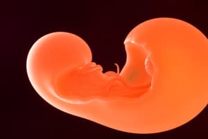
Nervous System Development
- The nervous system develops from embryonic ectoderm through a complex sequential process.
- The process involves five stages:
- Formation of Neural Tube (3-4 weeks gestation)
- Development of forebrain from neural tube (2-3 months gestation)
- Neuronal proliferation and migration (3-5 months gestation)
- Formation of network connections and synapses (5 months gestation to many years postnatal)
- Myelination (birth to many years postnatal)
Neural Tube Development
- The neural tube develops from the ectoderm germ layer.
- The neural plate folds to form the neural crest and neural groove, which then fuses dorsally to form the neural tube.
- The neural tube closes first in the cervical region and then "zippers" in two directions - cranially and caudally.
- The caudal end forms the brain and the rest develops into the spinal cord.
Neural Tube Defects
- Neural tube defects (NTDs) are the most common anomaly of the CNS.
- NTDs originate during the first month of embryonic development when the neural tube fails to close completely.
- Maternal folate deficiency is associated with a significant risk of NTDs.
- Other risk factors include maternal diabetes, maternal use of some seizure medications like valproic acid.
- Spina Bifida is the most common NTD.
Types of Spina Bifida
- Spina Bifida Occulta:
- Small opening in the vertebrae, but the defect is fully covered by skin and there is no visible exposure of meninges or neural tissues.
- Common, occurs in 10%-25% of infants.
- Usually causes no neurologic deficits because the spinal cord and spinal nerves are normal.
- Meningocele:
- Sac-like cyst of meninges filled with spinal fluid that protrudes through a defect in the posterior arch of the vertebra.
- Vertebral defect does not involve the spinal cord or nerve roots.
- Occurs with equal frequency in cervical, thoracic, and lumbar spine.
- Myelomeningocele:
- Herniation of sac-like cyst containing meninges, spinal fluid, and a portion of the spinal cord with its nerves through a defect in the posterior arch of a vertebra.
- Incidence: 0.5-1.0 per 1000 pregnancies.
- 80% are in lumbar and lumbosacral regions.
- Spinal cord and nerve roots may be malformed distal to the lesion, which can result in loss of motor, sensory, reflex, and autonomic function.
- Hydrocephalus occurs in 85% of infants with myelomeningocele.
Arnold Chiari Malformation
- Complex malformation of brainstem and cerebellum.
- Cerebellar tonsils are displaced downward into the cervical spinal canal, the upper medulla and lower pons are elongated and thin, and the medulla is displaced downward and sometimes has a kink.
- Type II:
- Associated with hydrocephalus from pressure that blocks the flow of CSF.
- Cognitive and motor defects are common.
- Requires surgical decompression or placement of CSF shunts.
Anencephaly
- Soft, bony component of the skull and part of the brain are missing.
- Incidence: 1 in 4895 live births in the US per year.
- Infants are stillborn or die within the first few days of life.
- Pathologic mechanism is unknown.
Prenatal Screening for NTD
- All pregnant people are offered screening for NTDs.
- 90-95% of cases occur in pregnancies without any risk factors.
- Screening tests include:
- Maternal serum alpha-fetoprotein (MSAFP):
- A fetal-specific globulin that is synthesized by the yolk sac, GI tract, and liver.
- Elevated MSAFP level is associated with neural tube defects.
- Testing is done ideally at 16-18 weeks gestation.
- Fetal ultrasound:
- Detection of NTDs on ultrasound examination depends on the size and location of the defect, the position of the fetus, the volume of amniotic fluid, maternal body habitus, and the skill and equipment of the sonographer/sonologist.
- Overall, very effective at detecting NTDs.
- Maternal serum alpha-fetoprotein (MSAFP):
Nervous System Development
- Forms from embryonic ectoderm through a complex sequential process
- Stages of development:
- Formation of neural tube (3-4 weeks gestation)
- Development of forebrain from neural tube (2-3 months gestation)
- Neuronal proliferation and migration (3-5 months gestation)
- Formation of network connections and synapses (5 months gestation to many years postnatal)
- Myelination (birth to many years postnatal)
Neural Tube Development
- Develops from ectoderm germ layer derived from epiblast
- Formation begins between week 3 and 4 of gestation
- Process:
- Neural plate folds to form neural crest and neural groove
- Neural crest fuses dorsally to form the neural tube
- Neural tube closes first in the cervical region and then "zippers" in two directions - cranially and caudally
- Caudal end forms the brain and the rest develops into the spinal cord
Neural Tube Defects (NTDs)
- Most common anomaly of the CNS
- Originate during the first month of embryonic development when the neural tube fails to close completely
- Types of NTDs:
- Spina Bifida
- Anencephaly
Spina Bifida
- Most common NTD
- Types:
- Spina Bifida Occulta
- Small opening in the vertebrae, but defect is fully covered by skin
- Usually causes no neurologic deficits
- Meningocele
- Saclike cyst of meninges filled with spinal fluid that protrudes through a defect in the posterior arch of the vertebra
- Occurs with equal frequency in cervical, thoracic, and lumbar spine
- Myelomeningiocele
- Herniation of saclike cyst containing meninges, spinal fluid, and a portion of the spinal cord with its nerves through a defect in the posterior arch of a vertebra
- Spinal cord and nerve roots may be malformed distal to the lesion, resulting in loss of motor, sensory, reflex, and autonomic function
- Hydrocephalus occurs in 85% of infants with myelomeningocele
- Almost always associated with Type II Arnold Chiari malformation
- Spina Bifida Occulta
Arnold Chiari Malformation
- Complex malformation of brainstem and cerebellum
- Types:
- Type I
- Does not involve the brainstem and may be asymptomatic
- Type II
- Associated with hydrocephalus from pressure that blocks the flow of CSF
- Cognitive and motor defects are common
- Requires surgical decompression or placement of CSF shunts
- Type III
- The brainstem or cerebellum extends into a high cervical myelomeningocele
- Type IV
- Cerebellum does not develop
- Type I
Anencephaly
- Soft, bony component of the skull and part of the brain are missing
- Infants are stillborn or die within first few days of life
Prenatal Screening for NTDs
- All pregnant people are offered screening for NTDs
- Screening tests:
- Maternal serum alpha fetoprotein (MSAFP)
- Fetal specific globulin synthesized by the yolk sac, GI tract, and liver
- Elevated MSAFP level is associated with neural tube defects
- Testing is done ideally at 16-18 weeks gestation
- Fetal ultrasound
- Detection of NTDs on ultrasound examination depends on the size and location of the defect, the position of the fetus, the volume of amniotic fluid, maternal body habitus, and the skill and equipment of the sonographer/sonologist
- Maternal serum alpha fetoprotein (MSAFP)
Studying That Suits You
Use AI to generate personalized quizzes and flashcards to suit your learning preferences.