Podcast
Questions and Answers
Which germ layer gives rise to the nervous system?
Which germ layer gives rise to the nervous system?
- Endoderm
- Blastoderm
- Ectoderm (correct)
- Mesoderm
During neurulation, the margins of the neural plate elevate along a longitudinal furrow called the _______.
During neurulation, the margins of the neural plate elevate along a longitudinal furrow called the _______.
neural groove
What is regulated by BMP4?
What is regulated by BMP4?
- Differentiation of the Neural Crest and Neural Plate (correct)
- Differentiation of the Notochord
- Closure of the Posterior Neuropore
- Elongation of the Neural Groove
Which of the following best describes the origin and differentiation of glial cells in the central nervous system (CNS)?
Which of the following best describes the origin and differentiation of glial cells in the central nervous system (CNS)?
The marginal layer of the neural tube eventually develops into the gray matter of the spinal cord.
The marginal layer of the neural tube eventually develops into the gray matter of the spinal cord.
What type of cells migrate from the mantle layer to the surface to form the strata of the cerebral cortex?
What type of cells migrate from the mantle layer to the surface to form the strata of the cerebral cortex?
Failure of the lateral hemispheres to form during development results in a condition called ______.
Failure of the lateral hemispheres to form during development results in a condition called ______.
Match the layer of the isocortex with its description.
Match the layer of the isocortex with its description.
Which of the following is NOT a component of the white matter in the spinal cord?
Which of the following is NOT a component of the white matter in the spinal cord?
Lateral horns, containing cell bodies of preganglionic sympathetic neurons, are present throughout the entire length of the spinal cord.
Lateral horns, containing cell bodies of preganglionic sympathetic neurons, are present throughout the entire length of the spinal cord.
The cerebellum is responsible for coordinating voluntary movements and maintaining normal ________.
The cerebellum is responsible for coordinating voluntary movements and maintaining normal ________.
The three layers of the cerebellar cortex are, molecular layer, Purkinje neurons layer, and ________ layer.
The three layers of the cerebellar cortex are, molecular layer, Purkinje neurons layer, and ________ layer.
In the molecular layer of the cerebellar cortex, which type of neuron has axons that form a 'basket' around Purkinje cells?
In the molecular layer of the cerebellar cortex, which type of neuron has axons that form a 'basket' around Purkinje cells?
Purkinje cells are characterized by having multiple layers of pear-shaped neurons within the Purkinje cells layer.
Purkinje cells are characterized by having multiple layers of pear-shaped neurons within the Purkinje cells layer.
In the granular layer, the dendrites of granule neurons resemble the ________ of a bird.
In the granular layer, the dendrites of granule neurons resemble the ________ of a bird.
Match the following components of the somatic nerve system reflex arc with their description:
Match the following components of the somatic nerve system reflex arc with their description:
Which of the following structures are derived from the neural crest?
Which of the following structures are derived from the neural crest?
The myelencephalon gives rise to the pons and cerebellum.
The myelencephalon gives rise to the pons and cerebellum.
What two main structures does the neural tube differentiate into?
What two main structures does the neural tube differentiate into?
Neural crest cells migrate in two streams to support the development of the face and the ______ branchial arch.
Neural crest cells migrate in two streams to support the development of the face and the ______ branchial arch.
Match the primary brain vesicles with their corresponding adult brain structures:
Match the primary brain vesicles with their corresponding adult brain structures:
Which layer of the developing neural tube gives rise to the gray matter of the spinal cord?
Which layer of the developing neural tube gives rise to the gray matter of the spinal cord?
Neuroblasts originate in the mantle layer of the neural tube.
Neuroblasts originate in the mantle layer of the neural tube.
What type of cells characterize the ependymal layer (ventricular zone) of the neural tube?
What type of cells characterize the ependymal layer (ventricular zone) of the neural tube?
Which layer of the cerebral cortex contains mostly fibers running parallel to the surface and relatively few cells?
Which layer of the cerebral cortex contains mostly fibers running parallel to the surface and relatively few cells?
Cerebral columns consist of cells that respond to multiple peripheral stimuli.
Cerebral columns consist of cells that respond to multiple peripheral stimuli.
What is the origin of the three envelopes that cover the spinal cord and cerebrum?
What is the origin of the three envelopes that cover the spinal cord and cerebrum?
The dura mater consists of fibrous __________ tissue and contains a large number of sensory nerves and blood vessels.
The dura mater consists of fibrous __________ tissue and contains a large number of sensory nerves and blood vessels.
Match the layers of the cerebral cortex with their primary cell types:
Match the layers of the cerebral cortex with their primary cell types:
Which type of nerve fibers in the cerebral cortex are formed by intermediate, comissural, and projection fibers?
Which type of nerve fibers in the cerebral cortex are formed by intermediate, comissural, and projection fibers?
From which layer of the cerebral cortex do axons of pyramidal neurons pass to the white matter?
From which layer of the cerebral cortex do axons of pyramidal neurons pass to the white matter?
Which of the following is the middle layer of the meninges surrounding the brain?
Which of the following is the middle layer of the meninges surrounding the brain?
Which of the following is NOT a component of the synaptic glomerulus of the cerebellum?
Which of the following is NOT a component of the synaptic glomerulus of the cerebellum?
Nerve impulses reach Purkinje cells exclusively via climbing fibers.
Nerve impulses reach Purkinje cells exclusively via climbing fibers.
Name the two main types of afferent fibers that reach the cerebellar cortex.
Name the two main types of afferent fibers that reach the cerebellar cortex.
The axons of granular neurons in the cerebellar cortex are also known as ______ fibers.
The axons of granular neurons in the cerebellar cortex are also known as ______ fibers.
Match the following cell types found in the cerebrum with their description:
Match the following cell types found in the cerebrum with their description:
Which layer of the cerebellar cortex contains Purkinje cells?
Which layer of the cerebellar cortex contains Purkinje cells?
Which of the following is a characteristic of Betz cells?
Which of the following is a characteristic of Betz cells?
The study of the distribution and cellular structure of the cerebral cortex is known as ______.
The study of the distribution and cellular structure of the cerebral cortex is known as ______.
Flashcards
Spinal Cord White Matter
Spinal Cord White Matter
Contains myelinated and unmyelinated nerve fibers, fibrous astrocytes, oligodendrocytes, and blood vessels.
Lateral Horns of Spinal Cord
Lateral Horns of Spinal Cord
Located between anterior and posterior horns; from (C8) T1 to L2-3. Contain cell bodies of preganglionic sympathetic neurons.
White Matter Bundles
White Matter Bundles
Posterior, lateral, and anterior bundles of nerve fibers in the spinal cord.
Somatic Reflex Arc Parts
Somatic Reflex Arc Parts
Signup and view all the flashcards
Brain Divisions
Brain Divisions
Signup and view all the flashcards
Cerebellum Function
Cerebellum Function
Signup and view all the flashcards
Cerebellar Cortex Layers
Cerebellar Cortex Layers
Signup and view all the flashcards
Molecular Layer Neurons
Molecular Layer Neurons
Signup and view all the flashcards
Molecular Layer
Molecular Layer
Signup and view all the flashcards
Purkinje Cells
Purkinje Cells
Signup and view all the flashcards
Granular Layer
Granular Layer
Signup and view all the flashcards
Mossy Fibers
Mossy Fibers
Signup and view all the flashcards
Synaptic Glomerulus
Synaptic Glomerulus
Signup and view all the flashcards
Climbing Fibers
Climbing Fibers
Signup and view all the flashcards
Cytoarchitectonics
Cytoarchitectonics
Signup and view all the flashcards
Myeloarchitectonics
Myeloarchitectonics
Signup and view all the flashcards
Ectoderm
Ectoderm
Signup and view all the flashcards
Notochord's Role
Notochord's Role
Signup and view all the flashcards
Neurulation
Neurulation
Signup and view all the flashcards
Neural Crest Cells
Neural Crest Cells
Signup and view all the flashcards
Brain Development
Brain Development
Signup and view all the flashcards
Mantle Layer Cells
Mantle Layer Cells
Signup and view all the flashcards
Spongioblasts
Spongioblasts
Signup and view all the flashcards
Marginal Layer Formation
Marginal Layer Formation
Signup and view all the flashcards
Isocortex
Isocortex
Signup and view all the flashcards
Holoprosencephaly
Holoprosencephaly
Signup and view all the flashcards
Inner Granular Layer
Inner Granular Layer
Signup and view all the flashcards
Inner Pyramidal Layer
Inner Pyramidal Layer
Signup and view all the flashcards
Outer Granular Layer
Outer Granular Layer
Signup and view all the flashcards
Outer Pyramidal Layer
Outer Pyramidal Layer
Signup and view all the flashcards
Polymorphic Layer
Polymorphic Layer
Signup and view all the flashcards
Tangential Bands
Tangential Bands
Signup and view all the flashcards
Dura Mater
Dura Mater
Signup and view all the flashcards
Neural Tube
Neural Tube
Signup and view all the flashcards
Neural Crest
Neural Crest
Signup and view all the flashcards
Neural Crest Cell Migration
Neural Crest Cell Migration
Signup and view all the flashcards
Primary Brain Vesicles
Primary Brain Vesicles
Signup and view all the flashcards
Rhombencephalon Divisions
Rhombencephalon Divisions
Signup and view all the flashcards
Prosencephalon Divisions
Prosencephalon Divisions
Signup and view all the flashcards
Neuroepithelial Cell Derivatives
Neuroepithelial Cell Derivatives
Signup and view all the flashcards
Neural Tube Layers
Neural Tube Layers
Signup and view all the flashcards
Study Notes
- These notes discuss cells of the nervous tissue, development of the nervous system, and the structure of the CNS.
Nervous System Division
- The nervous system is divided into the central nervous system (CNS) and the peripheral nervous system (PNS).
- The CNS consists of the brain and spinal cord.
- The PNS comprises peripheral nerves, nerve plexi, ganglia, and nerve endings.
Spinal Cord
- The outer part of the spinal cord consists of white matter, which contains ascending and descending nerve fibers.
- The inner part of the spinal cord consists of gray matter, forming an H or butterfly-shaped pattern around the central canal.
- Gray matter extends throughout the spinal cord's length, forming dorsal (posterior), anterior (ventral), and lateral (intermediate) horns.
Central Canal
- Cerebrospinal fluid circulates within the central canal of the spinal cord.
- The central canal has a lining of ependymal cells.
- Columnar ependymal cells line the central canal.
- Vascular ependymal cells secrete cerebrospinal fluid, with this occurring in the choroid plexus.
- Some ependymal cells feature cilia.
- Tanycytes, a type of ependymal cell, may be present in the third ventricle wall and play a role in regulating metabolism.
Gray Matter Composition
- Neurons, nerve cells with both myelinated and unmyelinated processes, the gray matter
- Neuroglia: protoplasmic astrocytes, oligodendrocytes and microglia
- Blood vessels are present.
- Spinal cord neurons are generally multipolar.
- These neurons are either motor neurons or interneurons.
- Nuclei are groups of cells performing the same function.
Oligodendrocytes
- Most common glial cell type
- Forms myelin sheaths around CNS neuron axons
- Analogous to Schwann cells in the PNS
- Form a supportive network around CNS neurons
- Have fewer processes than astrocytes and feature a round or oval cell body
Microglia
- Small cells located near blood vessels
- Role is phagocytic, clearing away dead cells.
- Protect the CNS from disease by phagocytizing microbes.
- Migrate to injury sites to clear debris from injured cells, but they may also kill healthy cells.
Astrocytes
- Largest and most numerous of glial cells
- Star-shaped with multiple processes projecting from cell body
- Help form and maintain the blood-brain barrier
- Offer structural support to neurons
- Maintain a chemical environment for nerve impulse/action potential generation
- Regulate nutrient concentrations for neuron survival
- Regulate ion concentrations that facilitate the generation of action potentials by neurons
- Take up excess neurotransmitters
- Aid in neuronal migration during brain development
- Perform repairs to stabilize tissue
Neurons
- Neurons have electrical excitability, an ability to produce action potentials or impulses in response to stimuli.
Cell Body or Soma
- Contains a single nucleus with a prominent nucleolus.
- Nissl bodies are present, with rough ER & free ribosomes for protein synthesis, that replace neuronal cellular components for growth and damaged axon repair in the PNS.
Dendrites
- Receiving or input portion of the neuron
- Short, tapering, and highly branched
- Specialized surfaces for contact with other neurons
- Cytoplasm contains Nissl bodies and mitochondria
Axons
- Transfer impulses away from the cell body and propagate nerve impulses to other neurons
- Long, thin, cylindrical processes containing mitochondria and microtubules
- Lacks an ER and does not feature protein synthesis
- Joins soma at a cone-shaped elevation known as the axon hillock
- The first part of the is the initial segment
- Most impulses originate at the junction of the axon hillock and the initial segment, the trigger zone
- Axoplasm is cytoplasm
- Axolemma is a plasma membrane
- Collaterals are side branches
Motor Neurons
- All such neurons are multipolar.
- They are divided into somatic, neurons of muscle spindle, and autonomic types.
- Somatic motor neurons are multipolar and located in the anterior horns.
- They have both large α neurons and small γ neurons
- Axons of these neurons exit through the anterior roots and innervate skeletal muscle
Motor Units
- Each skeletal fiber has only one neuromuscular junction (NMJ).
- A motor unit (MU) is a somatic neuron plus all the skeletal muscle fibers it innervates.
- The number and size of motor units indicate the precision of muscle control.
- A muscle twitch is a single momentary contraction or a response to a single stimulus.
- Motor unit activation follows the all-or-none theory, contracting completely or not at all.
Posterior Horns
- Contains zones such as the zona marginalis, substantia gelatinosa, n. proprius, and n. thoracicus
- Interneurons, glial cells, and processes of neurons are found here.
- Afferent fibers of pseudounipolar neurons bring sensory information to the posterior horns, switch on interneurons, which may convey the impulse to the anterior horns or to the brain.
- Ventral roots (anterior, efferent) join with the afferent fibers of the dorsal roots to form a mixed spinal nerve.
White matter
- Composed of myelinated and unmyelinated nerve fibers, fibrous astrocytes, oligodendrocytes, and blood vessels on each side
- Divided into 3 bundles: posterior, lateral, and anterior.
Lateral Horns
- Located between the anterior and posterior horns in the spinal cord's specific segments (C8-L2/3).
- Contain cell bodies of preganglionic sympathetic neurons
Reflex Arcs
- There are three parts in this arch: afferent/sensory (pseudounipolar neurons of spinal ganglion, afferent neurons), interneurons, and motor neurons (efferent neurons of anterior horns).
Brain
- Divided into the brain stem, cerebellum, and cerebrum.
Cerebellum
- Coordinates voluntary movements and muscle function for normal posture maintenance.
- Is covered in gray matter (cortex) and white matter
Cerebellum Cortex
- Cytoarchitectonics distributed in 3 layers: molecular, Purkinje neurons layer, and granular layer.
Molecular Layer
- Basket neurons, large stellate cells, and small stellate cells are found in the molecular layer of the cerebellar cortex.
- Axons of basket cells pass parallel to the surface, with branches that branch around Purkinje cells forming a basket of nerve fibers; dendrites branch in the molecular layer.
- Axons of stellate cells connect with the dendrites of Purkinje cells, with dendrites of stellate cells branching in the molecular layer.
Purkinje Cells Layer
- The single layer consists of pear-shaped neurons, where dendrites branch intensively in one plane and axons continue into the white matter via the granular layer.
Granular Layer
- Two types of cells are found in this layer: granule, and major stellate (Golgi).
- Granule neuron dendrites are short, branched, resemble bird feet, and participate in cerebellar glomerulus composition.
- The axon of a granule cell passes to the molecular layer, branches there into two branches that run parallel to the surface of the cerebellum and make parallel nerve fibers
- Large stellate (Golgi) cells have dendrites that pass to the molecular layer and synapse with parallel fibers, while axons make contacts with the granule cells dendrites.
Layers of Cerebellar Cortex
- Consists of the molecular layer, Purkinje cells, Granule neurons , and Golgi cells and also contains oligodendrocytes and astrocytes.
- Afferent fibers that reach are mossy (come to the granular layer), climbing, and parallel (axons of granular neurons).
- Mossy fibers reach the granular layer and communicate with the dendrites of granular cells, forming synaptic glomeruli of the cerebellum.
- The synaptic glomerulus consists of the mossy fiber, granule cell dendrites, terminal branches of stellate neuron axons, and dendrites of stellate neurons.
- Climbing fibers pass through the granular layer into the molecular layer and surround the dendrites of the Purkinje cells.
- Nerve impulses that reach are transferred to the Purkinje cells directly by climbing fibers are transferred and nerve impulses which arrive via mossy fibers pass to the Purkinje cells indirectly via granular neurons.
Cerebrum
- Cerebral cortex is composed of 10-18 billion neurons, their processes, and neuroglia.
- Cytoarchitectonics is distribution and cellular structure of cerebral cortex and myeloarchitectonics is the distribution and pattern of nerve fibers' arrangement.
- In the cerebral cortex, there are effector cells (pyramidal/spindle), and associative cells (granular/horizontal/T-shaped neurons).
Pyramidal Cells
- Can be small (10 µm), medium (20 µm), or large (40 µm)
Cerebrum Cortex
- A shape of a cone is seen in pyramidal cells, oriented towards the surface of the brain, where dendrites (from the apex) pass towards the surface and the axon passes towards the white matter.
- Middle-sized spindle cells, body lies perpendicular to the surface of the cortex with an axon that reaches the white matter
- There are six layers of cortex: molecular, outer granular, outer pyramidal, inner granular, inner pyramidal, polymorphic cells layer.
- Tangential bands/fibers run parallel to the surface in molecular, outer granular, inner granular, and inner pyramidal layers.
- Radial bands of nerve fibers are divided into intermediate, comissural, and projections types. These fibers compose radial bands of nerve fibers and reach the third layer of the cortex outer pyramidal layer.
- Cerebral columns (or modules) are vertical columns running through the depth of the cortex where all cells respond to a single peripheral stimulus and run vertically to the surface but vary in width (50 µm to the 500 µm in the visual cortex), depending on the stimulus location.
Meninges of the brain
- Spinal cord and cerebrum are covered with three envelopes of ecto-mesenchymal origin
- Dura mater -outermost
- Arachnoidea -middle
- Pia mater -innermost
- Dura mater consists of fibrous connective tissue containing a large number of sensory nerves and blood vessels.
- The deep surface is separated from the arachnoid by the subdural space, which appears when there is no cerebrospinal fluid (CSF) in the subarachnoid cavity.
- Around the brain, the cerebral dura adheres to inner periosteal lining of the skull bones, meaning that there is normally no epidural space, except bleeding leads to an epidural hematoma
- The dura mater surrounding brain and spinal cord is continuous through the foramen magnum, while the dura mater surrounding the spinal cord (spinal dura) is separated by an epidural space from the inner periosteum-lined surface of the vertebrae.
- The epidural space contains blood vessels veins and fat.
- Arachnoid is composed of two parts: a thin layer of connective tissue with plane epithelial cells plus trabeculae that connect with the pia mater.
- Space between dura mater and is subdural and the space between and pia mater is subarachnoidal.
- Pia mater is close to the brain, it intervenes into sulci and follows blood vessels.
- Has two plates with loose connective tissue between them and is connected with outer limiting glial membrane
- Choroid plexus forms pia mater intervenes into sulci of cerebrum and cerebellum along with vesicles of the brain
- This plexus is a loose connective tissue with ependymocytes that produces cerebrospinal fluid and contains numerous blood vessels
- There are two barriers in the brain: between blood and cerebrospinal fluid; and between blood and brain.
- Blood-cerebrospinal fluid barrier is cerebrospinal, separates substances and made of endothelial cells of fenestrated capillaries, basal lamina, and ependymal cells.
- Blood–brain barrier: endothelial cells of the capillaries, where plasma components are not fenestrated. There is a basement membrane, and astrocytes that surround the capillaries. The barrier is not present in regions of the hypothalamus, where certain things are monitored, in the posterior pituitary which releases hormones or in the choroid plexus (where cerebrospinal fluid is produced).
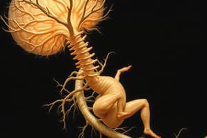
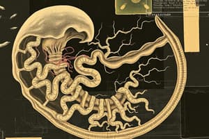
Development of the nervous system
- At the end of gastrulation, the established notochord induces the neural plate differentiation.
- The margins of the neural plate get elevated along a longitudinal furrow (neural groove).
- Neurulation (neural tube formation) is also associated with neural crest cells' delamination and migration
- The brain develops from the anterior (upper) part of the neural tube, and the spinal cord develops from the lower part (caudal end) of the neural tube.
- (BMP4 regulates the differentiation of neural plate and neural crest)
- The brain is divided into the Rhombencephalon, Mesencephalon, Prosencephalon. Neural crest forms the Peripheral nervous system, ganglionic nerve cells, glia cells, adrenal medulla Melanocytes, odontoblasts connective tissue of the head skull bones and cranial muscles.
Development of pharyngeal
- During branchial development from neural crest cells from the region of the future midbrain and 2 rhombomeres of hindbrain there is also migration
- Migration is in two streams, forwards to support the development of the face and other neural structure towards first branchial arch.
Brain vesicles
- In this developmental stage, the Rhombencephalon becomes the Myelencephalon and Metencephalon where as the Prosencephalon turns into the Diencephalon and Telencephalon.
Spinal cord and brain development
- Neural tube differentiates to produce neuroepithelial cells, which give rise to neuroblasts and glioblasts (spongioblasts).
- Neuroblasts, primitive nerve cells, form the layer and it later becomes the gray matter. The outer layer of the spinal cord is layer, a layer of nerve fibers, originating from neuroblasts for white matter. Neuroblasts divide, migrate and finally form horns of the spinal cord, which give rise to the marginal layer
- In early development, the ependymal layer (also called ventricular zone), is characterized by mitotic activity where neuro- originate along with gliablast cells
- Cells of the mantle layer (aka intermediate zone) will arise in the ependymal layer, migrate, and differentiate into neuroblasts and spongioblasts
- Extending from the cells in the mantle layer will composed of the of the neural tube, later it will develop into the white matter of the spinal cord, and molecular layer in the brain cortex.
- When the neuroblasts of the migrate to the surface, strata of the Cerebral cortex develops
- Isocortex: Six layers: Molecular, outer, granular, outer pyramidal, inner granular, inner pyramidal and .multiform while there is also allacortex
- Different developmental defects include such as different types of spina bifida and holoprosencephaly
- Holoprosencephaly – failure of the formation of lateral hemispheres from genetic mutations (Pax3, Sonic hedgehog, and openbrain), dietary factors affecting folic acid and blockage of folate receptors, certain vitamin deficiency along with overdosage (cholesterol reducing medications)
Studying That Suits You
Use AI to generate personalized quizzes and flashcards to suit your learning preferences.