Podcast
Questions and Answers
During neurulation, a population of cells separates from the tips of the neural folds. What do these cells become?
During neurulation, a population of cells separates from the tips of the neural folds. What do these cells become?
- Schwann cells
- Endocardial cushions
- Adrenal medulla
- All of the above (correct)
Which of the following best describes the sequence of primary brain vesicle formation during neural tube development?
Which of the following best describes the sequence of primary brain vesicle formation during neural tube development?
- Rhombencephalon, Mesencephalon, Prosencephalon
- Prosencephalon, Mesencephalon, Rhombencephalon (correct)
- Mesencephalon, Prosencephalon, Rhombencephalon
- Prosencephalon, Rhombencephalon, Mesencephalon
Spina bifida occulta is often characterized by which of the following?
Spina bifida occulta is often characterized by which of the following?
- Elevated alpha-fetoprotein levels during pregnancy
- Protrusion of the meninges through the vertebral defect
- A small tuft of hair over the defect (correct)
- Protrusion of both the meninges and spinal cord through the vertebral defect
On what day of fetal development does the cranial neural pore typically close?
On what day of fetal development does the cranial neural pore typically close?
Which of the following teratogens is most associated with increasing the risk of neural tube defects?
Which of the following teratogens is most associated with increasing the risk of neural tube defects?
Failure of the neural tube to close at the cranial end results in which condition?
Failure of the neural tube to close at the cranial end results in which condition?
If an ultrasound during pregnancy reveals polyhydramnios and elevated alpha-fetoprotein levels, which neural tube defect is most likely?
If an ultrasound during pregnancy reveals polyhydramnios and elevated alpha-fetoprotein levels, which neural tube defect is most likely?
Which of the following secondary brain vesicles does the mesencephalon develop into?
Which of the following secondary brain vesicles does the mesencephalon develop into?
In which type of spina bifida does the spinal cord protrude externally, without skin covering the defect?
In which type of spina bifida does the spinal cord protrude externally, without skin covering the defect?
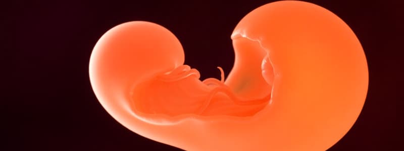
The development of the nervous system begins during which week of gestation?
The development of the nervous system begins during which week of gestation?
Flashcards
Neurolation
Neurolation
The process of neural tube formation, developing into the spinal cord and brain.
Primary Neuralation
Primary Neuralation
Begins in the 3rd week of gestation and forms the functional central nervous system.
Neural Plate
Neural Plate
Ectoderm thickens to form this, induced by the central notochord.
Neural Folds
Neural Folds
Signup and view all the flashcards
Neural Groove
Neural Groove
Signup and view all the flashcards
Neural Crest Cells
Neural Crest Cells
Signup and view all the flashcards
Neuropores
Neuropores
Signup and view all the flashcards
Neural Tube Defects
Neural Tube Defects
Signup and view all the flashcards
Anencephaly
Anencephaly
Signup and view all the flashcards
Spina Bifida
Spina Bifida
Signup and view all the flashcards
Study Notes
- Neurolation is the process where the neural tube forms and develops into the spinal cord and brain.
- Complications during neurolation can lead to neural tube defects.
- The nervous system's development starts in the third week of gestation.
- Primary neurulation forms the functional central nervous system.
Development of the Embryonic Nervous System
- By day 18, the ectodermal germ layer is disc-shaped with cranial and caudal ends.
- Cross-sections reveal three germ layers: ectoderm, mesoderm, and endoderm.
- The central notochord induces the ectoderm to thicken, forming the neural plate.
- By the end of the third week, neural folds extend upwards from the neural plate edges.
- The depressed mid-region becomes the neural groove.
- Neural folds fuse at the midline, converting the neural groove into a closed neural tube.
- Neural crest cells separate from the neural folds and migrate throughout the body.
- Neural crest cells form Schwann cells, meninges, endocardial cushions, parafollicular cells, and the adrenal medulla.
- Before full fusion, the cephalic and caudal ends communicate with the amniotic cavity through cranial and caudal neuropores.
- The cranial neuropore closes around day 25.
- The cranial end develops into three primary vesicles: the forebrain (prosencephalon), midbrain (mesencephalon), and hindbrain (rhombencephalon).
- These three develop into five secondary vesicles: telencephalon, diencephalon, mesencephalon, metencephalon, and myelencephalon.
- The caudal neuropore closes around day 27.
- Failure of the neural tube to close properly results in neural tube defects.
- Neural tube defects can occur as part of syndromes, with chromosomal disorders, or due to environmental exposure.
- Folic acid deficiency during pregnancy and folic acid antagonists increase the risk of neural tube defects.
Neural Tube Defects
- Anencephaly is the failure of the neural tube to close at the cranial end, preventing the brain from developing and is incompatible with life.
- Anencephaly results in high alpha-fetoprotein levels and polyhydramnios.
- Spina bifida is the failure of the neural tube to close at the caudal end.
- In spina bifida, the vertebra overlying the defect do not fully develop, leaving the vertebral arch open.
- Spina bifida occulta is an asymptomatic defect where the vertebral arch fails to fuse at the midline.
- Spina bifida occulta's only evidence may be a small tuft of hair over the defect.
- In spina bifida occulta, the spinal cord remains intact, and alpha-fetoprotein levels are normal during pregnancy.
- In spina bifida with meningocele, the meninges protrude through the vertebral defect, but the spinal cord does not.
- In spina bifida with meningomyelocele, both the meninges and spinal cord protrude through the vertebral defect, and is usually associated with Chiari II malformation and hydrocephalus.
- Alpha-fetoprotein levels are increased in spina bifida with meningomyelocele.
- In myeloschisis, the spinal cord is exposed because the skin does not cover the defect.
- Myeloschisis is the most severe type of spina bifida, and alpha-fetoprotein levels are increased during pregnancy.
Studying That Suits You
Use AI to generate personalized quizzes and flashcards to suit your learning preferences.