Podcast
Questions and Answers
What is the primary function of the diaphragm during breathing?
What is the primary function of the diaphragm during breathing?
- To increase lung volume by contracting downward. (correct)
- To decrease thoracic pressure by relaxing upward.
- To assist in maintaining intrapleural pressure without movement.
- To limit airflow by contracting during expiration.
Which of the following muscle groups actively contributes to forced expiration?
Which of the following muscle groups actively contributes to forced expiration?
- Internal intercostals and abdominal muscles. (correct)
- Sternocleidomastoid and scalene muscles.
- Diaphragm and external intercostals.
- Accessory muscles and diaphragm.
What happens to the intrapleural pressure during normal inspiration?
What happens to the intrapleural pressure during normal inspiration?
- It increases, causing the lungs to deflate.
- It remains constant throughout the breathing cycle.
- It decreases, promoting lung expansion. (correct)
- It fluctuates randomly with no significant pattern.
In the respiratory cycle, when does the cessation of inspiration typically occur?
In the respiratory cycle, when does the cessation of inspiration typically occur?
The generation of respiratory rhythm is primarily located in which part of the brain?
The generation of respiratory rhythm is primarily located in which part of the brain?
What physiological change occurs when air is introduced into the pleural cavity?
What physiological change occurs when air is introduced into the pleural cavity?
Which pressure change is primarily responsible for airflow during normal breathing?
Which pressure change is primarily responsible for airflow during normal breathing?
What role do accessory muscles of inspiration play in breathing?
What role do accessory muscles of inspiration play in breathing?
What is the primary role of the diaphragm during inspiration?
What is the primary role of the diaphragm during inspiration?
Which muscles are primarily responsible for forced expiration?
Which muscles are primarily responsible for forced expiration?
How are the lung and chest wall coupled in the human body?
How are the lung and chest wall coupled in the human body?
What happens to the chest wall during passive expiration?
What happens to the chest wall during passive expiration?
What is the functional relationship between the lung and chest wall modeled as?
What is the functional relationship between the lung and chest wall modeled as?
What is one consequence of the internal intercostal muscles contracting during expiration?
What is one consequence of the internal intercostal muscles contracting during expiration?
What is the role of accessory muscles during inspiration?
What is the role of accessory muscles during inspiration?
Which pleura covers the thoracic wall and superior face of the diaphragm?
Which pleura covers the thoracic wall and superior face of the diaphragm?
During forced expiration, what is the primary function of the rectus abdominis muscle?
During forced expiration, what is the primary function of the rectus abdominis muscle?
Which pressure is primarily responsible for the expansion of the lungs during inspiration?
Which pressure is primarily responsible for the expansion of the lungs during inspiration?
What is the primary function of the inspiratory muscles during breathing?
What is the primary function of the inspiratory muscles during breathing?
Which muscle is primarily responsible for active expiration during heavy breathing?
Which muscle is primarily responsible for active expiration during heavy breathing?
Where is the respiratory rhythm primarily generated in the body?
Where is the respiratory rhythm primarily generated in the body?
Which motoneurons innervate the diaphragm?
Which motoneurons innervate the diaphragm?
What type of pressure gradient primarily drives air flow during ventilation?
What type of pressure gradient primarily drives air flow during ventilation?
What is the role of the phrenic nerve in respiration?
What is the role of the phrenic nerve in respiration?
During heavy exercise, which of the following muscles becomes actively involved in expiration?
During heavy exercise, which of the following muscles becomes actively involved in expiration?
Which of the following statements best describes the role of the external intercostal muscles?
Which of the following statements best describes the role of the external intercostal muscles?
What happens to the intrapleural pressure during inspiration as the diaphragm and external intercostal muscles contract?
What happens to the intrapleural pressure during inspiration as the diaphragm and external intercostal muscles contract?
What effect does increased intrapleural pressure have on the respiratory system?
What effect does increased intrapleural pressure have on the respiratory system?
How is transpulmonary pressure calculated?
How is transpulmonary pressure calculated?
What effect does the expansion of the lungs during inspiration have on the alveolar pressure compared to atmospheric pressure?
What effect does the expansion of the lungs during inspiration have on the alveolar pressure compared to atmospheric pressure?
What initiates the airflow into the lungs at the start of inspiration?
What initiates the airflow into the lungs at the start of inspiration?
What occurs at the end of expiration regarding the alveolar pressure?
What occurs at the end of expiration regarding the alveolar pressure?
What occurs during the uncoupling of the lung and chest wall?
What occurs during the uncoupling of the lung and chest wall?
What is the functional residual capacity (FRC)?
What is the functional residual capacity (FRC)?
What is the typical intrapleural pressure at functional residual capacity (FRC)?
What is the typical intrapleural pressure at functional residual capacity (FRC)?
According to Boyle’s law, how is pressure related to volume?
According to Boyle’s law, how is pressure related to volume?
What results from the increase in volume in the intrapleural space at FRC?
What results from the increase in volume in the intrapleural space at FRC?
What happens to atmospheric pressure when measuring intrapleural pressure?
What happens to atmospheric pressure when measuring intrapleural pressure?
How is the pressure across the chest wall (Pcw) calculated?
How is the pressure across the chest wall (Pcw) calculated?
What is the outcome when the lung and chest wall are uncoupled due to injury?
What is the outcome when the lung and chest wall are uncoupled due to injury?
What defines the residual volume (RV) in terms of lung function?
What defines the residual volume (RV) in terms of lung function?
What happens to pressure in the intrapleural space when lung volume increases?
What happens to pressure in the intrapleural space when lung volume increases?
Flashcards are hidden until you start studying
Study Notes
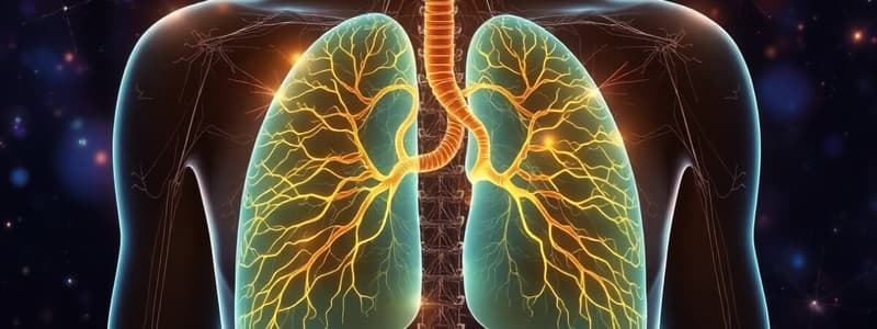
Neural Innervation of Respiratory Muscles
- The respiratory rhythm is generated in the medulla by pacemaker cells and groups of neurons.
- These neurons send signals to the spinal cord, where they synapse with phrenic and intercostal motoneurons.
- The phrenic nerve innervates the diaphragm, originating from motoneurons in the C3-C5 spinal segments.
- The external intercostal nerves innervate the external intercostal muscles, originating from motoneurons in the T1-T12 spinal segments.
- Expiratory muscles like the rectus abdominus and internal intercostals are activated during increased ventilation and are innervated by motoneurons in the upper lumbar and T1-T12 segments.
Respiratory Muscle Activity
- Inspiration involves contraction of the diaphragm and external intercostal muscles, pulling the ribs upward and outward.
- Passive expiration relies on the relaxation of inspiratory muscles and elastic recoil of the chest wall.
- Forced expiration involves contraction of the internal intercostal muscles, pulling the ribs downward and inward, along with the rectus abdominus and external oblique muscles.
Coupling of the Lung and Chest Wall
- The lung and chest wall are coupled by the visceral and parietal pleura, forming a thin, double-layered serosa.
- Parietal pleura covers the thoracic wall and diaphragm, while visceral pleura covers the lung surface.
- The coupling of the lung and chest wall creates a resting position at functional residual capacity (FRC), the end of expiration/beginning of inspiration.
Mechanics of the Lungs and Chest Wall
- When uncoupled, the lung contracts towards residual volume (RV) and the chest wall expands towards total lung capacity (TLC).
- At FRC, the chest wall's tendency to expand and the lung's tendency to contract creates negative intrapleural pressure, typically around -5 cmH2O.
- This negative pressure is relative to atmospheric pressure, meaning the absolute pressure in the intrapleural space is lower.
Lung Pressures and Pressure Gradients
- Pressure differences are measured from inside to outside a structure.
- Pressure across the chest wall (Pcw) is the difference between intrapleural pressure and atmospheric pressure.
- Transpulmonary pressure is the difference between alveolar pressure (intrapulmonary pressure) and intrapleural pressure.
- When there is no airflow, alveolar pressure equals atmospheric pressure.
- The pressure gradient across the respiratory system is the difference between alveolar pressure and atmospheric pressure.
Pressure Changes during Respiration
- During inspiration, contraction of inspiratory muscles increases intrapleural space volume, leading to more negative intrapleural pressure.
- This increased negative pressure creates a positive transpulmonary pressure, expanding the lungs.
- Lung expansion decompresses gas, dropping alveolar pressure below atmospheric pressure, generating airflow into the lungs.
- At the end of inspiration, airflow stops when alveolar pressure equals atmospheric pressure.
- During expiration, relaxation of the diaphragm and elastic recoil of the respiratory system compresses lung gas, increasing alveolar pressure above atmospheric pressure, generating airflow out of the lungs.
Studying That Suits You
Use AI to generate personalized quizzes and flashcards to suit your learning preferences.


