Podcast
Questions and Answers
What characterizes the broad cephalic portion of the central nervous system?
What characterizes the broad cephalic portion of the central nervous system?
- The formation of brain vesicles (correct)
- The presence of a spinal cord
- The closure of the neural tube
- The migration of neural crest cells
Which statement about neural crest cells (NCC) is correct?
Which statement about neural crest cells (NCC) is correct?
- NCC do not participate in any birth defects.
- NCC contribute to the craniofacial skeleton and other tissues. (correct)
- NCC are derived exclusively from mesoderm.
- NCC only migrate through the dorsal pathway.
What transition do neural crest cells undergo as they leave the neuroectoderm?
What transition do neural crest cells undergo as they leave the neuroectoderm?
- Epithelial proliferation
- Neuron differentiation
- Epithelial-to-mesenchymal transition (correct)
- Mesenchymal-to-epithelial transition
Where do trunk region neural crest cells migrate to form melanocytes?
Where do trunk region neural crest cells migrate to form melanocytes?
What is one significant evolutionary role of neural crest cells?
What is one significant evolutionary role of neural crest cells?
What is a primary outcome of neural crest cells' migration from cranial neural folds?
What is a primary outcome of neural crest cells' migration from cranial neural folds?
Which type of connective tissue do neural crest cells contribute to?
Which type of connective tissue do neural crest cells contribute to?
Which of the following statements about NCC is incorrect?
Which of the following statements about NCC is incorrect?
What component does each somite contribute to the body?
What component does each somite contribute to the body?
Which areas of the body does the intermediate mesoderm form excretory units?
Which areas of the body does the intermediate mesoderm form excretory units?
From which layer does the dermis of the body wall and limbs originate?
From which layer does the dermis of the body wall and limbs originate?
What are the two ways in which blood vessels form?
What are the two ways in which blood vessels form?
What does the visceral layer of lateral plate mesoderm form?
What does the visceral layer of lateral plate mesoderm form?
What is the role of mesothelial membranes formed by parietal layer mesoderm cells?
What is the role of mesothelial membranes formed by parietal layer mesoderm cells?
What is the primary function of the nephrogenic cord formed by the intermediate mesoderm?
What is the primary function of the nephrogenic cord formed by the intermediate mesoderm?
Where do the first blood islands appear during development?
Where do the first blood islands appear during development?
Which structure is NOT derived from the ectodermal germ layer?
Which structure is NOT derived from the ectodermal germ layer?
What is the most common site for spina bifida to occur?
What is the most common site for spina bifida to occur?
How can neural tube defects be prevented?
How can neural tube defects be prevented?
During which week does the paraxial mesoderm begin to organize into somitomeres?
During which week does the paraxial mesoderm begin to organize into somitomeres?
What happens if the neural tube fails to close in the cranial region?
What happens if the neural tube fails to close in the cranial region?
Which layer forms tissue surrounding the amnion?
Which layer forms tissue surrounding the amnion?
What structure does the lateral plate mesoderm give rise to?
What structure does the lateral plate mesoderm give rise to?
At what stage do cells of the mesodermal germ layer begin to form a thickened plate known as paraxial mesoderm?
At what stage do cells of the mesodermal germ layer begin to form a thickened plate known as paraxial mesoderm?
What significant event occurs during the fourth week of embryonic development involving the oropharyngeal membrane?
What significant event occurs during the fourth week of embryonic development involving the oropharyngeal membrane?
Which structure separates the upper part of the anal canal from the proctodeum?
Which structure separates the upper part of the anal canal from the proctodeum?
What is primarily indicated as a measurement for the age of the embryo during the second month?
What is primarily indicated as a measurement for the age of the embryo during the second month?
What major morphological change occurs at the end of the fourth week as indicated by the presence of approximately 28 somites?
What major morphological change occurs at the end of the fourth week as indicated by the presence of approximately 28 somites?
What role does the yolk sac primarily play during early embryonic development?
What role does the yolk sac primarily play during early embryonic development?
At what stage does the cloacal membrane break down to create the anus opening?
At what stage does the cloacal membrane break down to create the anus opening?
Which embryonic structures are found as paddle-shaped buds during the beginning of the fifth week?
Which embryonic structures are found as paddle-shaped buds during the beginning of the fifth week?
What is the primary developmental function of the allantois during embryonic growth?
What is the primary developmental function of the allantois during embryonic growth?
What is the main structural change that occurs in somites by the beginning of the fourth week?
What is the main structural change that occurs in somites by the beginning of the fourth week?
How can the age of an embryo be determined during the early stages of development?
How can the age of an embryo be determined during the early stages of development?
Which somite pair is specifically stated to disappear during development?
Which somite pair is specifically stated to disappear during development?
What do the cells at the dorsomedial and ventrolateral edges of the somite primarily give rise to?
What do the cells at the dorsomedial and ventrolateral edges of the somite primarily give rise to?
What is formed from the cells of the sclerotome during somite differentiation?
What is formed from the cells of the sclerotome during somite differentiation?
What role do the cells from the ventrolateral edge of the somite play in body development?
What role do the cells from the ventrolateral edge of the somite play in body development?
What happens to the somitomeres in the head region during development?
What happens to the somitomeres in the head region during development?
Which body structures are primarily derived from cells in the dermomyotome?
Which body structures are primarily derived from cells in the dermomyotome?
What are hemangioblasts a precursor for?
What are hemangioblasts a precursor for?
Where do definitive hematopoietic stem cells originate?
Where do definitive hematopoietic stem cells originate?
When does the liver become the major hematopoietic organ during development?
When does the liver become the major hematopoietic organ during development?
What is the primary organ system derived from the endodermal germ layer?
What is the primary organ system derived from the endodermal germ layer?
What results from the failure of the lateral body folds to close the body wall?
What results from the failure of the lateral body folds to close the body wall?
Which region of the gut tube communicates with the yolk sac?
Which region of the gut tube communicates with the yolk sac?
What is the consequence of the growth and closure of the lateral body wall folds?
What is the consequence of the growth and closure of the lateral body wall folds?
Which membrane temporarily bounds the foregut at its cephalic end?
Which membrane temporarily bounds the foregut at its cephalic end?
Flashcards
Neural Crest Cells (NCC)
Neural Crest Cells (NCC)
Cells at the lateral border of the neuroectoderm that detach and migrate during neural tube closure.
Epithelial-to-mesenchymal Transition (EMT)
Epithelial-to-mesenchymal Transition (EMT)
The process by which NCC undergo a change from tightly bound epithelial cells to loosely organized mesenchymal cells.
NCC Migration Pathways
NCC Migration Pathways
NCC migrate along two pathways: dorsal, forming melanocytes, and ventral, contributing to sensory ganglia, sympathetic and enteric neurons, Schwann cells, and adrenal medulla cells.
Cranial NCC Migration
Cranial NCC Migration
Signup and view all the flashcards
Mesoderm
Mesoderm
Signup and view all the flashcards
Mesenchyme
Mesenchyme
Signup and view all the flashcards
NCC as a 'Fourth Germ Layer'
NCC as a 'Fourth Germ Layer'
Signup and view all the flashcards
NCC in Birth Defects and Cancers
NCC in Birth Defects and Cancers
Signup and view all the flashcards
Ectoderm Function
Ectoderm Function
Signup and view all the flashcards
Ectoderm: CNS
Ectoderm: CNS
Signup and view all the flashcards
Ectoderm: PNS
Ectoderm: PNS
Signup and view all the flashcards
Ectoderm: Sensory Epithelium
Ectoderm: Sensory Epithelium
Signup and view all the flashcards
Ectoderm: Epidermis
Ectoderm: Epidermis
Signup and view all the flashcards
Ectoderm: Subcutaneous Glands
Ectoderm: Subcutaneous Glands
Signup and view all the flashcards
Ectoderm: Mammary Glands
Ectoderm: Mammary Glands
Signup and view all the flashcards
Ectoderm: Pituitary Gland
Ectoderm: Pituitary Gland
Signup and view all the flashcards
Somites
Somites
Signup and view all the flashcards
Somite Differentiation
Somite Differentiation
Signup and view all the flashcards
Sclerotome
Sclerotome
Signup and view all the flashcards
Dermatome
Dermatome
Signup and view all the flashcards
Myotome
Myotome
Signup and view all the flashcards
Epithelial-to-Mesenchymal Transition (EMT) in Somites
Epithelial-to-Mesenchymal Transition (EMT) in Somites
Signup and view all the flashcards
Somite Formation Rate
Somite Formation Rate
Signup and view all the flashcards
Somite Innervation
Somite Innervation
Signup and view all the flashcards
Angiogenesis
Angiogenesis
Signup and view all the flashcards
Blood Islands
Blood Islands
Signup and view all the flashcards
Vasculogenesis
Vasculogenesis
Signup and view all the flashcards
Lateral Plate Mesoderm Layers
Lateral Plate Mesoderm Layers
Signup and view all the flashcards
Lateral Body Wall Folds
Lateral Body Wall Folds
Signup and view all the flashcards
Visceral Serous Membranes
Visceral Serous Membranes
Signup and view all the flashcards
Parietal Serous Membranes
Parietal Serous Membranes
Signup and view all the flashcards
Serous Membranes
Serous Membranes
Signup and view all the flashcards
Blood cell origin in the yolk sac
Blood cell origin in the yolk sac
Signup and view all the flashcards
Where do definitive blood stem cells come from?
Where do definitive blood stem cells come from?
Signup and view all the flashcards
Liver's role in blood formation
Liver's role in blood formation
Signup and view all the flashcards
Bone marrow takes over blood production
Bone marrow takes over blood production
Signup and view all the flashcards
Endoderm's role in forming the gut
Endoderm's role in forming the gut
Signup and view all the flashcards
Sections of the developing gut tube
Sections of the developing gut tube
Signup and view all the flashcards
Yolk sac connection to the midgut
Yolk sac connection to the midgut
Signup and view all the flashcards
Oropharyngeal membrane: A temporary barrier
Oropharyngeal membrane: A temporary barrier
Signup and view all the flashcards
Oropharyngeal Membrane
Oropharyngeal Membrane
Signup and view all the flashcards
Cloacal Membrane
Cloacal Membrane
Signup and view all the flashcards
Rupturing of the Oropharyngeal Membrane
Rupturing of the Oropharyngeal Membrane
Signup and view all the flashcards
Rupturing of the Cloacal Membrane
Rupturing of the Cloacal Membrane
Signup and view all the flashcards
Distal Allantois
Distal Allantois
Signup and view all the flashcards
Crown-rump Length (CRL)
Crown-rump Length (CRL)
Signup and view all the flashcards
Forelimb and Hind Limb Buds
Forelimb and Hind Limb Buds
Signup and view all the flashcards
Pericardial Swelling
Pericardial Swelling
Signup and view all the flashcards
Study Notes
Embryology L4-P1: The Embryonic Period
- The embryonic period is also known as the period of organogenesis
- It occurs from the third to eighth weeks of development
- During this time, each of the three germ layers (ectoderm, mesoderm, and endoderm) develops into specific tissues and organs
- By the end of the second month, the major features of the external body form are recognizable
- The majority of birth defects occur between the third and eighth weeks
- Environmental or genetic insults during this period can lead to spontaneous abortion, but not all cases result in loss
- Defects that occur between the third and eighth week are more likely to result in a viable fetus than when they occur earlier
Derivatives of the Ectodermal Germ Layer
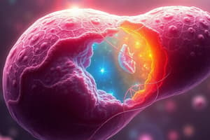
- At the beginning of the third week, the ectodermal germ layer has a disc shape, broader cephalically than caudally
- The notochord and prechordal mesoderm induce the overlying ectoderm to thicken, forming the neural plate
- The neural plate cells become neuroectoderm
- This induction is the initial event in neurulation
Neurulation
- Neurulation is the process where the neural plate forms the neural tube
- The process is regulated by the planar cell polarity pathway
- Key events include lengthening of the neural plate and lateral-to-medial cell movement in the ectoderm and mesoderm planes
- Neural folds elevate to form neural folds
- Neural groove formation is a result of folding
- Neural folds fuse in the midline to form the neural tube
- Cephalic and caudal neuropores initially connect the neural tube to the amniotic cavity
- These neuropores close during the process (cranial closes at day 25; caudal closes at day 28)
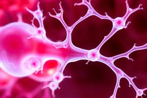
Neural Crest Cells
- Cells on the lateral border/crest of the neuroectoderm detach from their neighbors
- The cells undergo an epithelial-to-mesenchymal transition to move into the underlying mesoderm
- A dorsal pathway might migrate through the dermis to cells in the ectoderm
- A ventral pathway might migrate through the anterior half of each somite to become various structures
- These cells are critically important in the formation of multiple tissues/organs and about one-third of birth defects and cancers
Embryonic Period - P2: Derivatives of the Mesodermal Germ Layer
- Initially, mesodermal cells form a thin sheet on either side of the midline
- By day 17, cells close to the midline proliferate, forming the thickened paraxial mesoderm
- Cells further out from the midline remain as thin lateral plate mesoderm
- Lateral plate mesoderm divides into somatic and splanchnic layers
- During differentiation, intercellular cavities form in the lateral plate and divide into layers
- Somatic (parietal) mesoderm lines the amnion
- Splanchnic (visceral) mesoderm covers the yolk sac
- The intraembryonic cavity is formed with the appearance of the layers
- Intermediate mesoderm connects paraxial and lateral mesoderm structures
Paraxial Mesoderm
- By the third week, paraxial mesoderm segments into somitomeres
- These segments first appear in the cephalic region and form in a head-to-tail manner
- Somitomeres are specialized mesodermal cells to differentiate into somites
- In the head region, somitomeres form in association with the segmentation of the neural plate and form mesenchyme
- Caudally, somitomeres form somites
- The first pair of somites arises in the occipital region and occurs by day 20 of development
- Approximately 3 pairs of somites arise per day until about 42 or 44 pairs (of various types) are present at the end of the fifth week
Somite Differentiation
- Somites form from presomitic mesoderm
- They exhibit a donut shape
- Somite cells undergo epithelization with a thin lumen
- Ventral and medial cells lose characteristics of epithelial cells to become mesenchymal cells
- Collectively, these form the sclerotome, differentiating into vertebrae and ribs
- Dorsomedial and ventrolateral edges differentiate into precursors for muscle cells, whereas the area between forms the dermatome
- Muscle precursors become mesenchymal to migrate under the dermatome to form the dermomyotome
Intermediate Mesoderm
- Intermediate mesoderm temporarily connects paraxial and lateral plate mesoderm
- It forms urogenital structures (including the nephrogenic cord and the gonads, for the kidneys and reproductive organs)
- Forms from segmental cell clusters or an unsegmented structure
- Cervical and thoracic regions form segmental cell clusters
- Caudally, the intermediate mesoderm forms an unsegmented mass known as the nephrogenic cord
Lateral Plate Mesoderm
- Splits into parietal (somatic) and visceral (splanchnic) layers
- These layers line the intraembryonic cavity and surround organs
- Parietal layer with overlying ectoderm forms the body wall folds
- The parietal layer forms the dermis, bones, connective tissue of the limbs, and sternum
- Cells of the parietal layer migrate to form costal cartilages, limb, and body wall muscles
- Visceral layer plus embryonic endoderm form the gut tube
- Mesoderm cells from the parietal layer secrete fluid to form membranes along the cavity surfaces
Blood and Blood Vessels
- Blood cells and blood vessels originate from mesoderm
- Vasculogenesis is the formation of blood vessels; it also begins from an existing vessel system
- Blood islands first appear in the yolk sac and lateral plate mesoderm around 3 weeks of gestation
- Hemangioblasts are a precursor to blood and vessel formation
- Definitive hematopoietic stem cells develop from the aorta-gonad-mesonephros (AGM) region, near the mesonephric kidney
- Liver becomes the major hematopoietic organ from weeks 2 to 7
Derivatives of the Endodermal Germ Layer
- Endoderm forms the epithelial and parenchymal regions of gastrointestinal organs
- The endoderm originally covers the ventral surface of the embryo and forms the roof of the yolk sac
- Growth of the brain vesicles makes the embryo bulge into the amniotic cavity and takes a fetal position
- Two lateral folds close to enclose the ventral body wall
- The gut tube forms three regions: foregut, hindgut, and midgut
- The midgut connects to the yolk sac by the vitelline duct
- The foregut connects to the oropharyngeal membrane
- The hindgut connects to the cloacal membrane
- Various derivatives result from the interactions of those structures
External Appearance during the Second Month
- By the fourth week, the main external features are visible, including somites and pharyngeal arches
- The age of the embryo is typically given in terms of somites, but later, is described via crown-rump length (CRL) measured in millimeters
- The second month marks increased growth in the head, limb formation, and the development of facial features, ears, eyes, nose, etc
Studying That Suits You
Use AI to generate personalized quizzes and flashcards to suit your learning preferences.