Podcast
Questions and Answers
Which cell type is formed from the dental papilla during the development of teeth?
Which cell type is formed from the dental papilla during the development of teeth?
- Ameloblasts
- Odontoblasts (correct)
- Ectomesenchymal cells
- Stellate reticulum
What structure forms as the oral epithelium thickens in a band during tooth development?
What structure forms as the oral epithelium thickens in a band during tooth development?
- Cervical loop
- Dental follicle
- Dental sac
- Dental lamina (correct)
At what point does the enamel organ change its configuration to initiate odontogenesis?
At what point does the enamel organ change its configuration to initiate odontogenesis?
- When cells start to differentiate (correct)
- When the dental lamina is fully formed
- When the dental follicle surrounds the dental papilla
- When the outer enamel epithelium thickens
Which cells are indicative of the inner layer of the enamel organ?
Which cells are indicative of the inner layer of the enamel organ?
Which structure surrounds the dental papilla during the development of teeth?
Which structure surrounds the dental papilla during the development of teeth?
What is the main function of neural crest cells during embryonic development?
What is the main function of neural crest cells during embryonic development?
Which embryonic structure forms the basis for the development of the face?
Which embryonic structure forms the basis for the development of the face?
Which of the following correctly associates a branchial arch with its respective innervation?
Which of the following correctly associates a branchial arch with its respective innervation?
Which component is derived from the second branchial arch?
Which component is derived from the second branchial arch?
During which time frame does the neural tube begin to form in embryogenesis?
During which time frame does the neural tube begin to form in embryogenesis?
What is the clinical significance of cleft lip and palate?
What is the clinical significance of cleft lip and palate?
Which embryonic structure is responsible for forming the primary palate?
Which embryonic structure is responsible for forming the primary palate?
What major vessel arises from the third branchial arch during development?
What major vessel arises from the third branchial arch during development?
What triggers pre-ameloblasts to differentiate into ameloblasts?
What triggers pre-ameloblasts to differentiate into ameloblasts?
When does the lifecycle of the ameloblast conclude?
When does the lifecycle of the ameloblast conclude?
Which correctly describes the lifecycle of odontoblasts?
Which correctly describes the lifecycle of odontoblasts?
When is the formation of pulp complete?
When is the formation of pulp complete?
What is the primary source of vascular supply during amelogenesis?
What is the primary source of vascular supply during amelogenesis?
Which stage directly follows the laying down of pre-dentine?
Which stage directly follows the laying down of pre-dentine?
What is a consequence of stellate reticulum and stratum intermedium cells disappearing?
What is a consequence of stellate reticulum and stratum intermedium cells disappearing?
Where does dentinogenesis primarily begin?
Where does dentinogenesis primarily begin?
Which cells play a crucial role in the formation of the enamel matrix during tooth development?
Which cells play a crucial role in the formation of the enamel matrix during tooth development?
What is the role of cementoblasts in tooth development?
What is the role of cementoblasts in tooth development?
Which process does NOT contribute to the development of the periodontal ligament?
Which process does NOT contribute to the development of the periodontal ligament?
Cementogenesis is primarily associated with which cells originating from ectomesenchyme?
Cementogenesis is primarily associated with which cells originating from ectomesenchyme?
Dentinogenesis of the tooth root is triggered by contact with which specific matrix?
Dentinogenesis of the tooth root is triggered by contact with which specific matrix?
The formation of enamel pearls is primarily a result of disruptions in the processes involving which type of cells?
The formation of enamel pearls is primarily a result of disruptions in the processes involving which type of cells?
Which structure is instrumental in the mapping out of the root's shape during periodontal ligament formation?
Which structure is instrumental in the mapping out of the root's shape during periodontal ligament formation?
What important structure consists of remnants of Hertwig's epithelial root sheath that may contribute to regeneration and cyst formation?
What important structure consists of remnants of Hertwig's epithelial root sheath that may contribute to regeneration and cyst formation?
What role does the ameloblast play once amelogenesis is completed?
What role does the ameloblast play once amelogenesis is completed?
When does the eruption of teeth typically begin?
When does the eruption of teeth typically begin?
What is the primary function of the reduced enamel epithelium during tooth eruption?
What is the primary function of the reduced enamel epithelium during tooth eruption?
Which of the following accurately describes the three phases of eruption?
Which of the following accurately describes the three phases of eruption?
What is the function of the dento-gingival junction?
What is the function of the dento-gingival junction?
What sequence of events characterizes the advanced bell stage of tooth development?
What sequence of events characterizes the advanced bell stage of tooth development?
The stellate reticulum plays which role in the enamel organ during development?
The stellate reticulum plays which role in the enamel organ during development?
Which stage of tooth development is characterized by both the initiation of root formation and ameloblast activity?
Which stage of tooth development is characterized by both the initiation of root formation and ameloblast activity?
Study Notes
Embryonic Origins
- Ectoderm: forms the skin, nervous system, and sensory organs, among other tissues.
- Mesoderm: forms muscle, bone, cartilage, blood, and the circulatory system.
- Endoderm: forms the lining of the digestive and respiratory tracts, as well as some glands.
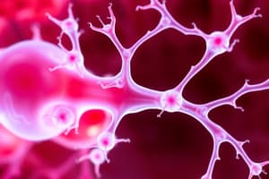
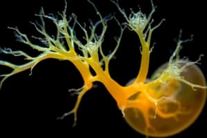
Neural Crest Cells
- Neural Crest Cells (NCC) are a unique group of cells that arise from the neuroectoderm during embryonic development.
- They migrate extensively throughout the embryo, giving rise to a diverse array of structures.
- NCC contribute to the formation of the: peripheral nervous system, skull bones, melanocytes, connective tissue of the head and neck, and parts of the heart.
Embryonic Development
- Zygote, Morula, Blastocyst: The zygote is the fertilized egg cell. The morula is a solid ball of cells formed by repeated cell division. The blastocyst is a fluid-filled sphere with an inner cell mass that will form the embryo.
- Trilaminar disc: This is a three-layered structure that forms during gastrulation. The three layers are:
- Ectoderm: forms the skin and nervous system.
- Mesoderm: forms muscle, bone, and connective tissue.
- Endoderm: forms the lining of the digestive and respiratory tracts.
- Neural Tube and Neuroectoderm: The neural tube develops from the neuroectoderm and eventually forms the brain and spinal cord.
- Stomodeum: The primary oral cavity forms from the stomodeum, which is an invagination of the ectoderm.
- Branchial Arches: Form during the 4th week of development, these arches give rise to the structures of the face, neck, and head.
- Face: The face develops by the fusion of five facial prominences derived from the branchial arches.
- Palate and Secondary Palate: The palate forms by the fusion of two processes, the palatine shelves, which fuse with the nasal septum and primary palate.
- Tongue: The tongue develops from the first branchial arch (mandibular arch).
- Cleft lip and palate: Occur when the facial prominences or the palatine shelves fail to fuse properly during development.
Branchial Arches
| Branchial Arches | Innervation | Muscles | Artery | Cartilage or bones |
|---|---|---|---|---|
| I | Trigeminal nerve | Muscles of mastication | Aortic arch 1 | Meckel's cartilage |
| II | Facial nerve | Muscles of facial expression | Aortic arch 2 | Reichert's cartilage |
| III | Glossopharyngeal nerve | Stylopharyngeus muscle | Aortic arch 3 | Hyoid bone |
| IV | Vagus nerve | Muscles of the larynx | Aortic arch 4 | Hyoid bone |
| V | Vagus nerve | Muscles of the larynx | Aortic arch 5 | Incus |
| VI | Vagus nerve | Muscles of the larynx | Aortic arch 6 | Stapes |
Embryology of Oral Tissues
- Dental Lamina: Forms as a thickening of the oral epithelium, marking the future location of teeth.
- Enamel Organ: Develops from the dental lamina, and determining the shape of the tooth. The cells differentiate to prepare for odontogenesis, the formation of teeth.
- Dental Papilla: The dental papilla is the underlying ectomesenchyme that will give rise to dentin and pulp.
- Dental Follicle: Surrounding the dental papilla, the dental follicle will form cementum, periodontal ligament.
Embryology of the Crown
Odontogenesis Pathway:
- Outer Enamel Epithelium (OEE)
- Stellate Reticulum
- Stratum Intermedium
- Inner Enamel Epithelium (IEE)
- Pre-Ameloblasts
- Ameloblasts
- Dental Papilla
- Pre-Odontoblasts
- Odontoblasts
- Pulpal cells
Crown Stages:
- Stage 1: The inner enamel epithelial cells differentiate into pre-ameloblasts.
- Stage 2: The outer periphery cells of the dental papilla differentiate into pre-odontoblasts.
- Stage 3: Pre-ameloblasts are triggered by dentine matrix to differentiate into ameloblasts.
- Stage 4: Pre-odontoblasts are triggered by pre-ameloblasts to differentiate into odontoblasts.
- Stage 5: Dentinogenesis starts at the cusp tips.
- Stage 6: Amelogenesis starts at the cusp tips.
- Stage 7: Stellate reticulum cells deflate.
- Stage 8: Stellate reticulum and stratum intermedium cells disappear.
- Stage 9: Enamel matrix is laid down.
- Stage 10: Pre-dentine is laid down.
- Stage 11: The dentine matrix is mineralized.
- Stage 12: The enamel matrix is mineralized.
Amelogenesis
- Amelogenesis is a process in two stages:
- Secretion: Ameloblasts deposit enamel protein matrix.
- Maturation: The enamel matrix mineralizes.
Ameloblasts
- Lifecycle: Ameloblast activity is limited to the time that crown formation is complete.
- Differentiation: Ameloblasts differentiate from the inner enamel epithelium.
Odontoblasts
- Lifecycle: The odontoblasts continue their activity throughout the life of the tooth, forming dentin throughout root development.
- Differentiation: Odontoblasts differentiate from the outer periphery cells of the dental papilla.
Pulp Formation
- Formation: The pulp develops during crown formation and continues through root formation.
Amelogenesis vs Dentinogenesis
| Features | Amelogenesis | Dentinogenesis |
|---|---|---|
| Cell | Ameloblast | Odontoblast |
| Location it begins | Cusp tips | Cusp tips |
| Pattern of formation | Appositional | Appositional |
| Direction in relation to dental papilla | Outward | Inward |
| When is it complete? | Crown formation | Root formation |
| Source of vascular supply | Dental follicle | Dental papila |
| Outcome of cell after crown formation complete | Degeneration | Maintain activity |
Embryology of Root and Supporting Structures
- Dental Sac: This structure surrounds the tooth germ and gives rise to the periodontal ligament, cementum, and alveolar bone.
- Epithelial Root Sheath (HERS): HERS guides the formation of the root and determines its shape.
- Cementoblasts: These cells, derived from the dental sac, lay down cementum.
- Fibroblasts: Fibroblasts, also derived from the dental sac, produce collagen fibers that form the periodontal ligament.
Stages of Root Formation
- Stage 1: HERS plays a role in shaping the root by breaking down and fusing with the dental follicle.
- Stage 2: Dentinogenesis continues as the root forms.
- Stage 3: Cementogenesis occurs, with cementum being laid down by cementoblasts.
- Stage 4: The periodontal ligament is formed from collagen fibers produced by fibroblasts from the dental sac.
- Stage 5: Alveolar bone formation occurs.
Eruption and Exfoliation
- Eruption: The process by which a tooth moves from its position in the jawbone to its functional place in the mouth.
- Exfoliation: The shedding of a primary tooth. The process is initiated by the formation of a resorption space on the root of the primary tooth.
Stages of Eruption
- Pre-eruptive: The tooth is developing within the jawbone. This involves a period of growth and development of the tooth crown, followed by root formation.
- Intraosseous: The tooth moves through the alveolar bone, a slow and methodical process in which the tooth gradually migrates towards the oral cavity.
- Supraosseous: The final stage where the tooth erupts from the alveolar bone and into the oral cavity.
Reduced Enamel Epithelium (REE)
- REE is a protective layer that forms over the erupting tooth.
- The REE originates from the ameloblasts.
Dento-Gingival Junction
- This junction is the area where the tooth meets the gingiva.
- It forms the gingival sulcus and helps to protect the tooth root from bacteria.
Studying That Suits You
Use AI to generate personalized quizzes and flashcards to suit your learning preferences.
Related Documents
Description
Explore the fascinating world of embryonic development, focusing on the three germ layers: ectoderm, mesoderm, and endoderm. Understand the role of neural crest cells and their contributions to various structures in the body. This quiz covers key stages like zygote, morula, and blastocyst.