Podcast
Questions and Answers
What is the earliest sign of eye development seen in the embryo?
What is the earliest sign of eye development seen in the embryo?
- Closure of the neural tube
- Pair of shallow grooves on each side of the forebrain (correct)
- Formation of the lens vesicle
- Development of the optic cup
From which sources is the optic cup and lens vesicle derived?
From which sources is the optic cup and lens vesicle derived?
- Neuroectoderm, surface ectoderm, mesoderm, and neural crest cells (correct)
- Fibrous and vascular coats of the eye
- Ectoderm of the body and mesoderm
- Neural crest cells only
What happens during the invagination of the optic vesicles?
What happens during the invagination of the optic vesicles?
- They induce changes in the surface ectoderm (correct)
- They develop into the optic nerve
- They become a single-walled structure
- They migrate to form the fibrous coat of the eye
Which embryonic structure eventually develops into the retina?
Which embryonic structure eventually develops into the retina?
What occurs to the intraretinal space as development progresses?
What occurs to the intraretinal space as development progresses?
What triggers the dissociation of neural crest cells from their neighboring cells?
What triggers the dissociation of neural crest cells from their neighboring cells?
What is a primary consequence of defective neural crest cell migration in the digestive system?
What is a primary consequence of defective neural crest cell migration in the digestive system?
Which of the following structures is derived from neural crest cells?
Which of the following structures is derived from neural crest cells?
Which embryological abnormality can result from improper development of the eye?
Which embryological abnormality can result from improper development of the eye?
What role do neural crest cells play in the formation of cranial nerve ganglia?
What role do neural crest cells play in the formation of cranial nerve ganglia?
Which condition is NOT associated with failure of neural crest cell migration?
Which condition is NOT associated with failure of neural crest cell migration?
What is the main result of the epithelial-to-mesenchymal transition of neural crest cells?
What is the main result of the epithelial-to-mesenchymal transition of neural crest cells?
The parasympathetic ganglia of the gastrointestinal tract are derived from which type of cells?
The parasympathetic ganglia of the gastrointestinal tract are derived from which type of cells?
What structure does the hyaloid artery and vein eventually become after development?
What structure does the hyaloid artery and vein eventually become after development?
During which week do the lips of the choroid fissure fuse?
During which week do the lips of the choroid fissure fuse?
Which part of the optic cup develops into the inner layer of the iris?
Which part of the optic cup develops into the inner layer of the iris?
What is formed from the inner layer of mesenchyme surrounding the eye primordium?
What is formed from the inner layer of mesenchyme surrounding the eye primordium?
What is the role of the optic stalk during development?
What is the role of the optic stalk during development?
Which structure is responsible for the formation of the ciliary body and iris?
Which structure is responsible for the formation of the ciliary body and iris?
What happens to the lens vesicle during the 5th week of development?
What happens to the lens vesicle during the 5th week of development?
The substantia propria of the cornea originates from which layer?
The substantia propria of the cornea originates from which layer?
What is a characteristic of the optic nerve regarding myelination?
What is a characteristic of the optic nerve regarding myelination?
Which type of cells are responsible for myelinating the optic nerve?
Which type of cells are responsible for myelinating the optic nerve?
Which condition results from the failure of closure of the choroid fissure?
Which condition results from the failure of closure of the choroid fissure?
Which structure is derived from the first pharyngeal cleft?
Which structure is derived from the first pharyngeal cleft?
What do the malleus and incus originate from?
What do the malleus and incus originate from?
What forms the connective tissue surrounding the inner ear structures?
What forms the connective tissue surrounding the inner ear structures?
Which component is responsible for maintaining balance in the inner ear?
Which component is responsible for maintaining balance in the inner ear?
Which statement is true about the optic nerve post-transection?
Which statement is true about the optic nerve post-transection?
What is the origin of the definitive eardrum?
What is the origin of the definitive eardrum?
Which structures develop from the first and second pharyngeal arches?
Which structures develop from the first and second pharyngeal arches?
Which of the following is NOT a congenital malformation of the ear?
Which of the following is NOT a congenital malformation of the ear?
What is the role of neural crest cells in the pharyngeal arches?
What is the role of neural crest cells in the pharyngeal arches?
Which pharyngeal cleft contributes to the formation of the external auditory meatus?
Which pharyngeal cleft contributes to the formation of the external auditory meatus?
What comprises each pharyngeal arch?
What comprises each pharyngeal arch?
Which of the following is a derivative of the first pharyngeal arch?
Which of the following is a derivative of the first pharyngeal arch?
Which statement about auricular anomalies is true?
Which statement about auricular anomalies is true?
What developmental structure does the neural crest primarily differentiate into during eye formation?
What developmental structure does the neural crest primarily differentiate into during eye formation?
Which embryonic structure forms the lens of the eye during development?
Which embryonic structure forms the lens of the eye during development?
During eye development, what is the significance of the optic vesicles?
During eye development, what is the significance of the optic vesicles?
Which of the following statements best describes the fate of the intraretinal space during eye development?
Which of the following statements best describes the fate of the intraretinal space during eye development?
What is the primary source of the retina and optic nerve in developing eyes?
What is the primary source of the retina and optic nerve in developing eyes?
What is the primary consequence of neural crest cells failing to migrate into the bowel walls in Hirschsprung's disease?
What is the primary consequence of neural crest cells failing to migrate into the bowel walls in Hirschsprung's disease?
Which of the following structures is NOT a derivative of neural crest cells?
Which of the following structures is NOT a derivative of neural crest cells?
What abnormality can occur as a result of improper development of the eye due to issues with neural crest cells?
What abnormality can occur as a result of improper development of the eye due to issues with neural crest cells?
Congenital deafness can be attributed to issues during the development of which structure?
Congenital deafness can be attributed to issues during the development of which structure?
Which condition is primarily characterized by delayed passage of meconium and involves issues with neural crest cells?
Which condition is primarily characterized by delayed passage of meconium and involves issues with neural crest cells?
Failure of which neural crest derivative can lead to congenital megacolon?
Failure of which neural crest derivative can lead to congenital megacolon?
Which embryological abnormality specifically involves the formation of an incomplete optic fissure?
Which embryological abnormality specifically involves the formation of an incomplete optic fissure?
What structure forms the iridopupillary membrane during development?
What structure forms the iridopupillary membrane during development?
Which part of the developing optic cup gives rise to the ciliary body?
Which part of the developing optic cup gives rise to the ciliary body?
During which week does the choroid fissure close?
During which week does the choroid fissure close?
Which structures are formed from the mesoderm present between the optic cup and surface epithelium?
Which structures are formed from the mesoderm present between the optic cup and surface epithelium?
What does the outer layer of the mesenchyme surrounding the eye primordium differentiate into?
What does the outer layer of the mesenchyme surrounding the eye primordium differentiate into?
What developmental structure is primarily involved in forming the optic nerve?
What developmental structure is primarily involved in forming the optic nerve?
Which layer of the optic cup is responsible for forming the pigment layer of the retina?
Which layer of the optic cup is responsible for forming the pigment layer of the retina?
The anterior chamber of the eye develops through which process?
The anterior chamber of the eye develops through which process?
What is a characteristic of the optic nerve regarding its ability to regenerate?
What is a characteristic of the optic nerve regarding its ability to regenerate?
Which structure forms from the neural crest surrounding the inner ear?
Which structure forms from the neural crest surrounding the inner ear?
Which of the following statements accurately describes the components of the external ear?
Which of the following statements accurately describes the components of the external ear?
What type of cells are responsible for myelination of the optic nerve?
What type of cells are responsible for myelination of the optic nerve?
Which condition is indicated by the absence of the lens?
Which condition is indicated by the absence of the lens?
How is the tympanic membrane primarily formed?
How is the tympanic membrane primarily formed?
Which of the following statements is true regarding the development of the cochlear duct?
Which of the following statements is true regarding the development of the cochlear duct?
What are the derivatives of the first and second pharyngeal arches related to the ear?
What are the derivatives of the first and second pharyngeal arches related to the ear?
What is the primary embryonic origin of the definitive eardrum?
What is the primary embryonic origin of the definitive eardrum?
Which of the following auricular anomalies is characterized by a complete absence of the external ear?
Which of the following auricular anomalies is characterized by a complete absence of the external ear?
Which pharyngeal cleft is known to contribute to the formation of the external auditory meatus?
Which pharyngeal cleft is known to contribute to the formation of the external auditory meatus?
What type of cells migrate into the pharyngeal arches to contribute to the skeletal components of the face?
What type of cells migrate into the pharyngeal arches to contribute to the skeletal components of the face?
What structures primarily originate from the mesenchymal tissue of the pharyngeal arches?
What structures primarily originate from the mesenchymal tissue of the pharyngeal arches?
Which condition results from congenital malformation associated with the first and second pharyngeal arches?
Which condition results from congenital malformation associated with the first and second pharyngeal arches?
Which of the following is a main component of each pharyngeal arch?
Which of the following is a main component of each pharyngeal arch?
What is the fate of the epithelial lining of the first pharyngeal cleft?
What is the fate of the epithelial lining of the first pharyngeal cleft?
Flashcards
Neural Crest Cells Origin
Neural Crest Cells Origin
Specialized cells found at the border of the developing neural tube
Neural Crest Cell Migration
Neural Crest Cell Migration
Neural crest cells leave the neuroectoderm and travel to different parts of the developing embryo to make different tissues
Hirschsprung's Disease Cause
Hirschsprung's Disease Cause
Failure of neural crest cells to migrate properly to the bowel wall
Hirschsprung's Disease Effect
Hirschsprung's Disease Effect
Signup and view all the flashcards
Neural Crest Derivatives (face)
Neural Crest Derivatives (face)
Signup and view all the flashcards
Neural Crest Derivatives (autonomic nervous system)
Neural Crest Derivatives (autonomic nervous system)
Signup and view all the flashcards
Coloboma/Cataract Development
Coloboma/Cataract Development
Signup and view all the flashcards
Congenital Deafness Cause
Congenital Deafness Cause
Signup and view all the flashcards
Eye development sources
Eye development sources
Signup and view all the flashcards
Earliest sign of eye development
Earliest sign of eye development
Signup and view all the flashcards
Optic Vesicles formation
Optic Vesicles formation
Signup and view all the flashcards
Optic cup formation
Optic cup formation
Signup and view all the flashcards
Lens vesicle role
Lens vesicle role
Signup and view all the flashcards
Choroid fissure
Choroid fissure
Signup and view all the flashcards
Hyaloid artery/vein
Hyaloid artery/vein
Signup and view all the flashcards
Lens placode
Lens placode
Signup and view all the flashcards
Lens vesicle
Lens vesicle
Signup and view all the flashcards
Choroid
Choroid
Signup and view all the flashcards
Sclera
Sclera
Signup and view all the flashcards
Anterior chamber
Anterior chamber
Signup and view all the flashcards
Optic nerve (CN II)
Optic nerve (CN II)
Signup and view all the flashcards
Optic nerve myelination
Optic nerve myelination
Signup and view all the flashcards
Optic nerve regeneration
Optic nerve regeneration
Signup and view all the flashcards
Coloboma iridis
Coloboma iridis
Signup and view all the flashcards
Aniridia
Aniridia
Signup and view all the flashcards
Ear development origins
Ear development origins
Signup and view all the flashcards
Ear parts
Ear parts
Signup and view all the flashcards
Membranous labyrinth
Membranous labyrinth
Signup and view all the flashcards
Tympanic membrane derivation
Tympanic membrane derivation
Signup and view all the flashcards
Eardrum Development
Eardrum Development
Signup and view all the flashcards
Auricle Development
Auricle Development
Signup and view all the flashcards
Congenital Deafness Types
Congenital Deafness Types
Signup and view all the flashcards
Auricular Anomalies
Auricular Anomalies
Signup and view all the flashcards
Pharyngeal Arch Composition
Pharyngeal Arch Composition
Signup and view all the flashcards
First Pharyngeal Arch Derivatives
First Pharyngeal Arch Derivatives
Signup and view all the flashcards
Pharyngeal Pouch Function
Pharyngeal Pouch Function
Signup and view all the flashcards
First Pharyngeal Cleft Function
First Pharyngeal Cleft Function
Signup and view all the flashcards
Neural Crest Cells
Neural Crest Cells
Signup and view all the flashcards
Hirschsprung's Disease
Hirschsprung's Disease
Signup and view all the flashcards
Eye Development
Eye Development
Signup and view all the flashcards
Coloboma
Coloboma
Signup and view all the flashcards
Ear Development
Ear Development
Signup and view all the flashcards
Congenital Deafness
Congenital Deafness
Signup and view all the flashcards
Pharyngeal Arches
Pharyngeal Arches
Signup and view all the flashcards
What are the four sources of eye development?
What are the four sources of eye development?
Signup and view all the flashcards
What is the earliest sign of eye development?
What is the earliest sign of eye development?
Signup and view all the flashcards
What is the function of the lens vesicle?
What is the function of the lens vesicle?
Signup and view all the flashcards
What is the optic cup?
What is the optic cup?
Signup and view all the flashcards
What is the difference between the inner and outer walls of the optic cup?
What is the difference between the inner and outer walls of the optic cup?
Signup and view all the flashcards
Hyaloid vessels
Hyaloid vessels
Signup and view all the flashcards
Central artery and vein of the retina
Central artery and vein of the retina
Signup and view all the flashcards
What is the role of pharyngeal pouches?
What is the role of pharyngeal pouches?
Signup and view all the flashcards
Function of the First Pharyngeal Cleft
Function of the First Pharyngeal Cleft
Signup and view all the flashcards
Study Notes
Neural Crest, Eye and Ear
- The class is Year 2, Semester 1
- The lecturer is Dr. Vijayal Akshmi S B, from the Department of Anatomy
- The date of the lecture is 26-11-2024
Learning Outcomes
- Describe the origin and migration of neural crest cells
- Explain how failure of normal neural crest migration can cause pathologies, like Hirschsprung's disease
- Describe the development of the eye, and explain how embryological abnormalities like coloboma or cataract formation occur
- Describe the development of the ear, and explain how problems like congenital deafness occur
- Describe the pharyngeal arches and their derivatives
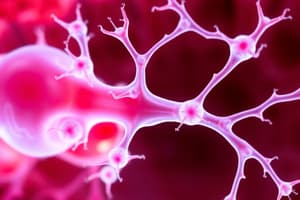
Neural Crest Cells
- Neural crest cells are a specialized cell population situated at the lateral border/crest of the neuroectoderm
- These cells detach from their neighbours as neural folds elevate and fuse to form the neural tube
- Neural crest cells undergo an epithelial-to-mesenchymal transition
- They migrate extensively to various locations in the body, contributing to diverse structures
Neural Crest Derivatives
- Connective tissue and bones of the face and skull
- Cranial nerve ganglia
- C cells of the thyroid gland
- Conotruncal septum in the heart
- Odontoblasts
- Dermis in face and neck
- Spinal (dorsal root) ganglia
- Sympathetic chain and pre-aortic ganglia
- Parasympathetic ganglia of the gastrointestinal tract
- Adrenal medulla
- Schwan cells
- Glial cells
- Meninges (forebrain)
- Melanocytes
- Smooth muscle cells to blood vessels of the face and forebrain
Hirschsprung's Disease
- Failure of neural crest cells to migrate into the bowel wall
- Defective parasympathetic plexus (Auerbach's and Meissner's)
- Aganglionic segment, particularly the sigmoid and rectum
- Delayed passage of meconium
- Constipation, vomiting
- Abdominal distension
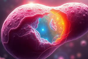
Development of Eye
- The earliest sign of eye development is seen in the 22-day embryo as shallow grooves on each side of the forebrain
- Closure of the neural tube forms outpocketings called optic vesicles
- Four sources: neuroectoderm of the forebrain, surface ectoderm of the head, mesoderm between these two tissues, and neural crest cells.
- Neuroectoderm gives rise to the retina, posterior layers of iris and optic nerve
- Surface ectoderm creates the lens of the eye and the corneal epithelium
- Mesoderm between neuroectoderm and surface ectoderm forms the fibrous and vascular coats(choroid, sclera) of the eye
- Neural crest cells migrate, contributing to the choroid, sclera, and corneal endothelium
- Optic vesicles form the optic cup, the lens placode invaginates to create the lens vesicle.
- Choroid fissure allows the hyaloid vessels during development, the lumen then disappears.
Optic Cup and Lens Vesicle
- Lens vesicle contacts the surface ectoderm, inducing changes
- Optic vesicles invaginate, forming the double-walled optic cup
- Inner and outer walls of the cup are initially separated by an intraretinal space; this space disappears during development as the walls come together
- Surface ectoderm contacting the optic vesicle forms lens placodes, which invaginate into lens vesicles.
Development of Ear
- The ear (inner, middle, external) develops from the thickened ectodermal placode located at the rhombencephalon (hindbrain) and first and second pharyngeal arches.
- The otic placode induces vesicles, the dorsal vestibular region and ventral cochlear region.
- The otic vesicles create the cochlear duct and semicircular canals and the endolymphatic duct.
- Neural crest forms surrounding connective tissue (mesenchyme) which becomes cartilaginous and then ossifies
- The first arch is responsible for the malleus and incus, and the second arch is responsible for the stapes
Pharyngeal Arches and Derivatives
- Develop during the 4th and 5th weeks
- Consist of arch (mesoderm), groove/clefts (ectoderm), and pouches (endoderm)
- Arches contribute to facial muscles, bones (malleus, incus, stapes), and the hyoid bone and laryngeal cartilages
Fate of Pharyngeal Arch
- Maxillary prominence (smaller) : maxilla, zygomatic, and temporal bones
- Mandibular prominence (larger): lower face structures
Derivatives of Pharyngeal Arches
- First arch cartilages (Meckel's cartilage): middle ear ossicles (malleus, incus), sphenomandibular ligament, mandible
- Second arch cartilages (Reichert's cartilage): stapes, stylopharyngeus, stylohyoid ligament, lesser and greater horn of the hyoid bone
- Third arch cartilage: inferior part of hyoid bone, greater horn of hyoid bone
- Fourth and sixth arches cartilages: laryngeal cartilages (cricoid, thyroid, etc.)
Pharyngeal Pouches
- Endodermal outpouchings from the pharynx
- First pouch: auditory tube, tympanic cavity, mastoid antrum
- Second pouch: palatine tonsils, tonsillar crypts, superior parathyroid gland
- Third pouch: inferior parathyroid glands, thymus
- Fourth pouch: superior parathyroid glands
- Fifth pouch: ultimobranchial body, parafollicular cells of thyroid
Pharyngeal Clefts
- Ectodermal grooves on the outside of the pharynx
- First cleft: external auditory meatus
- Other clefts contribute to the cervical sinus, which normally disappears.
Clinical Correlates
- Coloboma iridis: due to failure of closure of choroid fissure
- Persistence of iridopupillary membrane: causes a lack of iris tissues
- Aniridia: absence of iris tissue
- Congenital Aphakia: absence of the lens
- Congenital Cataracts: opaque lens
- Synophthalmia/Cyclopia: fused or single eye
1st Arch Defects
- Mandibulofacial dysostosis: autosomal dominant genetic disorder
- Micrognathia: underdevelopment of the mandible
- Cleft palate
- Glossoptosis: tongue posteriorly placed
- Robin sequence: a syndrome characterized by several developmental issues
- Treacher Collins syndrome
3rd & 4th Pouch Syndrome
- Absence of thymus
- Absence of parathyroid glands
- Persistent truncus arteriosus
- Abnormal external ear
- Micrognathia - underdevelopment of the mandible (some cases)
Studying That Suits You
Use AI to generate personalized quizzes and flashcards to suit your learning preferences.