Podcast
Questions and Answers
Which of the following best describes the role of the nervous system in response to environmental changes?
Which of the following best describes the role of the nervous system in response to environmental changes?
- It solely focuses on internal changes within the body.
- It receives information about external and internal changes and regulates appropriate functions. (correct)
- It only reacts to changes that pose an immediate threat to the body.
- It ignores external stimuli to maintain homeostasis.
A neuron is defined as a single nerve cell, excluding any of its processes or parts.
A neuron is defined as a single nerve cell, excluding any of its processes or parts.
False (B)
What is the primary function of dendrites in a neuron?
What is the primary function of dendrites in a neuron?
- To produce neurotransmitters
- To transmit impulses away from the cell body
- To carry impulses towards the cell body (correct)
- To insulate the axon
How do Golgi type-I neurons differ from Golgi type-II neurons?
How do Golgi type-I neurons differ from Golgi type-II neurons?
Match the division of the nervous system with its function.
Match the division of the nervous system with its function.
The autonomic nervous system requires conscious direction to regulate bodily processes.
The autonomic nervous system requires conscious direction to regulate bodily processes.
Which of the following responses is characteristic of the sympathetic nervous system?
Which of the following responses is characteristic of the sympathetic nervous system?
What is the general function of meninges?
What is the general function of meninges?
The dura mater is the thinnest and innermost layer of the meninges.
The dura mater is the thinnest and innermost layer of the meninges.
Which of the following is the correct order of meningeal layers, from outer to inner?
Which of the following is the correct order of meningeal layers, from outer to inner?
What is the primary function of arachnoid villi?
What is the primary function of arachnoid villi?
The space beneath the arachnoid mater, called the ______ space, is filled with cerebrospinal fluid.
The space beneath the arachnoid mater, called the ______ space, is filled with cerebrospinal fluid.
What is the role of the pia mater?
What is the role of the pia mater?
The epidural space is located between the dura mater and the arachnoid mater.
The epidural space is located between the dura mater and the arachnoid mater.
Which space is considered a potential space that may fill with fluid or blood after a trauma?
Which space is considered a potential space that may fill with fluid or blood after a trauma?
Cerebrospinal fluid is primarily produced by the ______ in the ventricles of the brain.
Cerebrospinal fluid is primarily produced by the ______ in the ventricles of the brain.
Ventricles of the brain do not contain cerebrospinal fluid (CSF).
Ventricles of the brain do not contain cerebrospinal fluid (CSF).
What is the main function of cerebrospinal fluid (CSF)?
What is the main function of cerebrospinal fluid (CSF)?
Match the following components of the CSF flow with their location.
Match the following components of the CSF flow with their location.
During brain embryology, which secondary brain vesicle does the cerebrum develop from?
During brain embryology, which secondary brain vesicle does the cerebrum develop from?
What are the three major parts that the fully developed brain is divided into?
What are the three major parts that the fully developed brain is divided into?
The size of the brain has a direct relationship to the intelligence of an animal.
The size of the brain has a direct relationship to the intelligence of an animal.
Clusters of neuron cell bodies located deep within the brain or spinal cord are referred to as ______.
Clusters of neuron cell bodies located deep within the brain or spinal cord are referred to as ______.
What is the name of the bundles of nerve fibers that connect corresponding regions of the left and right hemispheres of the brain?
What is the name of the bundles of nerve fibers that connect corresponding regions of the left and right hemispheres of the brain?
Gyri are grooves, while sulci are ridges on the surface of the cerebral hemispheres.
Gyri are grooves, while sulci are ridges on the surface of the cerebral hemispheres.
What landmark divides the semi-ovoid cerebral hemispheres from each other?
What landmark divides the semi-ovoid cerebral hemispheres from each other?
Name the part of the ventral brain view that includes the midbrain, pons, and MO?
Name the part of the ventral brain view that includes the midbrain, pons, and MO?
The cranial part of the ventral view of the brain includes the forebrain, particularly the hypothalamus, and the crossing formation known as the optic ______.
The cranial part of the ventral view of the brain includes the forebrain, particularly the hypothalamus, and the crossing formation known as the optic ______.
Cranial nerves mainly originate from which view ?
Cranial nerves mainly originate from which view ?
In white matter, signals are transmitted faster due to the absence of myelin.
In white matter, signals are transmitted faster due to the absence of myelin.
Match the brain matter with the structures they contain.
Match the brain matter with the structures they contain.
Cerebral hemispheres are part of which specific part of the brain?
Cerebral hemispheres are part of which specific part of the brain?
Name at least three of the four parts of the Diencephalon.
Name at least three of the four parts of the Diencephalon.
The ______ or epiphysis cerebri, is called the "third eye".
The ______ or epiphysis cerebri, is called the "third eye".
Which hormone does the endocrine gland secrete?
Which hormone does the endocrine gland secrete?
Which part of the diencephalon is the largest part, and consists of the dorsal and sub regions.
Which part of the diencephalon is the largest part, and consists of the dorsal and sub regions.
Hypothalamus forms the roof of the third ventricle.
Hypothalamus forms the roof of the third ventricle.
Match the description with the following: Metathalamus parts
Match the description with the following: Metathalamus parts
______is a gray matter on telencephalon
______is a gray matter on telencephalon
Name a component of the midbrain
Name a component of the midbrain
Where does the oculomotor nerve emerge from?
Where does the oculomotor nerve emerge from?
Flashcards
What is Neurology?
What is Neurology?
The study of the nervous system.
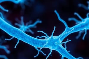
What is a neuron?
What is a neuron?
A nerve cell with all its processes.
What is an Axon?
What is an Axon?
A long process that carries impulses away from the cell body.
What are Dendrites?
What are Dendrites?
Signup and view all the flashcards
What are the Meninges?
What are the Meninges?
Signup and view all the flashcards
What is Cerebrospinal Fluid (CSF)?
What is Cerebrospinal Fluid (CSF)?
Signup and view all the flashcards
What is the Choroid Plexus?
What is the Choroid Plexus?
Signup and view all the flashcards
What are the Ventricles of the Brain?
What are the Ventricles of the Brain?
Signup and view all the flashcards
What is Grey Matter?
What is Grey Matter?
Signup and view all the flashcards
What is White Matter?
What is White Matter?
Signup and view all the flashcards
What are Forebrain, Midbrain, and Hindbrain?
What are Forebrain, Midbrain, and Hindbrain?
Signup and view all the flashcards
What is the Thalamus?
What is the Thalamus?
Signup and view all the flashcards
What is the Amygdaloid body?
What is the Amygdaloid body?
Signup and view all the flashcards
What is the Tectum?
What is the Tectum?
Signup and view all the flashcards
What is the Medulla Oblongata?
What is the Medulla Oblongata?
Signup and view all the flashcards
What is the Pons?
What is the Pons?
Signup and view all the flashcards
What is the Cerebellum?
What is the Cerebellum?
Signup and view all the flashcards
What is the Medulla Oblongata?
What is the Medulla Oblongata?
Signup and view all the flashcards
What is the trapezoid body?
What is the trapezoid body?
Signup and view all the flashcards
What is the Pons?
What is the Pons?
Signup and view all the flashcards
What is the Cerebellum?
What is the Cerebellum?
Signup and view all the flashcards
What is the Spinal cord?
What is the Spinal cord?
Signup and view all the flashcards
What is the Pia Mater?
What is the Pia Mater?
Signup and view all the flashcards
What is the Arachnoid Mater?
What is the Arachnoid Mater?
Signup and view all the flashcards
What is the Dura Mater?
What is the Dura Mater?
Signup and view all the flashcards
Study Notes
- Neurology studies the nervous system
- The nervous system processes internal and external environmental changes, regulating appropriate bodily functions in response
Nervous System Details
- The neuron is the functional unit
- A neuron consists of an axon, dendrites/dendrons, and a cell body
- Axons carry impulses away from the cell body
- Dendrites/dendrons carry impulses toward the cell body
- The cell body contains a nucleus, neuroplasm, neurofibrils, Nissl bodies, mitochondria, Golgi apparatus, ER, ribosomes, inclusions, neurosecretory materials, and a cell membrane
Neuron Classification
By Number of Processes
- Apolar neurons have no processes
- Unipolar neurons have one process
- Bipolar neurons have two processes
- Pseudo-unipolar neurons are a subtype
- Multipolar neurons have multiple processes
By Relative Lengths
- Golgi type-I neurons have short, numerous dendrites and long axons, found in peripheral locations
- Golgi type-II neurons have short axons, morphologically similar to dendrites, confined to gray matter for synaptic connections
- Amacrine neurons, unique to the retina, contain terminal fibers of axons and dendrons but lack axons
The Nervous System
- The central nervous system (CNS) is the body's master control unit
- The peripheral nervous system (PNS) links the body to the outside world
- The CNS includes the spinal cord and brainstem
- The brainstem connects the brain to the spinal cord
- The brain is divided into the hindbrain, midbrain, and forebrain
- The PNS consists of the autonomic and somatic nervous systems
- The autonomic NS regulates involuntary bodily processes
- The autonomic NS includes functions like regulating heart rate, respiration, digestion, and pupil contraction, operating automatically without conscious direction
- The somatic NS carries sensory information to the CNS, relays motor commands to muscles, and controls voluntary movements
- The craniospinal NS consists of the cranial and spinal nerves
- The sympathetic NS prepares the body for action and stress ("fight or flight")
- The parasympathetic NS calms the body and helps conserve energy
Brain Meninges
- Meninges are protective membranes surrounding the brain and spinal cord, providing structural integrity, cushioning, and infection defense
- Cerebrospinal fluid (CSF) are present in the brain
- From outermost to innermost the meninges layers are: dura mater, arachnoid mater, and pia mater
Dura Mater
- The dura mater is the toughest and outermost meningeal layer
- Made of dense, fibrous connective tissue which gives mechanical protection to the brain
- Has two layers: periosteal and meningeal
- The periosteal layer attaches to the inner surface of the skull
- The meningeal layer is closer to the brain and continuous with the dura mater of the spinal cord
- Structures formed include: falx cerebri, tentorium cerebelli, venous sinuses, and diaphragm sellae
Arachnoid Mater
- The arachnoid mater is a thin, web-like membrane located beneath the dura mater
- Has a spiderweb-like appearance due to delicate fibers that extend downwards
- The subarachnoid space beneath it is filled with CSF
- Arachnoid villi or granulations protrude into dural venous sinuses and allow CSF to drain into the bloodstream
Pia Mater
- The pia mater is the thinnest and innermost layer, attached directly to the brain's and spinal cord's surface
- Closely follows brain contours including sulci and gyri
- Contains rich vascularization to provide oxygen and nutrients
Meningeal Spaces
- The epidural space is located between the skull and dura mater (in the spinal cord)
- Contains fat and blood vessels
- The subdural space lies between the dura and arachnoid mater
- A potential space that may fill with fluid or blood during trauma
- The subarachnoid space is between the arachnoid and pia mater
- Filled with CSF, which cushions the brain and spinal cord from mechanical shocks
- Houses blood vessels that supply the brain
Cerebrospinal Fluid (CSF)
- CSF is a clear, colorless fluid surrounding the brain and spinal cord
- CSF is produced primarily by the choroid plexus via capillaries located in the brain ventricles
- Ependymal cells of the choroid plexus secrete CSF through active filtration of blood plasma
Brain Ventricles
- The brain consists of network of cavities filled with cerebrospinal fluid (CSF)
CSF Flow
- CSF forms in the lateral ventricle
- The CSF flows through the foramen of Monro
- It then progresses to aqueductus Sylvius
- CSF then goes from the third ventricle to the fourth ventricle
- CSF flows through the foramen of Magendie and foramen of Luschka
- Then proceeds through the cisterna magna and cisterna lateralis
- The fluid flows through central canal
- CSF moves to Subarachnoid spaces
- The CSF then passes through the cerebral hemispheres and spinal cord
Brain Embryology
- During brain development, there are three primary vesicles
- The primary brain vesicles are the prosencephalon (forebrain), mesencephalon (midbrain) and rhombencephalon (hindbrain)
Brain Morphology
- Brains are irregularly shaped vs. the regular shape of the spinal cord
- Brain size does not correspond to animal size
- Average brain weighs:
- 500 grams in ox
- 1.5 kg in humans
- 750 grams in horse
- 60 grams in dog
Brain Terminologies
- Nuclei are clusters of neuron cell bodies located deep within the brain or spinal cord
- Ganglia are collections of neuron cell bodies outside the central nervous system, in the peripheral nervous system (PNS)
- Tracts are bundles of axons (nerve fibers) within the central nervous system
- Nerves are bundles of axons (nerve fibers) inside the peripheral nervous system
- Commissures are bundles of nerve fibers connecting corresponding regions of the left and right hemispheres, enabling communication
Brain Views
- The cerebral hemisphere and cerebellum are visible from a dorsal view
- The medulla oblongata is partially visible
- Semi-ovoid cerebral hemispheres are bisected by a deep longitudinal fissure and separated from the cerebellum by a transverse fissure
- Ridges (gyri) and grooves (sulci) create a distinct pattern on each hemisphere
- Caudal parts, medulla oblongata, pons, and midbrain are visible in Ventral Views
- The forebrain, including the hypothalamus, is part of the cranial section
- The crossing/chiasm formed by the optic nerve, and round piriform lobes are also part of the cranial section
- Superficial origins of all cranial nerves except the trochlear are visible
Brain Matter
- Gray matter contains neuron cell bodies, dendrites, and unmyelinated axons
- Gray matter mainly integrates information and processes
- Gray matter is found in butterfly shaped structures in the inner spinal region
- Gray matter is slower at signal transmission
- White matter contains myelinated axons (nerve fibers)
- White matter transmits signals
- White matter lies beneath the gray matter
Brain Sections
- The Brain is split into: Forebrain, Midbrain and Hindbrain
- The Forebrain splits into: Diencephalon and Telecephalon
- Diencephalon contains: epithalamus, thalamus, metathalamus, & hypothalamus
- Telecephalon contains: cerebral hemispheres
- Midbrain components is mesencephalon
- Hindbrain contains: myelencephalon and metencephalon
- Myelencephalon: medulla oblongata
- Metencephalon: pons and cerebellum
The Forebrain
- Split into two main parts :
- Diencephalon
- Telecephalon
- In Diencephalon, following components are present :
- Epithalamus
- Thalamus
- Sub thalamus (dorsal)
- Metathalamus
- Hypothalamus
- Following part present in Telecephalon :hemispheres
Epithalamus
- Main portion of the epithalamus is the pineal gland.
- Contains a small ovoid gland, positioned dorsally between thalami and corpora quadrigemina.
- Positioned in posterosuperior the third Ventricle
- Hollow cavity which is a small recess of the Third ventricle
- The gland helps regulate behavior, and ovarian activity in animals(sheep, horse).
Thalamus
- Largest portion of the Diencephalon
- Positioned in the the walls of the 3rd ventricle
- Dorsal thalamus is composed of a large number of nuclei
- Subthalamus is a continuation of mesencephalon.
Hypothalamus
- Makes floor and walls up of 3rd Ventricle
- Optic chiasm is present in the front, near the optic nerves
- Near the mammillary body, which is connected to the the pituitary glands
Metathalamus
- Consists of lateral and medial bodies. Lateral geniculate body: receives fibers from optic tract
- Medial geniculate body: Relays acoustic signals to the cortex
Telecephalon
- Main component is Hemisphere
- Hemisphere Structure Information:
- The largest portion is the brain with symmetrical halves.
- It is connected by commisural fibers.
- The corpus callosum, and spreads radial in the white matter
- Has high concentration of cerebral cortex matters
- Has underlying white matter
- Holds accumulations of base Nuclei
Pallium/ Cerebral cortex/ Surface grey matter:
The pallium components include three sections, as divided up by matter:
- Paleopallium is limited to the brains base
- Neopallium: Large portion of telencephalon contains high concentration of gyri and sulci
- Archipallium contains the hippocampus.
Rhinencephalon
- Encompasses the olfactory bulb, tract, and striae, trigonium olfactorium, and piriform lobe
- Structures elongate and curve upwards in front of the frontal pole
- Olfactory Bulbs : The convex exterior is fit into the ethmoid fossa
- Olfactory Tracts: Have wide range of white matter
- The nerves are contained on the lateral cavities.
- The two types of Olfactory Striae:
- Lateral: larger than medial
- Medial both join at the piriform lobe
Limbic system/Visceral brain:
- Controls emotional stimuli
- Contains olfactory and piriform lobes
- Houses the hypothalmus.
- Amygdaloid Structure:
- Almond shaped, concentrated near the base.
- The hippocampus works as a memory center by transfer of cerebral cortex
- Balances emotion along the cerebral cortex The limbic system is also concerned with emotion, behavior, endocrine and somatic functions
Midbrain
-
Contains three layers: dorsal, middle, ventral crura/cerebral peduncles.
-
Midbrain Structures include : the forebrain, midbrain, and hindbrain .
-
The dorsal tectum consists of paired rostral and caudal swelliings called colliculi.
-
Caudal/ inferior colliculi relays center called the auditiory pathway
-
Midbrain contains the mesencephalic/cerebal part of 3rd and fourth ventricles
Cerebral Crura Two-fold Structure
- Two main peduncles make up for the structure and connect to the spiral
- Consists of ascending and descending pieces of spiral cord.
- The tuber is the hollow with tuber cinerum
- Covered by mammillary body/corpus albicans
Substantia Nigra
- Contains an abundance of pigmented cells
- Has a large concentration of the midbrain
Hindbrain
- The Hind brain is a large mass spanning from the pons to the medulla and cerebullem
- Medulla oblongata, continuation of spinal cord and extends from the foramen magnum to caudal margin of pons
- Ventral surface: marked by median fissure, continues with that of spinal cord, flanked by longitudinal ridges c/a pyramids
Trapezoid body/ Pyramidal decussation Functions
- Consists of ascending.descsnding fibers and the 5-12th craneal nerves.
- Regulates the heart, respiration and regulation of body temperature
POONS - Nuclei Structure Information
- Consists of Nuclei, affects coordination ,cerebellum controlled by vitals and repiration
- Contains a trgeminal nerve from the frontal lobe
- Located beetween the mo the mb
- Contain trnsverse grooves
The Cerrebelum Structures
-
Located in the middle of the poons to medulla, covers 3 lobes, anterior posterrior
-
Each lobe contains longitudinal fissure
-
Center zone is called vermis
-
Outer zone ir irregular
-
Surface of the brain stem is covered by multiple curves and curves of fold and folia.
-
The tissue gives tertiary laminae which look like arbors = arborvitae/ tree of life.
Cerebellum Three Pundicules
Structures:
- Superior: connects cerrebelum with mind brain
- Middle: comes from the spons 4, Inferier comes from MO to cerebrum Functions Controls posture and stabiility receives inputs from auditirory and visual systems Cerebellum has three peduncles
Spinal Cord Structure Information
- a medulla spinalis
- Protected via cns Located near the vertebral collumn Cylindricaly divided into two halves with dorsal and longitudinal fissurers. Cranial end Continuous with the medulla, around foramen magnum.
- Has a caudial end in many parts mostly near domestic vertebrae to the middle sacrum. Spinal cord is divided into cervical thoracic lumsacral and coccygeal.
Spinal cord structure
-
Divided into cervical, thoracic, lumbar, sacral and coccygeal
- Spinal consists of gray Matter(inner), and outer matter (outer Part) -Has dorsal/vetral root forming spinal -
Functions, composed of Neuronal synapse
. The spinal cord also composed of motor trats.
Spinal cord composition
- Divided in thee sets/horns, with Gray Matter
Spinal matter is devided into thrree horns
- Contains sensory that reeive the sensory
Studying That Suits You
Use AI to generate personalized quizzes and flashcards to suit your learning preferences.