Podcast
Questions and Answers
In neurons, what would be the consequence of a mutation that disrupts the function of the Na+/K+ ATPase pump, particularly concerning its role in maintaining the resting membrane potential?
In neurons, what would be the consequence of a mutation that disrupts the function of the Na+/K+ ATPase pump, particularly concerning its role in maintaining the resting membrane potential?
- The neuron's ability to generate action potentials would be severely compromised or abolished. (correct)
- The neuron would be unaffected due to compensatory mechanisms by other ion channels.
- The resting membrane potential would become more negative as potassium efflux is enhanced.
- The neuron would hyperpolarize due to an excess of intracellular positive charge.
Considering the complexities of neurotransmission, what is the most plausible effect of a drug that inhibits acetylcholinesterase at the neuromuscular junction, while also acting as a muscarinic acetylcholine receptor antagonist in the parasympathetic nervous system?
Considering the complexities of neurotransmission, what is the most plausible effect of a drug that inhibits acetylcholinesterase at the neuromuscular junction, while also acting as a muscarinic acetylcholine receptor antagonist in the parasympathetic nervous system?
- Unpredictable, oscillating muscle activity alternating between flaccidity and spasticity, alongside normal parasympathetic function.
- Muscle rigidity due to sustained acetylcholine levels at the neuromuscular junction, along with suppressed parasympathetic responses. (correct)
- No discernible effect, as the two mechanisms of action would negate each other.
- Enhanced muscle relaxation combined with increased parasympathetic activity.
Within the context of the blood-brain barrier (BBB), how would the disruption of astrocyte end-feet processes impact the selective permeability of the BBB?
Within the context of the blood-brain barrier (BBB), how would the disruption of astrocyte end-feet processes impact the selective permeability of the BBB?
- It would enhance the transport of all molecules across the BBB due to increased vasodilation.
- It would lead to increased selective permeability due to enhanced tight junction integrity.
- It would have no effect on the BBB, as astrocytes play a negligible role in its function.
- It would result in decreased selective permeability due to impaired regulation of endothelial tight junctions. (correct)
In the context of neuroplasticity and recovery after a stroke, what is the most accurate description of how Constraint-Induced Movement Therapy (CIMT) facilitates cortical reorganization?
In the context of neuroplasticity and recovery after a stroke, what is the most accurate description of how Constraint-Induced Movement Therapy (CIMT) facilitates cortical reorganization?
Considering the complexity of cranial nerve function, what would be the most likely presentation of a patient with selective damage to the solitary nucleus within the medulla oblongata?
Considering the complexity of cranial nerve function, what would be the most likely presentation of a patient with selective damage to the solitary nucleus within the medulla oblongata?
Given the intricacies of neuronal signaling, what characterizes the primary mechanism by which myelin sheaths increase the velocity of action potential propagation along an axon?
Given the intricacies of neuronal signaling, what characterizes the primary mechanism by which myelin sheaths increase the velocity of action potential propagation along an axon?
Considering the regional specialization of the cerebral cortex, what is the most plausible outcome of a highly selective lesion in the right parietal lobe, sparing other cortical areas?
Considering the regional specialization of the cerebral cortex, what is the most plausible outcome of a highly selective lesion in the right parietal lobe, sparing other cortical areas?
If a patient presents with an expressive aphasia characterized by non-fluent speech and impaired grammar, with preserved comprehension, what area of the cerebral cortex is most likely affected?
If a patient presents with an expressive aphasia characterized by non-fluent speech and impaired grammar, with preserved comprehension, what area of the cerebral cortex is most likely affected?
What would be the direct consequence of a mutation that impairs the function of gamma-aminobutyric acid (GABA) receptors in the central nervous system?
What would be the direct consequence of a mutation that impairs the function of gamma-aminobutyric acid (GABA) receptors in the central nervous system?
What is the most reliable mechanism by which astrocytes contribute to regulating synaptic transmission and preventing excitotoxicity in the central nervous system?
What is the most reliable mechanism by which astrocytes contribute to regulating synaptic transmission and preventing excitotoxicity in the central nervous system?
What characterizes the interaction between the sympathetic and parasympathetic nervous systems in the regulation of cardiovascular function?
What characterizes the interaction between the sympathetic and parasympathetic nervous systems in the regulation of cardiovascular function?
What effect does the basal ganglia have on motor control?
What effect does the basal ganglia have on motor control?
In a patient diagnosed with damage to the cerebellum, what is the most probable presentation of motor deficits?
In a patient diagnosed with damage to the cerebellum, what is the most probable presentation of motor deficits?
Which functional consequence would most clearly show that there is damage to the spinothalamic tract?
Which functional consequence would most clearly show that there is damage to the spinothalamic tract?
What is the impact of lesions in the amygdala on emotional and behavioral responses?
What is the impact of lesions in the amygdala on emotional and behavioral responses?
In the context of spinal cord anatomy, what functional difference distinguishes the anterior horn from the posterior horn?
In the context of spinal cord anatomy, what functional difference distinguishes the anterior horn from the posterior horn?
How would you characterise the distinctive role of microglia compared to other glial cells in the central nervous system?
How would you characterise the distinctive role of microglia compared to other glial cells in the central nervous system?
What is the sequence of meningeal layers from superficial to deep?
What is the sequence of meningeal layers from superficial to deep?
Considering the anatomy of spinal nerves, what accurately describes the composition and functional role of the dorsal root?
Considering the anatomy of spinal nerves, what accurately describes the composition and functional role of the dorsal root?
What is the predicted outcome of complete transection of the spinal cord at the thoracic level?
What is the predicted outcome of complete transection of the spinal cord at the thoracic level?
What is an accurate description of the oculovestibular reflex (caloric ice water test), and what does its altered response indicate?
What is an accurate description of the oculovestibular reflex (caloric ice water test), and what does its altered response indicate?
The Glasgow Coma Scale (GCS) assess what functions?
The Glasgow Coma Scale (GCS) assess what functions?
What would be the most appropriate intervention for identifying changes in neurological status?
What would be the most appropriate intervention for identifying changes in neurological status?
When performing a neurological examination, what is the most important step to assessing the patient's mental status effectively?
When performing a neurological examination, what is the most important step to assessing the patient's mental status effectively?
After administering painful stimuli to a patient, what observation is crucial to accurately determining the Glasgow Coma Scale (GCS) motor score?
After administering painful stimuli to a patient, what observation is crucial to accurately determining the Glasgow Coma Scale (GCS) motor score?
After a traumatic brain injury, a patient consistently exhibits decerebrate posturing. Where is the most probable location of neurological damage?
After a traumatic brain injury, a patient consistently exhibits decerebrate posturing. Where is the most probable location of neurological damage?
A doctor suspects a lesion on the spinal cord. Which clinical test is most appropriate for determining function of the spinal cord?
A doctor suspects a lesion on the spinal cord. Which clinical test is most appropriate for determining function of the spinal cord?
To accurately interpret the results of the Caloric Ice Water Test you must understand the pathology to the...
To accurately interpret the results of the Caloric Ice Water Test you must understand the pathology to the...
What is the typical rate of production of cerebrospinal fluid (CSF) in an adult, assuming normal physiological conditions?
What is the typical rate of production of cerebrospinal fluid (CSF) in an adult, assuming normal physiological conditions?
What structures are protected by both the Skull and CSF (Cerebrospinal Fluid)?
What structures are protected by both the Skull and CSF (Cerebrospinal Fluid)?
What is a key component of the spinal cord, in addition to grey and white matter?
What is a key component of the spinal cord, in addition to grey and white matter?
The facial nerve is a Mixed cranial nerve, but primarily is involved in controlling what?
The facial nerve is a Mixed cranial nerve, but primarily is involved in controlling what?
Both Sensory or Afferent and Motor or Efferent divisions are principal subdivisions of the Peripheral Nervous System, but what is a difference between the two divisions?
Both Sensory or Afferent and Motor or Efferent divisions are principal subdivisions of the Peripheral Nervous System, but what is a difference between the two divisions?
Damage to the cerebellum is most likely going to affect what?
Damage to the cerebellum is most likely going to affect what?
The Thalamus has some important roles related to sensory and memory, but what is its main role?
The Thalamus has some important roles related to sensory and memory, but what is its main role?
What is the most important role of the midbrain?
What is the most important role of the midbrain?
Flashcards
Nervous System
Nervous System
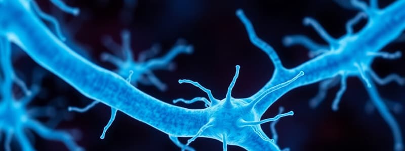
The organ system containing a network of specialized cells called neurons; it controls various bodily processes.
Dendrites
Dendrites
Threadlike structures of a neuron that convey incoming electrochemical messages toward the cell body.
Axon
Axon
A long projection of a neuron that generates impulses away from the cell body.
Cell Body
Cell Body
Signup and view all the flashcards
Synapses
Synapses
Signup and view all the flashcards
Myelin Sheath
Myelin Sheath
Signup and view all the flashcards
Nodes of Ranvier
Nodes of Ranvier
Signup and view all the flashcards
Neurotransmitters
Neurotransmitters
Signup and view all the flashcards
Amines group
Amines group
Signup and view all the flashcards
Acetylcholine
Acetylcholine
Signup and view all the flashcards
Norepinephrine
Norepinephrine
Signup and view all the flashcards
Dopamine
Dopamine
Signup and view all the flashcards
Serotonin
Serotonin
Signup and view all the flashcards
Amino Acid group
Amino Acid group
Signup and view all the flashcards
Peptide group
Peptide group
Signup and view all the flashcards
Substance P
Substance P
Signup and view all the flashcards
Supporting Cells
Supporting Cells
Signup and view all the flashcards
Astroglia/Astrocytes
Astroglia/Astrocytes
Signup and view all the flashcards
Ependymal cells
Ependymal cells
Signup and view all the flashcards
Microglia
Microglia
Signup and view all the flashcards
Oligodendroglia
Oligodendroglia
Signup and view all the flashcards
Central Nervous System
Central Nervous System
Signup and view all the flashcards
Cerebrum
Cerebrum
Signup and view all the flashcards
Gyri/Gyrus
Gyri/Gyrus
Signup and view all the flashcards
Sulci/Sulcus
Sulci/Sulcus
Signup and view all the flashcards
Fissures
Fissures
Signup and view all the flashcards
Frontal Lobe
Frontal Lobe
Signup and view all the flashcards
Broca's area
Broca's area
Signup and view all the flashcards
Parietal Lobe
Parietal Lobe
Signup and view all the flashcards
Temporal Lobe
Temporal Lobe
Signup and view all the flashcards
Occipital Lobe
Occipital Lobe
Signup and view all the flashcards
Diencephalon
Diencephalon
Signup and view all the flashcards
Thalamus
Thalamus
Signup and view all the flashcards
Hypothalamus
Hypothalamus
Signup and view all the flashcards
Epithalamus
Epithalamus
Signup and view all the flashcards
Brain Stem
Brain Stem
Signup and view all the flashcards
Midbrain
Midbrain
Signup and view all the flashcards
Pons
Pons
Signup and view all the flashcards
Medulla Oblongata
Medulla Oblongata
Signup and view all the flashcards
Cerebellum
Cerebellum
Signup and view all the flashcards
Study Notes
Nervous System Overview
- The nervous system includes a network of specialized cells known as neurons.
- It controls various bodily processes.
- The two main parts are the Central Nervous System (CNS) and the Peripheral Nervous System (PNS).
- Sensory input, processing and interpreting information, and motor output are key nervous system functions.
- Nervous system composition consists of conducting and supportive cells.
Cells of the Nervous System: Neurons
- Neurons, also known as neurones or nerve cells, are key to the nervous system.
- Dendrites are threadlike structures conveying incoming electrochemical messages to the cell body.
- An axon is a long projection that generates impulses away from the cell body.
- The cell body serves as the neuron's center and contains the nucleus.
- Synapses facilitates communication between neurons and other cells.
- The myelin sheath is a protective, fatty material covering long nerve fibers.
- The myelin sheath protects and insulates nerve fibers, increasing the rate of nerve impulses
- Schwann cells myelinate the axon.
- Nodes of Ranvier: Gaps or indentations between myelin sheaths.
Neurotransmitters
- Neurotransmitters are chemicals produced by neurons.
- Neurotransmitters either excite or inhibit a target cell's activity, modulating their action.
- Neurotransmitters act to to potentiate, terminate, or modulate a specific action.
- They communicate messages from one neuron to a specific target tissue.
- Amines are neurotransmitters containing molecules of carbon, hydrogen, and nitrogen.
- Acetylcholine is the most widely used neurotransmitter in the human body.
- Norepinephrine is a major neurotransmitter of the sympathetic nervous system.
- Dopamine is involved in muscle movement and controls the secretion of prolactin.
- Serotonin helps to control mood and sleep and also inhibits pain pathways.
- Amino acid group neurotransmitters are organic compounds with an amino group (NH2) and a carboxylic acid group (COOH).
- Glycine, glutamic acid, aspartic acid, and gamma-aminobutyric acid (GABA) are examples of amino acid group neurotransmitters.
- Peptide group neurotransmitters contain at least 2, and sometimes as many as 100 amino acids.
- Substance P influences the sensation of pain.
Supporting Cells: Neuroglia
- Neuroglia, also known as glial cells or non-neuronal cells, support neuron function.
- They cannot transmit impulses.
- Neuroglia are 50 times more abundant than neurons and make up 40% of the brain's bulk.
- Astroglia/astrocytes supply nutrients to neurons, help maintain their electrical potential, and assist in forming the blood-brain barrier.
- Ependymal cells help produce cerebrospinal fluid (CSF).
- Microglia are phagocytic cells that ingest and digest microorganisms and waste products from injured neurons.
- Oligodendroglia support and electrically insulate CNS axons by forming protective myelin sheaths.
Central Nervous System (CNS)
- The brain and spinal cord comprise the CNS.
- The CNS is surrounded and protected by bone, including the skull and vertebrae.
- Fluid and tissue also insulate the brain and spinal cord.
- The brain, part of the CNS contained within the cranium, consists of spongy pinkish gray nerve tissue.
- The brain accounts for around 2% of total body weight (TBW).
- Key brain regions include the cerebrum, cerebellum, diencephalon, and brainstem.
Cerebrum
- The cerebrum is the largest and most prominent part of the brain and governs higher mental processes.
- The cerebrum consists of right (R) and left (L) hemispheres separated by the great longitudinal fissure.
- The corpus callosum joins the lower portion of the fissure joining the two hemispheres.
- Gyri or Gyrus are brain convolutions.
- Sulci or Sulcus are shallow grooves.
- Fissures are deeper grooves in the brain.
- Layers include gray and white matter, with gray matter managing muscle control, sensory perception, memory, emotions, and speech.
- Frontal Lobe: The largest lobe, responsible for concentration, abstract thought, information storage, memory, motor function, Broca's area as well as affect, judgment, personality and inhibiting actions.
- Parietal Lobe: Primary sensory cortex to analyze sensory information, relays interpretation to thalamus, essential to awareness of body in space.
- Temporal Lobe: Contains auditory receptive areas, interpretive area for somatization, visual, auditory processing and Wernicke's area.
- Occipital Lobe: The posterior lobe and is primarily responsible for visual interpretation.
Diencephalon
- The diencephalon contains the Thalamus, Hypothalamus, and Epithalamus.
- The thalamus is an important relay station that processes all sensory, memory, and pain impulses
- The hypothalamus, anterior and inferior to the thalamus, contains nuclei that are very important for maintaining homeostasis, regulating visceral activities, and regulating pituitary secretion of hormones.
- The epithalamus is located superior and posterior to thalamus, involved in emotional/visceral responses to odor, and contains the pineal body.
Brain Stem
- The brain stem connects the brain to the spinal cord.
- Midbrain: Connects the pons and cerebellum with the cerebral hemispheres, acting as a message coordinator in and out of the brain to the spinal cord.
- Pons: Helps regulate breathing; houses the respiratory center.
- Medulla Oblongata: Is the most inferior part of the brainstem containing centres that control heart rate (HR), blood pressure (BP), sneezing, coughing, breathing, swallowing, and vomiting.
Cerebellum
- Posterior to the midbrain and pons, and below the occipital lobe.
- Also know as the "little brain".
- Responsible for balance, coordination, and muscle tone.
Brain Protection
- The skull, a bony structure, protects the brain from injury.
- Major bones protecting the brain include the frontal, temporal, parietal, and the occipital.
- Suture lines include the coronal, sagittal, lambdoidal, and squamosal.
-Meninges, a fibrous connective tissue, covers the brain and spinal cord, providing protection, support, and nourishment.
- Dura Mater: The outermost layer, tough, inelastic, fibrous, and gray.
- Epidural space is the potential space between the dura mater and skull in the cranium.
- Arachnoid Mater: The middle membrane, white, containing the choroid plexus, also has unique fingerlike projections, arachnoid villi .
- Pia Mater: The innermost membrane, thin, transparent layer that hugs the brain closely.
- Cerebrospinal Fluid (CSF) protects the central nervous system.
- It's clear, colorless and acts as a protective cushion.
- It is produced at about 500 ml per day.
- CSF is colorless and clear containing a normal specific gravity of 1.00, a minimal WBC count, glucose, and some electrolytes while having no red blood cells (RBC).
Spinal Cord
- Spinal cord runs along the dorsal side of the body, linking the brain to the rest of the body.
- Carries sensory and motor information to and from the brain.
- The bony vertebrae encase the spinal cord.
- Meninges also surround the spinal cord, with the average spinal cord being 45cm (18") long, about the thickness of a finger.
- Gray matter: the inner core, mostly of cell bodies and dendrites.
- White matter: made up of fiber bundles that are ascending and descending.
- Vertebral column: Consists of 7 cervical, 12 thoracic, 5 lumbar vertebrae, sacrum (fused mass of 5 vertebrae); coccyx.
- Lower portion is broader than upper portion and corresponds to the anterior horn
- Anterior Horn: Contains cells with fibers that form the anterior (motor) root end; essential for the voluntary and reflex activity of the muscles they innervate.
- Posterior Horn: Contains cells with fibers that enter over the posterior (sensory) root end, thus as a relay station in the sensory/ reflex pathway
Peripheral Nervous System (PNS)
- PNS has two principal subdivisions: The sensory and motor divisions.
- Sensory (afferent) division: Carries information from the outside world; sensory receptors are in different parts of the body such as somatic and visceral sensory fibers.
- Motor (efferent) division Transmits impulses from the brain & spinal cord to the muscles such as the voluntary nervous system or the autonomic nervous system.
- Cranial nerves: Carry impulses to and from the brain and contain 12 pairs emerging from the brain's lower surface.
- Spinal nerves: Carry impulses to and from the spinal cord, containing 31 pairs arising from the spinal cord, which pass out through the vertebrae.
- Consisting of 8 cervical, 12 thoracic, 5 lumbar, 5 sacral & 1 coccygeal.
- The above nerves communicates to lines of the body.
Autonomic Nervous System (ANS)
- The autonomic nervous system operates without conscious control, regulating the activities of internal organs.
- Maintenance and restoration of internal homeostasis is generally the ANS' responsibility and is regulated by centers in the spinal cord, in brain stem, and in the hypothalamus.
- The ANS is a major division along with two subsystems, SNS and PNS
- SNS; predominantly excitatory responses, also known as the (fight or flight response).
- Effects: bronchioles dilate, heart's contractions are stronger and faster, arteries to heart and voluntary muscles dilate, peripheral BV constrict
- PNS; predominant during quiet, non-stressful conditions.
- Each of these subsystems operates in the reverse of the other (antagonism).
Cranial Nerve Assessment
- I Olfactory: Test by identifying common odors.
- II Optic: Snellen chart and peripheral vision checks.
- III Oculomotor: Check for pupil constriction.
- IV Trochlear: Check for convergence as the object is brought near the eyes.
- V Trigeminal: Check for strength of lid closure, identify location of stimulus, assesses the ability to feel light, dull and sharp sensation on the face, check jaw strength, check corneal reflex.
- VI Abducens: Check for eye movement from left to right.
- VII Facial: Check for symmetry of facial expressions and muscle strength. Identify sweet, sour and salty tastes.
- VIII Acoustic: Weber and Rinne test for hearing loss, Romberg’s test for balance.
- IX: Identify sweet, sour and salty tastes and check gag and swallowing reflex.
- X - Ask the client to say “Ah” - uvula should rise midline and check ability to swallow
- XI - Client shrugs shoulders against resistance and turns the head to one side against the resistance of the hand
- XII - Client stick out tongue, observe for deviations or tremors, check strength of tongue movement presses against tongue blade
Glasgow Coma Scale
- Assesses eye-opening, verbal, and motor responses to determine brain injury severity.
- Eye response ranges from 4 (spontaneous) to 1 (no response).
- Verbal response ranges from 5 (oriented) to 1 (no response).
- Motor response rangers from 6 (obeys commands) to 1 (no response).
- Minor injury = 13-15, Moderate brain injury = 9-12
- Severe brain injury = 3-8.
- Decorticate posture means the arms are adducted and flexed, with the wrists and fingers flexed on the chest. The legs are stiffly extended and internally rotated, with plantar flexion of the feet.
- Decerebrate posture: Damage to the upper brain stem; arms are adducted and extended, wrists pronated, fingers flexed, and legs stiffly extended with plantar flexion of feet.
Levels of Consciousness
- Changes is the most sensitive indicator of neurologic function and is first assessment to change when there's neurologic injury.
- Level 1 - Highest level; conscious, alert, fully-awake, is oriented to person, place and time.
- Level 2 - Listless, lethargic, somnolent and obtunded and can be aroused from sleep but when stimulation ceases, tends to fall asleep again.
- Level 3 - Stuporous; Response can only be evoked from a strong and continuous stimuli, results in the inability to have the patient have elicit a strong and continuous stimuli (i.e., reflex withdrawal).
- Level 4 Semi-coma - no spontaneous movement but patient may demonstrate reflex activities like coughing, swallowing and vomiting or become incontinence. Level 5 - Coma/Deep Coma – absence of involuntary responses to any stimuli; Reflexes are absent as well may be constricted/dilated and does not react to light.
Examining Reflexes
- Reflexes are involuntary contractions of muscles or muscle groups in response to a stimulus.
- Classified as deep tendon, superficial or pathologic
- Assesses the intactness of the spinal reflex arc at various spinal cord levels.
- The limb should be relaxed while applying a short and snappy blow with a reflex hammer.
- Hold the hammer loosely in a relaxed manner, making a wrist action. Allow the hammer to bounce.
- Includes: Patellar, Achilles, Triceps, and Biceps
- Responses: 0 no response, 1+ is diminished, low normal, 2+ is average, normal, 3+ brisker than normal, and 4+ very brisk and hyperactive
- Oculocephalic Reflex (Doll’s Eye Phenomenon) done by holding the person’s eyelids open and rotating the head from side to side.
- The reflex is present if the eyes move in the opposite direction of the head movements.
- Oculovestibular Reflex (Caloric Ice Water Test) - Uses temperature differences to diagnose damage to the ear or brainstem.
- Cold water = FAST phase of nystagmus to the Opposite side from the cold water filled ear.
- Warm water = FAST phase of nystagmus to the Same side as the warm water filled ear
Pupillary Exam
- Changes can indicate abnormal intracranial pressure (ICP).
- Normal findings: PERRLA (Pupil, Equal, Round and Reactive to Light and Accommodation.
- Sizes: Pinpoint (miotic:), Dilated (mydriatic), Normal 2 - 6mm.
- Ovoid may indicate IICP.
- Keyhole: patients who had iridectomy. Reaction to light
- Brisk, Sluggish: Suggests Meningitis or Subarachnoid Hematoma, Non-Reactive Comparison of pupils: Isocoria meaning both are same
- Anisocoria is when there is unequal size of pupils.
- AVPU scale - quick and easy method for testing neurological response.
- A: Awake: Alert and oriented to person, place and time, Acting reasonably to stimuli, Patient is aware of you approaching him/her
- V: Verbal Stimulation (responds to): Responds to voice and is Unawake but will respond by speaking/moaning, with facial expressions, limb movement, eyelid blinking.
- P: Verbal Stimulation (responds to) - Patient will only respond to painful stimuli as: Includes Suborbital pressure, Pinching the fleshy part near the axilla, or by doing the Trapezius squeeze, Shaking the shoulders, Earlobe pinch, and Nail pinch
- U: Unresponsive to verbal and pain stimuli.
- Trapezeus Squeeze - Use the index finger and thumb to squeezes about 1.5 to 2 inches of the trapezius muscle, Increase pressure gradually for up to 10 seconds, note the patient's response
Studying That Suits You
Use AI to generate personalized quizzes and flashcards to suit your learning preferences.


