Podcast
Questions and Answers
What does the term 'preload' refer to in cardiac physiology?
What does the term 'preload' refer to in cardiac physiology?
- The resistance the left ventricle must overcome during systole
- The amount of blood pumped out of the ventricles with each beat
- The total volume of blood the heart pumps in one minute
- The volume of blood in the ventricles at the end of diastole (correct)
Which cardiac output range is considered normal?
Which cardiac output range is considered normal?
- 10-12 L/min
- 4-8 L/min (correct)
- 8-10 L/min
- 2-4 L/min
What is considered a critical level for cardiac troponin?
What is considered a critical level for cardiac troponin?
- 1.0 ng/mL
- 0.1 ng/mL
- 0.5 ng/mL
- >1.5 ng/mL (correct)
What does an increased afterload indicate regarding cardiac physiology?
What does an increased afterload indicate regarding cardiac physiology?
What normal range is found for the ejection fraction?
What normal range is found for the ejection fraction?
What shape is the heart described as?
What shape is the heart described as?
Where is the heart located within the human body?
Where is the heart located within the human body?
What is the function of the aorta?
What is the function of the aorta?
What does the fibrous pericardium do?
What does the fibrous pericardium do?
Which vessels carry deoxygenated blood from the right ventricle?
Which vessels carry deoxygenated blood from the right ventricle?
Which layer of the pericardium is responsible for creating a lubricated environment?
Which layer of the pericardium is responsible for creating a lubricated environment?
What is the role of coronary arteries?
What is the role of coronary arteries?
What do pulmonary veins do?
What do pulmonary veins do?
What role does the pericardium play in heart function?
What role does the pericardium play in heart function?
What is the primary function of the coronary arteries?
What is the primary function of the coronary arteries?
Which structure directs deoxygenated blood back to the right atrium?
Which structure directs deoxygenated blood back to the right atrium?
What is the apex of the heart described as?
What is the apex of the heart described as?
Which heart sound is associated with the closing of semilunar valves?
Which heart sound is associated with the closing of semilunar valves?
What do the auricles of the heart do?
What do the auricles of the heart do?
In which chamber of the heart does oxygenated blood enter from the lungs?
In which chamber of the heart does oxygenated blood enter from the lungs?
What might an S3 heart sound indicate?
What might an S3 heart sound indicate?
Flashcards are hidden until you start studying
Study Notes
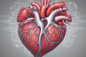
External Anatomy of the Heart
- The heart is approximately the size of a fist and has a cone-like shape.
- Located in the thoracic cavity, slightly left of the midline, behind the sternum.
- Surrounded by a double-walled sac called the pericardium.
Pericardium Structure
- Fibrous Pericardium: Tough outer layer prevents heart overfilling.
- Serous Pericardium: Inner layer consists of two parts:
- Parietal layer: Lines the fibrous pericardium.
- Visceral layer (epicardium): Adheres to the heart muscle.
Major Blood Vessels
- Aorta: Largest artery, carries oxygenated blood from the left ventricle to the body.
- Pulmonary Arteries: Transport deoxygenated blood from the right ventricle to the lungs.
- Pulmonary Veins: Return oxygenated blood from the lungs to the left atrium.
- Superior and Inferior Vena Cava: Bring deoxygenated blood from the body back to the right atrium.
- Coronary Arteries: Supply oxygen-rich blood to the heart muscle.
- Coronary Veins: Drain deoxygenated blood from the heart muscle back into the right atrium.
Heart Structure Details
- Apex: The pointed end of the heart, directed downward and to the left.
- Base: The broader upper portion, where major blood vessels enter and exit.
- Auricles: Small, ear-like pouches on atria that increase volume.
Physiology Related to External Features
- Pericardial Function: Anchors the heart and reduces friction during heartbeats.
- Coronary Circulation: Ensures heart muscle receives necessary oxygen and nutrients.
- Blood Flow Dynamics: Heart's orientation promotes efficient blood flow.
Blood Circulation Pathways
- Right Side: Pumps deoxygenated blood:
- Receives via Superior/Inferior Vena Cava → Right Atrium → Tricuspid Valve → Right Ventricle → Pulmonary Valve → Pulmonary Artery → Lungs.
- Left Side: Pumps oxygenated blood:
- Receives via Pulmonary Veins → Left Atrium → Mitral Valve → Left Ventricle → Aortic Valve → Aorta → Body.
Heart Sounds
- S1: Closing of atrioventricular valves; high-pitched, normal sound.
- S2: Closing of semilunar valves; high-pitched, normal sound.
- S3: Low-pitched; may indicate fluid overload, possibly normal.
- S4: Low-pitched; indicates ventricular resistance, considered abnormal.
- Murmurs: Low-pitched sounds that could indicate valve or wall defects, possibly abnormal.
Cardiac Terms
- Preload: Volume of blood in ventricles at the end of diastole.
- Afterload: Resistance the left ventricle must overcome during systole, impacting cardiac workload.
- Stroke Volume: Volume of blood pumped from the ventricles per beat; normal range is 60-100 ml/beat.
- Cardiac Output: Total blood pumped by the heart in one minute; calculated as CO = HR X SV, with a normal range of 4-8 L/min.
- Ejection Fraction: Percentage of blood expelled from the heart with each contraction; normal range is 50-70%.
Cardiac Biomarkers
- Troponin: Normal level is 0-0.4 ng/mL; elevated levels (≥1.5 ng/mL) indicate heart damage, particularly during acute myocardial infarction (MI).
- Creatine Kinase (CK): Normal level is 0-5 ng/mL; indicates muscle damage when elevated.
- Brain Natriuretic Peptide (BNP): Released when ventricles fill; provides insight into heart strain.
Studying That Suits You
Use AI to generate personalized quizzes and flashcards to suit your learning preferences.