Podcast
Questions and Answers
Which muscle is responsible for bringing the thumb across the palm?
Which muscle is responsible for bringing the thumb across the palm?
- Abductor Pollicis Brevis
- Flexor Pollicis Brevis
- Adductor Pollicis
- Opponens Pollicis (correct)
Which nerve innervates the superficial head of the flexor pollicis brevis?
Which nerve innervates the superficial head of the flexor pollicis brevis?
- Median Nerve (correct)
- Musculocutaneous Nerve
- Ulnar Nerve
- Radial Nerve
What action is performed by the abductor pollicis brevis?
What action is performed by the abductor pollicis brevis?
- Flexion of the thumb
- Opposition of the thumb
- Adduction of the thumb
- Abduction of the thumb (correct)
Which muscle is not classified as a thenar muscle, yet performs a related function?
Which muscle is not classified as a thenar muscle, yet performs a related function?
In terms of anatomical relationships, where is the opponens pollicis located in relation to the flexor pollicis brevis?
In terms of anatomical relationships, where is the opponens pollicis located in relation to the flexor pollicis brevis?
Which muscle of the hypothenar group specifically allows for the opposition of the little finger?
Which muscle of the hypothenar group specifically allows for the opposition of the little finger?
Which hypothenar muscle is responsible for flexing the proximal phalanx of the little finger?
Which hypothenar muscle is responsible for flexing the proximal phalanx of the little finger?
What is the specific nerve that innervates the hypothenar muscles?
What is the specific nerve that innervates the hypothenar muscles?
Which muscle originates from the pisiform bone?
Which muscle originates from the pisiform bone?
What is the anatomical insertion of the Opponens Digiti Minimi?
What is the anatomical insertion of the Opponens Digiti Minimi?
Which intrinsic muscle of the hand is responsible for flexing the fingers at the metacarpophalangeal joints and extending them at the interphalangeal joints?
Which intrinsic muscle of the hand is responsible for flexing the fingers at the metacarpophalangeal joints and extending them at the interphalangeal joints?
What is the primary nerve responsible for innervating the 3rd and 4th lumbricals in the hand?
What is the primary nerve responsible for innervating the 3rd and 4th lumbricals in the hand?
Where do the interossei muscles of the hand originate?
Where do the interossei muscles of the hand originate?
What is the role of intrinsic muscles in fine motor skills?
What is the role of intrinsic muscles in fine motor skills?
Which of the following muscles in the foot originates from the calcaneus?
Which of the following muscles in the foot originates from the calcaneus?
What is the primary action of the Biceps Brachii?
What is the primary action of the Biceps Brachii?
Which muscle originates from the upper eight or nine ribs?
Which muscle originates from the upper eight or nine ribs?
What is the insertion site of the Deltoid muscle?
What is the insertion site of the Deltoid muscle?
Which nerve is responsible for innervating the Pectoralis Major?
Which nerve is responsible for innervating the Pectoralis Major?
Which of the following muscles flexes the lumbar spine and compresses abdominal contents?
Which of the following muscles flexes the lumbar spine and compresses abdominal contents?
Which origin corresponds to the Latissimus Dorsi muscle?
Which origin corresponds to the Latissimus Dorsi muscle?
What is the insertion site for the Serratus Posterior Superior muscle?
What is the insertion site for the Serratus Posterior Superior muscle?
Which action is performed by the Rhomboid Major and Minor muscles?
Which action is performed by the Rhomboid Major and Minor muscles?
Which nerve innervates the Levator Scapulae muscle?
Which nerve innervates the Levator Scapulae muscle?
What are the nerve roots associated with the Trapezius muscle?
What are the nerve roots associated with the Trapezius muscle?
Which of the following muscles originates from the sacrum and coccyx?
Which of the following muscles originates from the sacrum and coccyx?
What is the insertion point of the Gluteus Medius muscle?
What is the insertion point of the Gluteus Medius muscle?
Which nerve is responsible for innervating the Gluteus Minimus muscle?
Which nerve is responsible for innervating the Gluteus Minimus muscle?
Identify the correct nerve root segments associated with the Gluteus Maximus muscle.
Identify the correct nerve root segments associated with the Gluteus Maximus muscle.
Which statement about the insertion of the Gluteus Maximus is correct?
Which statement about the insertion of the Gluteus Maximus is correct?
Which deep gluteal muscle originates from the ischial spine?
Which deep gluteal muscle originates from the ischial spine?
Which function is primarily associated with the deep gluteal muscles during dynamic activities?
Which function is primarily associated with the deep gluteal muscles during dynamic activities?
What is the nerve supply of the Quadratus Femoris muscle?
What is the nerve supply of the Quadratus Femoris muscle?
Which of the following muscles assists in the lateral rotation and abduction of the hip?
Which of the following muscles assists in the lateral rotation and abduction of the hip?
What is the primary function of the deep gluteal muscles in the context of movement?
What is the primary function of the deep gluteal muscles in the context of movement?
Which medial thigh muscle originates from the ischial tuberosity?
Which medial thigh muscle originates from the ischial tuberosity?
What is the primary action of the Gracilis muscle in the medial thigh?
What is the primary action of the Gracilis muscle in the medial thigh?
Which medial thigh muscle inserts at the trochanteric fossa of the femur?
Which medial thigh muscle inserts at the trochanteric fossa of the femur?
Which of the following muscles is innervated by both the obturator nerve and the tibial nerve?
Which of the following muscles is innervated by both the obturator nerve and the tibial nerve?
What are the nerve roots associated with the innervation of the Adductor Brevis muscle?
What are the nerve roots associated with the innervation of the Adductor Brevis muscle?
What is the primary origin of the Iliopsoas muscle?
What is the primary origin of the Iliopsoas muscle?
Which insertion corresponds to the Rectus Femoris muscle?
Which insertion corresponds to the Rectus Femoris muscle?
What action does the Sartorius muscle primarily perform?
What action does the Sartorius muscle primarily perform?
Which nerve innervates both the Iliacus and Rectus Femoris muscles?
Which nerve innervates both the Iliacus and Rectus Femoris muscles?
Which nerve root contributions primarily serve the Psoas Major muscle?
Which nerve root contributions primarily serve the Psoas Major muscle?
Flashcards are hidden until you start studying
Study Notes
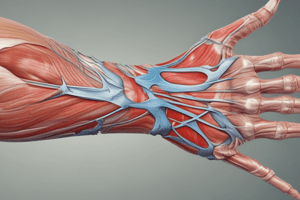
Muscle Anatomy
- Location: Thenar muscles are located at the base of the thumb on the palm side of the hand.
- Muscles Included:
- Abductor Pollicis Brevis: Abducts the thumb.
- Flexor Pollicis Brevis: Flexes the thumb. Has two heads:
- Superficial head
- Deep head
- Opponens Pollicis: Allows opposition of the thumb (bringing the thumb across the palm).
- Adductor Pollicis: Adducts the thumb. Not technically a thenar muscle but closely related in function.
Nerve Innervation
- Median Nerve:
- Supplies the abductor pollicis brevis, flexor pollicis brevis (superficial head), and opponens pollicis.
- Ulnar Nerve:
- Supplies the flexor pollicis brevis (deep head) and adductor pollicis.
Functional Movements
- Abduction: Moving the thumb away from the palm (abductor pollicis brevis).
- Flexion: Bending the thumb towards the palm (flexor pollicis brevis).
- Opposition: Thumb movement to touch other fingers (opponens pollicis).
- Adduction: Moving the thumb back towards the index finger (adductor pollicis).
Muscle Relations
- Superficial Relationships: The thenar muscles are situated beneath the skin and superficial fascia of the palm.
- Deep Relationships:
- Proximity to the carpal bones of the wrist.
- The opponens pollicis lies deep to the abductor and flexor pollicis brevis.
- Functional Interactions: Work synergistically with other intrinsic muscles of the hand (like the hypothenar muscles) to facilitate precise hand movements.
Muscle Anatomy
- Thenar muscles are situated at the base of the thumb on the palm side.
- Abductor Pollicis Brevis: Responsible for abducting the thumb away from the palm.
- Flexor Pollicis Brevis:
- Flexes the thumb toward the palm.
- Composed of two heads: superficial head and deep head.
- Opponens Pollicis: Facilitates opposition, allowing the thumb to touch other fingers across the palm.
- Adductor Pollicis: Adducts the thumb; while not a true thenar muscle, it plays a significant role in hand function.
Nerve Innervation
- Median Nerve:
- Innervates the abductor pollicis brevis, superficial head of the flexor pollicis brevis, and opponens pollicis.
- Ulnar Nerve:
- Innervates the deep head of the flexor pollicis brevis and adductor pollicis.
Functional Movements
- Abduction: Moving the thumb away from the palm, primarily by abductor pollicis brevis.
- Flexion: Bending the thumb towards the palm, primarily by flexor pollicis brevis.
- Opposition: The thumb moves to touch other fingers, mainly through opponens pollicis.
- Adduction: Moving the thumb back towards the index finger, facilitated by adductor pollicis.
Muscle Relations
- Superficial Relationships: The thenar muscles lie deep to the skin and superficial fascia of the palm.
- Deep Relationships:
- Positioned near the wrist's carpal bones.
- Opponens pollicis is located deep to both abductor and flexor pollicis brevis.
- Functional Interactions: Work in coordination with other intrinsic hand muscles, including hypothenar muscles, to enable intricate hand movements.
Hypothenar Muscles
- Located on the ulnar side of the hand; comprised of three muscles.
- Abductor Digiti Minimi:
- Originates from the pisiform bone.
- Inserts at the ulnar side of the proximal phalanx of the little finger.
- Flexor Digiti Minimi Brevis:
- Originates from the hook of the hamate bone and the flexor retinaculum.
- Inserts at the ulnar side of the proximal phalanx of the little finger.
- Opponens Digiti Minimi:
- Originates from the hook of the hamate bone and the flexor retinaculum.
- Inserts at the ulnar margin of the 5th metacarpal bone.
Muscle Functions
- Abductor Digiti Minimi: Responsible for abducting the little finger from the ring finger.
- Flexor Digiti Minimi Brevis: Flexes the proximal phalanx of the little finger.
- Opponens Digiti Minimi: Facilitates opposition of the little finger by bringing it across the palm towards the thumb.
Nerve Supply
- All hypothenar muscles are innervated by the ulnar nerve, spanning C8 to T1.
- The deep branch of the ulnar nerve specifically innervates these muscles.
Muscle Insertions
- Intrinsic muscles are located entirely within a specific body region, such as the hand or foot.
- In the hand, lumbricals insert on the extensor expansions of the fingers.
- In the foot, interossei muscles insert on the proximal phalanges.
Muscle Actions
- Intrinsic muscles facilitate fine motor control and maintain postural stability.
- Lumbricals flex fingers at the metacarpophalangeal joints while extending them at the interphalangeal joints.
- Interossei muscles enable abduction (dorsal) and adduction (palmar) of the fingers.
Innervation and Nerve Roots
- Lumbricals in the hand are innervated by the median nerve (1st and 2nd lumbricals) and the ulnar nerve (3rd and 4th lumbricals); origins are from C8-T1 nerve roots.
- Interossei muscles in the hand are solely innervated by the ulnar nerve, also associated with C8-T1 roots.
- Foot intrinsic muscles are primarily innervated by the lateral and medial plantar nerves, linked to S1-S2 nerve roots.
Muscle Origins
- Lumbricals in the hand originate from the tendons of the flexor digitorum profundus muscle.
- Interossei muscles originate from the sides of the metacarpals.
- In the foot, the abductor hallucis muscle originates from the calcaneus, as well as the flexor digitorum brevis muscle.
Functional Anatomy of Muscles
- Intrinsic muscles stabilize and control digit movements and allow for adaptable grip strength.
- They work synergistically with extrinsic muscles for precise and coordinated actions.
- Essential for tasks requiring dexterity, such as writing and gripping objects.
- Intrinsic muscles also maintain the arches of the foot and hand during various activities, supporting posture.
Muscle Origins
- Pectoralis Major: Has two heads; the clavicular head originates from the medial half of the clavicle, while the sternocostal head arises from the sternum and the first six ribs.
- Rectus Abdominis: Originates from the pubic symphysis and the pubic crest, providing support for the abdominal wall.
- Serratus Anterior: Arises from the upper eight or nine ribs, playing a crucial role in scapular movement.
- Deltoid: Composed of three parts with origins at the lateral third of the clavicle, acromion, and spine of scapula, facilitating shoulder movements.
- Biceps Brachii: Short head originates from the coracoid process of the scapula, while the long head comes from the supraglenoid tubercle, contributing to upper arm flexion.
Muscle Insertions
- Pectoralis Major: Inserts at the lateral lip of the intertubercular groove of the humerus, crucial for arm movement.
- Rectus Abdominis: Inserts at the costal cartilages of ribs 5-7 and the xiphoid process, aiding in trunk flexion.
- Serratus Anterior: Inserts on the medial border of the scapula, essential for scapular positioning.
- Deltoid: Inserts at the deltoid tuberosity of the humerus, allowing for diverse shoulder actions.
- Biceps Brachii: Inserts at the radial tuberosity and through the bicipital aponeurosis, facilitating elbow flexion and forearm supination.
Muscle Actions
- Pectoralis Major: Functions to adduct, medially rotate, and flex the arm at the shoulder joint.
- Rectus Abdominis: Primarily flexes the lumbar spine and compresses abdominal contents for stability.
- Serratus Anterior: Protracts and rotates the scapula, ensuring its stability against the thoracic wall during arm movements.
- Deltoid: Responsible for abducting, flexing, and extending the arm, important for shoulder mobility.
- Biceps Brachii: Flexes the elbow and supinates the forearm; also contributes weakly to shoulder flexion.
Nerve Root Functions
- Pectoralis Major: Innervated by both medial and lateral pectoral nerves, originating from spinal roots C5-T1, supporting arm movements.
- Rectus Abdominis: Innervated by the thoracoabdominal nerves (T7-T11), involved in abdominal and trunk motions.
- Serratus Anterior: Innervated by the long thoracic nerve (C5-C7), crucial for scapular function and stability.
- Deltoid: Innervated by the axillary nerve (C5-C6), vital for shoulder joint movement and stability.
- Biceps Brachii: Innervated by the musculocutaneous nerve (C5-C7), essential for flexing the elbow and forearm movements.
Muscle Origins
- Trapezius originates from the occipital bone, nuchal ligament, and spinous processes of C7 to T12.
- Latissimus Dorsi arises from the spinous processes of T7 to L5, iliac crest, and thoracolumbar fascia.
- Rhomboid Major starts from the spinous processes of T2 to T5.
- Rhomboid Minor originates from the spinous processes of C7 to T1.
- Levator Scapulae is derived from the transverse processes of C1 to C4.
- Serratus Posterior Superior originates from the spinous processes of C7 to T3.
- Serratus Posterior Inferior comes from the spinous processes of T11 to L2.
Muscle Insertions
- Trapezius inserts into the clavicle, acromion, and spine of the scapula.
- Latissimus Dorsi attaches to the intertubercular groove of the humerus.
- Rhomboid Major connects to the medial border of the scapula.
- Rhomboid Minor also attaches to the medial border of the scapula, positioned above the major.
- Levator Scapulae inserts at the superior angle of the scapula.
- Serratus Posterior Superior connects to ribs 2-5.
- Serratus Posterior Inferior attaches to ribs 9-12.
Muscle Actions
- Trapezius elevates, retracts, and rotates the scapula; extends the neck.
- Latissimus Dorsi extends, adducts, and medially rotates the arm.
- Rhomboid Major and Minor work together to retract and elevate the scapula.
- Levator Scapulae elevates the scapula and assists in downward rotation.
- Serratus Posterior Superior elevates upper ribs, aiding in inspiration.
- Serratus Posterior Inferior depresses lower ribs, facilitating expiration.
Innervation
- Trapezius innervated by the accessory nerve (CN XI).
- Latissimus Dorsi receives innervation from the thoracodorsal nerve.
- Rhomboid Major and Minor are innervated by the dorsal scapular nerve.
- Levator Scapulae is innervated by the dorsal scapular nerve and cervical nerves (C3-C4).
- Serratus Posterior Superior is innervated by intercostal nerves (T2-T5).
- Serratus Posterior Inferior is innervated by intercostal nerves (T9-T12).
Nerve Roots
- Trapezius is influenced by nerve roots C3 and C4 (proprioception) and CN XI.
- Latissimus Dorsi is associated with nerve roots C6, C7, and C8.
- Rhomboid Major and Minor connect to nerve roots C4 and C5.
- Levator Scapulae is tied to nerve roots C3, C4, and C5.
- Serratus Posterior Superior associates with nerve roots T1-T4.
- Serratus Posterior Inferior connects to nerve roots T9-T12.
Muscle Origins
- Gluteus Maximus: Originates from the posterior iliac crest, sacrum, coccyx, and sacrotuberous ligament, contributing to its function in hip extension and lateral rotation.
- Gluteus Medius: Arises from the external surface of the ilium, positioned between the anterior and posterior gluteal lines, playing a crucial role in hip stabilization during walking.
- Gluteus Minimus: Also originates from the external surface of the ilium but is situated between the anterior and inferior gluteal lines, assisting in hip abduction and medial rotation.
Muscle Insertions
- Gluteus Maximus: Inserts onto the iliotibial tract and the gluteal tuberosity of the femur, facilitating movements like climbing and running.
- Gluteus Medius: Attaches to the lateral surface of the greater trochanter of the femur, essential for maintaining the pelvis level during locomotion.
- Gluteus Minimus: Inserts into the anterior surface of the greater trochanter of the femur, supporting hip abduction and stabilization.
Innervation
- Gluteus Maximus: Innervated by the inferior gluteal nerve, which is crucial for its powerful extension and external rotation of the hip.
- Gluteus Medius and Gluteus Minimus: Both receive innervation from the superior gluteal nerve, vital for hip abduction and medial rotation.
Nerve Root Segments
- Gluteus Maximus: Governed by nerve root segments L5, S1, and S2, linking it to the lumbosacral plexus for movement coordination.
- Gluteus Medius and Gluteus Minimus: Both associated with nerve root segments L4, L5, and S1, which are critical for efficient lower limb function and stability during various activities.
Muscle Anatomy
- Deep gluteal muscles are located beneath the gluteus maximus, contributing to hip stability and movement.
- Piriformis:
- Originates from the anterior surface of the sacrum and inserts at the greater trochanter of the femur.
- Function: Primarily laterally rotates the hip, crucial for rotational movements.
- Gemellus Superior:
- Originates from the ischial spine and also inserts at the greater trochanter (with obturator internus).
- Function: Assists in lateral rotation and hip abduction.
- Obturator Internus:
- Originates from the pelvic surface of the obturator membrane and surrounding bones, inserting at the greater trochanter.
- Function: Laterally rotates the thigh and stabilizes the hip joint.
- Gemellus Inferior:
- Originates from the ischial tuberosity and inserts with obturator internus at the greater trochanter.
- Function: Aids in lateral rotation of the hip.
- Quadratus Femoris:
- Originates from the ischial tuberosity, inserting at the intertrochanteric crest of the femur.
- Function: Laterally rotates the hip and stabilizes the femur during movement.
Nerve Supply
- Piriformis: Innervated by the nerve to piriformis (S1, S2), essential for muscle function.
- Gemelli and Obturator Internus: Received innervation from the nerve to obturator internus (L5, S1).
- Quadratus Femoris: Innervated by the nerve to quadratus femoris (L5, S1).
- Importance of nerve supply lies in facilitating muscle function and coordination during movement.
Functional Movements
- Deep gluteal muscles play a crucial role in stabilizing the hip joint during dynamic activities like walking and running.
- These muscles engage primarily in laterally rotating the femur, which is vital for various physical activities.
- Assist with hip abduction, important for movements like side leg raises and lateral movements.
- Proper functioning of deep gluteal muscles is essential for a balanced gait, preventing excessive pelvic drop on the opposite side during walking.
- Common activities involving these muscles include sports with rotational movements (tennis, golf) and those requiring lateral movements (dancing, martial arts).
Medial Thigh Muscles
Muscle Anatomy
- The medial thigh primarily comprises five key muscles:
- Adductor Longus
- Adductor Brevis
- Adductor Magnus
- Gracilis
- Obturator Externus
Origin Points
- Adductor Longus originates from the pubis near the pubic symphysis.
- Adductor Brevis starts from the inferior pubic ramus.
- Adductor Magnus has dual origin points:
- Ischial tuberosity for the hamstring part
- Inferior pubic ramus and ischial ramus for the adductor part.
- Gracilis is derived from the inferior pubic ramus.
- Obturator Externus arises from the outer surface of the obturator membrane and surrounding bone.
Insertion Sites
- Adductor Longus inserts on the middle third of the femur's linea aspera.
- Adductor Brevis attaches to the pectineal line and proximal part of the linea aspera.
- Adductor Magnus connects to the entire length of the linea aspera and the adductor tubercle for its hamstring part.
- Gracilis inserts on the medial surface of the tibia, contributing to the pes anserinus.
- Obturator Externus attaches to the trochanteric fossa of the femur.
Muscle Actions
- Adductor Longus functions in hip adduction, flexion, and medial rotation.
- Adductor Brevis mainly aids in hip adduction and flexion.
- Adductor Magnus performs dual roles:
- Adductor part: hip adduction and flexion
- Hamstring part: hip extension.
- Gracilis is involved in hip adduction, knee flexion, and medial rotation of the leg.
- Obturator Externus specializes in lateral rotation of the hip.
Innervation and Nerve Roots
- Adductor Longus and Adductor Brevis are innervated by the obturator nerve with roots at L2 and L3.
- Adductor Magnus receives innervation from two sources:
- Adductor part: obturator nerve (L2, L3)
- Hamstring part: tibial nerve (L4).
- Gracilis is also innervated by the obturator nerve (L2, L3).
- Obturator Externus has innervation from the obturator nerve with nerve roots L3 and L4.
Muscle Origins
- Iliopsoas:
- Comprises Psoas Major from lumbar vertebrae (T12-L5) and Iliacus from the iliac fossa.
- Rectus Femoris:
- Originates from the anterior inferior iliac spine (AIIS) and the superior margin of the acetabulum.
- Sartorius:
- Starts at the anterior superior iliac spine (ASIS).
Muscle Insertions
- Iliopsoas:
- Inserts at the lesser trochanter of the femur.
- Rectus Femoris:
- Connects to the tibial tuberosity via the patellar ligament.
- Sartorius:
- Attaches to the medial aspect of the proximal tibia at the pes anserinus.
Muscle Actions
- Iliopsoas:
- Responsible for hip flexion and assists with lateral rotation of the thigh.
- Rectus Femoris:
- Facilitates hip flexion and knee extension.
- Sartorius:
- Engages in hip flexion, abduction, lateral rotation, and assists in knee flexion.
Nerve Innervation
- Iliopsoas:
- Psoas Major is innervated by the lumbar plexus (L2-L3); Iliacus is innervated by the femoral nerve (L2-L4).
- Rectus Femoris:
- Innervated by the femoral nerve (L2-L4).
- Sartorius:
- Also innervated by the femoral nerve (L2-L4).
Nerve Root Contributions
- Iliopsoas:
- Primarily L2-L3 for Psoas Major; L2-L4 for Iliacus.
- Rectus Femoris:
- Contributes from L2-L4.
- Sartorius:
- Involved nerve roots are L2-L4.
Studying That Suits You
Use AI to generate personalized quizzes and flashcards to suit your learning preferences.