Podcast
Questions and Answers
The medulla, pons, and cerebellum are primary components of the forebrain.
The medulla, pons, and cerebellum are primary components of the forebrain.
False (B)
The oculomotor nerve exits the brainstem at the level of the medulla.
The oculomotor nerve exits the brainstem at the level of the medulla.
False (B)
The abducens nerve (CN VI) exits the brainstem at the junction of the pons and medulla.
The abducens nerve (CN VI) exits the brainstem at the junction of the pons and medulla.
True (A)
The trochlear nerve exits the brainstem ventrally.
The trochlear nerve exits the brainstem ventrally.
The cerebello-pontine angle is located at the interface of the spinal cord, medulla and cerebellum.
The cerebello-pontine angle is located at the interface of the spinal cord, medulla and cerebellum.
The pre-olivary sulcus is where cranial nerve XII exits the medulla.
The pre-olivary sulcus is where cranial nerve XII exits the medulla.
The restiform body is part of the crus cerebri.
The restiform body is part of the crus cerebri.
The superior colliculi are responsible for auditory reflexes.
The superior colliculi are responsible for auditory reflexes.
The vagal trigone is located superior to the hypoglossal trigone
The vagal trigone is located superior to the hypoglossal trigone
The primary fissure separates the anterior and posterior lobes of the cerebellum.
The primary fissure separates the anterior and posterior lobes of the cerebellum.
The horizontal fissure separates the flocculus from the nodulus.
The horizontal fissure separates the flocculus from the nodulus.
The superior cerebellar peduncles connect the cerebellum to the spinal cord.
The superior cerebellar peduncles connect the cerebellum to the spinal cord.
In the Corticospinal Tract (CST), upper motor neurons originate in the cerebellum.
In the Corticospinal Tract (CST), upper motor neurons originate in the cerebellum.
The corticospinal tract, after decussating in the pyramidal decussation, becomes the anterior Corticospinal Tract.
The corticospinal tract, after decussating in the pyramidal decussation, becomes the anterior Corticospinal Tract.
The rubrospinal tract is responsible for decussation in the ventral tegmental area.
The rubrospinal tract is responsible for decussation in the ventral tegmental area.
The dorsal column medial lemniscus (DCML) pathway conveys tactile and proprioceptive information from the face.
The dorsal column medial lemniscus (DCML) pathway conveys tactile and proprioceptive information from the face.
The gracile fasiculus carries sensory information from the upper extremities.
The gracile fasiculus carries sensory information from the upper extremities.
The anterolateral system (ALS) decussates via the posterior white commissure.
The anterolateral system (ALS) decussates via the posterior white commissure.
The anterolateral system (ALS) carries information about pain and temperature, synapsing in the ventral posterolateral nucleus of the thalamus.
The anterolateral system (ALS) carries information about pain and temperature, synapsing in the ventral posterolateral nucleus of the thalamus.
The dorsal horn contains motor neurons.
The dorsal horn contains motor neurons.
The anterior funiculus contains the rubrospinal tract.
The anterior funiculus contains the rubrospinal tract.
The inferior cerebellar peduncle receives its blood supply from the posterior inferior cerebellar artery (PICA).
The inferior cerebellar peduncle receives its blood supply from the posterior inferior cerebellar artery (PICA).
Wallenberg syndrome, or lateral medullary syndrome, results from infarction of the anterior spinal artery.
Wallenberg syndrome, or lateral medullary syndrome, results from infarction of the anterior spinal artery.
The Anterior Inferior Cerebellar Artery or AICA supplies the basilar region of the brain.
The Anterior Inferior Cerebellar Artery or AICA supplies the basilar region of the brain.
In lateral medullary syndrome, vertigo results from damage to the vestibular nuclei.
In lateral medullary syndrome, vertigo results from damage to the vestibular nuclei.
Flashcards
Brainstem
Brainstem
The medulla, pons, and midbrain; contains cranial nerve nuclei and ascending/descending tracts.
Crus Cerebri
Crus Cerebri
Cerebral peduncles; contains descending tracts.
Tectum
Tectum
Includes the superior and inferior colliculi.
Pons
Pons
Signup and view all the flashcards
Medulla Oblongata
Medulla Oblongata
Signup and view all the flashcards
Restiform Body
Restiform Body
Signup and view all the flashcards
Posterior Medulla landmarks
Posterior Medulla landmarks
Signup and view all the flashcards
Cerebellar Peduncles
Cerebellar Peduncles
Signup and view all the flashcards
Primary Fissure
Primary Fissure
Signup and view all the flashcards
Basilar Artery
Basilar Artery
Signup and view all the flashcards
Wallenberg Syndrome
Wallenberg Syndrome
Signup and view all the flashcards
Wallenberg Syndrome signs
Wallenberg Syndrome signs
Signup and view all the flashcards
Dejerine's Syndrome
Dejerine's Syndrome
Signup and view all the flashcards
Medial Lemniscus
Medial Lemniscus
Signup and view all the flashcards
Anterolateral System (ALS)
Anterolateral System (ALS)
Signup and view all the flashcards
Brainstem Tegmentum
Brainstem Tegmentum
Signup and view all the flashcards
Inferior Olivary Nucleus
Inferior Olivary Nucleus
Signup and view all the flashcards
Corticospinal tract
Corticospinal tract
Signup and view all the flashcards
Lateral Medullary Syndrome
Lateral Medullary Syndrome
Signup and view all the flashcards
Dorsal horn
Dorsal horn
Signup and view all the flashcards
Lateral Funiculus
Lateral Funiculus
Signup and view all the flashcards
Cranial nerve nuclei
Cranial nerve nuclei
Signup and view all the flashcards
Cranial nerve nuclei damage
Cranial nerve nuclei damage
Signup and view all the flashcards
Cerebellar Penduncles
Cerebellar Penduncles
Signup and view all the flashcards
cranial nerve nuclei
cranial nerve nuclei
Signup and view all the flashcards
Study Notes
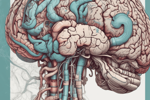
- Lecture covers the medulla, pons, and cerebellum, including external anatomy, fiber tracts, sections, major blood supply, and vascular syndromes of the brainstem.
Anatomical Overview
- Inferior brainstem anatomy includes the crus cerebri (cerebral peduncles), pons (brachium pontis), pre- and post-olivary sulci, olivary eminence, pyramids, and restiform body.
- Superior brainstem anatomy shows superior and inferior colliculi, the crus cerebri, restiform body, and gracile and cuneate tubercles.
- Key brainstem exit points are: Pons: IV (dorsal exit), V; Pons/medulla: VI, VII, VIII; Cerebello-pontine angle (interface of pons, medulla, and cerebellum): VII, VIII; Medulla: post olivary IX, X pre-olivary sulci XII.
Cerebellum Anatomy
- Cerebellum features include: Anterior lobe, Posterior lobe, Vermis, Intermediate hemisphere, Lateral hemisphere, and Tonsils.
- Key fissures of the cerebellum are: Primary fissure, Horizontal fissure, and Postero-lateral fissure.
- Flocculo-Nodular lobe includes the Nodulus and Flocculus.
Major Descending Pathways - Corticospinal Tract (CST)
- Cortical Upper Motor Neurons (UMNs) pass through the Internal Capsule and Crus Cerebri (cerebral peduncles).
- The CST descends via the Pontine CST and Medullary CST (Pyramids).
- The CST decussates and becomes the Lateral Corticospinal Tract (LCS) in the lateral funiculus; fibers that don't decussate become the Anterior Corticospinal Tract (ACS) in the anterior funiculus, and both innervate Ventral horn LMNs.
- The Rubrospinal Tract originates in the Red Nucleus, undergoes Ventral tegmental decussation in the midbrain, and descends in the Lateral funiculus to influence Proximal limb flexor LMNs.
Major Ascending Pathways - Dorsal Column Medial Lemniscus (DCML)
- DCML is responsible for tactile and proprioceptive sensation
- Two dorsal columns (fasciculi): Gracile (lower extremities) and Cuneate (upper extremities).
- Relays in two medullary nuclei: gracile and cuneate
- The DCML decussates via internal arcuate fibers.
- Travels in the medial lemniscus (white matter) to the Ventral Posterolateral Nucleus of the Thalamus (VPL) and then to the Somatic Sensory Cortex.
Antero Lateral System (ALS)
- Carries pain and temperature information.
- ALS consists of fibers from dorsal horn interneurons.
- Fibers decussate via the Anterior White Commissure (AWC) and ascend as dorsal and ventral spinothalamic tracts (= Antero Lateral System).
- ALS ascends and approaches the medial lemniscus, eventually reaching the VPL of the thalamus and projecting to the Somatic Sensory Cortex.
- Trigeminal System (head/face) uses both DCML and ALS pathways.
Spinal Cord Anatomy
- Structures in the rostral spinal cord include the Dorsal Horn, Spinal accessory nucleus, Spinal trigeminal tract, posteromarginal nucleus, and substantia gelatinosa.
- Intermediate Horn contains the Dorsal nucleus of Clarke.
- Anterior Horn consists of Lateral and Medial motor columns.
- Posterior Funiculus includes the Gracile and Cuneate fasiculus.
- Lateral Funiculus includes the Rubro-spinal tract, Lateral cortico-spinal tract, Anterolateral system, and Spinocerebellar tracts.
- Anterior funiculus contains the Anterior cortico-spinal tract and Medial longitudinal fasiculus, along with the Vestibulo-, Tecto-, reticulo-spinal tracts
Medulla
- Sectional anatomy highlights the Cerebellar vermis, Cerebellar tonsil, Foramen of Magendie, Inferior cerebellar peduncle, Cerebellar cortex, and Medial lemniscus.
- Sectional anatomy highlights Cortico-spinal tract, Medullary pyramids and Inferior olivary nucleus.
- Caudal medulla contains: Hypoglossal nucleus, Dorsal vagal motor nucleus, Nucleus ambiguus, Solitary nucleus and tract, Spinal trigeminal nucleus and tract.
- Other features of the Caudal medulla: Reticular formation, DCML, Internal arcuate fibers, MLF, ALS, Spinocerebellar tracts, Inferior olivary nucleus, and Corticospinal tract/pyramids.
- Rostral medulla contains: Hypoglossal nucleus*, Vestibular nuclei, Cochlear nuclei, Dorsal vagal motor nucleus*, Solitary nucleus, and tract, Nucleus ambiguus*, Spinal trigeminal nucleus, and tract.
Key Structures in Rostral Medulla
- Reticular formation
- Cerebellar peduncle (inferior)
- Medial Lemniscus
- MLF
- ALS
- Spinocerebellar tracts
- Inferior olivary nucleus
- Corticospinal tract / pyramids
Pons
- Sectional anatomy of the pontine region includes Superior medullary velum, 4th ventricle, Superior Cerebellar peduncle, Medial Lemniscus, Pontine Reticular formation, Middle Cerebellar peduncle, and Cortico-spinal Tract.
- Caudal pons: abducens nucleus, facial motor nucleus, vestibular & spinal trigeminal nuclei, dentate, interposed and fastigial cerebellar nuclei and also: cerebellar peduncles, reticular formation, medial and lateral lemniscus, MLF, ALS, spinocerebellar and superior olivary tracts, and also corticospinal, pontine and bulbar tracts.
- Mid-pons features: Principal sensory and Trigeminal motor nuclei, Mesencephalic nucleus and tract, Reticular formation, Superior cerebellar peduncles, Medial and Lateral Lemniscus, MLF, DLF, ALS, Spinocerebellar tracts, Superior olivary nuclei, and Corticospinal, pontine, bulbar tracts.
Brainstem Vascular Supply - General Concepts
- Vertebral artery feeds the Basilar artery
- The AICA, PICA and SCA all contribute to the blood supply.
- Basilar artery branches include: Paramedian branches and Long & short cirucumferential branches.
Vascular Syndromes of the Brainstem
- Wallenberg (lateral medullary) syndrome: common after an infarction of the brainstem, caused by vertebral & PICA artery occlusion
- Features Ipsilateral Vertigo (vestibular nuclei), decreased facial pain & temperature sensation (trigeminal nuclei/tract), decreased taste (solitary nucleus), dysphagia/hoarseness (nucleus ambiguus).
- Other features: Anterolateral system (ALS), ataxia (cerebellar peduncle)
- Ptosis and miosis indicate a disruption of sympathetic fibers.
- Medial Medullary Syndrome (Dejerine's): localizing sign is tongue weakness and contralateral hemiparesis/loss touch and proprioception.
- Brainstem Syndromes - Pons: Medial, Lateral & Superior
Medial (Foville Syndrome)
- Paramedian branches of basilar artery affect CN VI, paramedian pontine reticular formation (pprf*), and the medial longitudinal fasciculus that connect eyemotor nuclei.
- Horizontal gaze palsy and internuclear ophthalmoplegia can result.
Lateral (Gubler Syndrome)
- AICA or superior cerebellar artery
- Affects CN VII, cerebellar peduncle, CN V and spinothalamic tract
- Facial paralysis, ataxia, jaw weakness, facial and contralateral body pain/Temp loss.
- Also results in decreased taste, tinnitus, deafness, solitary and cochlear nuclei are affected
Studying That Suits You
Use AI to generate personalized quizzes and flashcards to suit your learning preferences.
Related Documents
Description
Lecture on the anatomy of the medulla, pons, and cerebellum with a focus on external anatomy, fiber tracts, and blood supply. Key areas include the crus cerebri, olivary eminence, pyramids, and cerebellar lobes and fissures. Vascular syndromes of the brainstem are also covered.