Podcast
Questions and Answers
What is the primary function of the pleuropericardial folds during development?
What is the primary function of the pleuropericardial folds during development?
- To separate respiratory and digestive cavities
- To delineate the boundaries of abdominal and thoracic cavities (correct)
- To provide structural support to the heart
- To facilitate blood flow from the veins to the heart
At what stage do myoblasts migrate into the septum transversum?
At what stage do myoblasts migrate into the septum transversum?
- Week 5-6 (correct)
- Week 3-4
- Week 8-9
- Week 6-7
The septum transversum is initially located at which cervical level?
The septum transversum is initially located at which cervical level?
- T3
- T1
- C1 (correct)
- C5
What is the main effect of the anterior and lateral expansion during embryonic development?
What is the main effect of the anterior and lateral expansion during embryonic development?
What anatomical structures do the pleuropericardial folds contain?
What anatomical structures do the pleuropericardial folds contain?
How does the position of the septum transversum change by week 8 of development?
How does the position of the septum transversum change by week 8 of development?
What is the significance of the two open passageways left by the septum transversum?
What is the significance of the two open passageways left by the septum transversum?
What direction does the pleuropericardial folds grow during embryonic development?
What direction does the pleuropericardial folds grow during embryonic development?
What is the primary role of somitomeres and somites during embryonic development?
What is the primary role of somitomeres and somites during embryonic development?
Which embryonic structure remains open after the closure of the ventral body wall?
Which embryonic structure remains open after the closure of the ventral body wall?
What aids in the closure of the ventral body wall?
What aids in the closure of the ventral body wall?
At what stage does the vitelline duct degenerate during gestation?
At what stage does the vitelline duct degenerate during gestation?
What type of epithelium makes up the serous membranes?
What type of epithelium makes up the serous membranes?
Which layers of the mesoderm are involved in forming the serous membranes?
Which layers of the mesoderm are involved in forming the serous membranes?
Which body cavity is NOT mentioned in the context of embryonic development?
Which body cavity is NOT mentioned in the context of embryonic development?
What happens during the closure of the gut tube?
What happens during the closure of the gut tube?
What characterizes the ventral mesentery?
What characterizes the ventral mesentery?
Which statement about the dorsal mesentery is true?
Which statement about the dorsal mesentery is true?
Which layers comprise the structure of the mesenteries?
Which layers comprise the structure of the mesenteries?
What is the primary function of the mesenteries in the abdominal cavity?
What is the primary function of the mesenteries in the abdominal cavity?
At which location do the lateral plate mesoderm connect with each other?
At which location do the lateral plate mesoderm connect with each other?
What is the role of the septum transversum?
What is the role of the septum transversum?
Which statement accurately describes the relationship between the visceral and parietal layers of the serous membrane?
Which statement accurately describes the relationship between the visceral and parietal layers of the serous membrane?
Which of the following is NOT a characteristic of the peritoneal cavity?
Which of the following is NOT a characteristic of the peritoneal cavity?
What is characterized by a markedly reduced lung volume and hypertrophy of smooth muscle in the pulmonary arteries?
What is characterized by a markedly reduced lung volume and hypertrophy of smooth muscle in the pulmonary arteries?
Which mesodermal layer consists of parietal and visceral layers?
Which mesodermal layer consists of parietal and visceral layers?
What may result from inadequate branching morphogenesis during lung development?
What may result from inadequate branching morphogenesis during lung development?
What anatomical structure prevents normal lung development by compressing it?
What anatomical structure prevents normal lung development by compressing it?
Which condition is likely inconsistent with life due to extreme lung hypoplasia?
Which condition is likely inconsistent with life due to extreme lung hypoplasia?
What is NOT associated with the parietal layer of the lateral plate mesoderm?
What is NOT associated with the parietal layer of the lateral plate mesoderm?
What structure is notably affected in cases of lung hypoplasia due to congenital conditions?
What structure is notably affected in cases of lung hypoplasia due to congenital conditions?
What is the primary role of the phrenic nerves in relation to the diaphragm?
What is the primary role of the phrenic nerves in relation to the diaphragm?
What may characterize the pericardioperitoneal canals during embryonic development?
What may characterize the pericardioperitoneal canals during embryonic development?
During which week does the developing diaphragm reposition to the level of thoracic somites?
During which week does the developing diaphragm reposition to the level of thoracic somites?
What contributes to the shift of the common cardinal veins toward the midline?
What contributes to the shift of the common cardinal veins toward the midline?
What structure eventually forms the fibrous pericardium in adults?
What structure eventually forms the fibrous pericardium in adults?
At what level do some of the dorsal bands of the diaphragm originate by the beginning of the third month?
At what level do some of the dorsal bands of the diaphragm originate by the beginning of the third month?
What type of tissue contributes to the most peripheral part of the diaphragm?
What type of tissue contributes to the most peripheral part of the diaphragm?
What changes occur to the pleuropericardial membranes during development?
What changes occur to the pleuropericardial membranes during development?
How are the pleural cavities defined during the development of the thoracic cavity?
How are the pleural cavities defined during the development of the thoracic cavity?
Study Notes
Embryonic Development Overview
- Paraxial mesoderm differentiates into somitomeres and somites, crucial for forming skull and vertebrae.
- By the end of week four, lateral body wall folds migrate to the midline and fuse, closing the ventral body wall.
Body Cavity Development
- Growth of head and tail regions causes embryo to curve into the fetal position.
- Ventral body wall closure is almost complete, except for the connecting stalk (future umbilical cord).
- Closure of the gut tube is also nearly complete, leaving the vitelline duct connection from the midgut to the yolk sac.
Serous Membranes
- Serous membranes consist of simple cuboidal epithelium, forming parietal and visceral layers.
- These layers are continuous at the junction of the gut tube with the posterior body wall.
- Dorsal mesentery suspends the gut tube from the posterior body wall and extends from the foregut to the hindgut.
Mesenteries
- Ventral mesentery is present only from the caudal foregut to the duodenum, resulting from thinning of the septum transversum.
- Mesenteries are double layers of peritoneum providing pathways for blood vessels, nerves, and lymphatics to organs.
Diaphragm Formation
- The septum transversum is a mesodermal thick plate between thoracic cavity and body stalk.
- Initially opposite cervical segments, the diaphragm descends to thoracic somite level by week six.
- Rapid dorsal growth of the embryo leads to repositioning of the diaphragm during development.
Phrenic Nerve and Innervation
- The phrenic nerves supply motor and sensory innervation to the diaphragm, originating from cervical ventral rami (C3, C4, C5).
- The continuity of the pleuropericardial membranes evolves into the fibrous pericardium in adults.
Lung Development and Hypoplasia
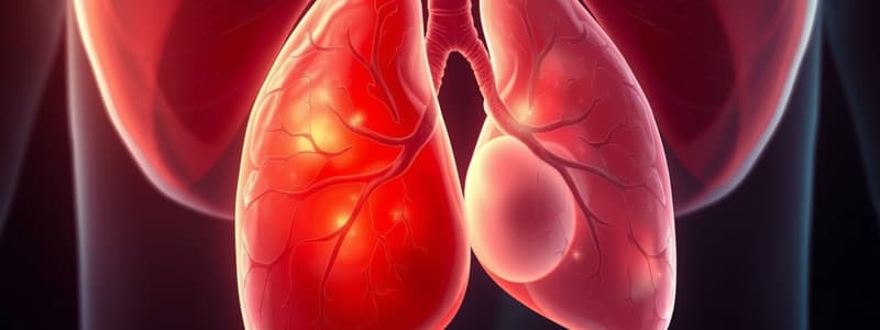
- Lung hypoplasia results from reduced lung volume and smooth muscle hypertrophy in pulmonary arteries.
- Often caused by congenital diaphragmatic hernias or congenital heart disease, extreme hypoplasia is typically not compatible with life.
Congenital Diaphragmatic Hernias
- Abnormal positioning of abdominal viscera compresses the developing lung, hindering normal lung development.
Studying That Suits You
Use AI to generate personalized quizzes and flashcards to suit your learning preferences.
Related Documents
Description
This quiz explores the concepts surrounding lung hypoplasia, including the parietal layer of the lateral plate mesoderm and its implications on heart and vascular development. Dive deep into understanding the pathology and mechanisms that lead to reduced lung volume in affected individuals.


