Podcast
Questions and Answers
What is the normal duration of the P wave in an ECG?
What is the normal duration of the P wave in an ECG?
- 0.16 sec
- 0.06 sec
- 0.10 sec (correct)
- 0.25 sec
Which component of the ECG is primarily associated with ventricular depolarization?
Which component of the ECG is primarily associated with ventricular depolarization?
- T wave
- QRS complex (correct)
- U wave
- P wave
What is the amplitude of a normal T wave in an ECG?
What is the amplitude of a normal T wave in an ECG?
- 0.4 mV
- 0.2 mV (correct)
- 1 mV
- 0.1 mV
How is the heart rate calculated using the number of large boxes between two consecutive beats?
How is the heart rate calculated using the number of large boxes between two consecutive beats?
In which condition is the PR interval most likely prolonged?
In which condition is the PR interval most likely prolonged?
What does a prolonged PR interval indicate?
What does a prolonged PR interval indicate?
What does the U wave in an ECG typically indicate?
What does the U wave in an ECG typically indicate?
What does Lead II in an ECG record?
What does Lead II in an ECG record?
What is the standard speed of ECG paper?
What is the standard speed of ECG paper?
Which unipolar lead is assigned to the left leg?
Which unipolar lead is assigned to the left leg?
Where is the exploring electrode placed for lead V1?
Where is the exploring electrode placed for lead V1?
Which leads are used to assess the inferior surface of the heart?
Which leads are used to assess the inferior surface of the heart?
What does lead aVR primarily examine?
What does lead aVR primarily examine?
What is the purpose of checking voltage calibration in ECG interpretation?
What is the purpose of checking voltage calibration in ECG interpretation?
Which leads look at the right ventricle?
Which leads look at the right ventricle?
Which of the following is NOT a step in the commonly followed sequence of ECG interpretation?
Which of the following is NOT a step in the commonly followed sequence of ECG interpretation?
What is the role of capillaries in the circulatory system?
What is the role of capillaries in the circulatory system?
Which property of cardiac muscle allows it to initiate beats independently of nervous signals?
Which property of cardiac muscle allows it to initiate beats independently of nervous signals?
What is the formula for calculating Stroke Volume (SV)?
What is the formula for calculating Stroke Volume (SV)?
How much blood is typically pumped by each ventricle per beat at rest?
How much blood is typically pumped by each ventricle per beat at rest?
What is the primary function of the cardiovascular system?
What is the primary function of the cardiovascular system?
What characteristic of cardiac muscle allows it to spread electrical impulses throughout the heart?
What characteristic of cardiac muscle allows it to spread electrical impulses throughout the heart?
During which phase do all four chambers of the heart relax?
During which phase do all four chambers of the heart relax?
What is considered the steady-state volume of blood pumped by the heart per minute at rest?
What is considered the steady-state volume of blood pumped by the heart per minute at rest?
What does the equation CO = HR X SV represent?
What does the equation CO = HR X SV represent?
What does preload represent in cardiac function?
What does preload represent in cardiac function?
Which statement regarding electrocardiography (ECG) is correct?
Which statement regarding electrocardiography (ECG) is correct?
What is the normal range for ejection fraction?
What is the normal range for ejection fraction?
Einthoven's Triangle is used to illustrate what concept in ECG?
Einthoven's Triangle is used to illustrate what concept in ECG?
Which process occurs first in the cardiac cycle?
Which process occurs first in the cardiac cycle?
What is the primary function of the left ventricle in the cardiovascular system?
What is the primary function of the left ventricle in the cardiovascular system?
What does afterload signify in the context of heart function?
What does afterload signify in the context of heart function?
The electrical activity that precedes cardiac contraction is primarily due to what ion influx?
The electrical activity that precedes cardiac contraction is primarily due to what ion influx?
Which component of the heart controls blood flow from the atrium to the ventricle?
Which component of the heart controls blood flow from the atrium to the ventricle?
Which sequence accurately describes the systemic circulation pathway?
Which sequence accurately describes the systemic circulation pathway?
What is the role of the right ventricle in the cardiovascular system?
What is the role of the right ventricle in the cardiovascular system?
In the context of blood vessels, which statement is correct?
In the context of blood vessels, which statement is correct?
What term describes the two circuits of blood flow in the cardiovascular system?
What term describes the two circuits of blood flow in the cardiovascular system?
Which statement accurately describes the function of semilunar valves?
Which statement accurately describes the function of semilunar valves?
What does the term 'Einthoven triangle' refer to?
What does the term 'Einthoven triangle' refer to?
Flashcards
Cardiovascular System
Cardiovascular System
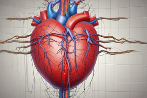
The heart, the main pressure pump, and the blood vessels, including arteries, arterioles, capillaries, venules, and veins.
Heart Chambers
Heart Chambers
The right and left sides of the heart, each containing an atrium and a ventricle. The atrium receives blood, and the ventricle pumps it out.
Atrioventricular Valves
Atrioventricular Valves
Valves in the heart that control the direction of blood flow from the atria to the ventricles.
Semilunar Valves
Semilunar Valves
Signup and view all the flashcards
Aorta
Aorta
Signup and view all the flashcards
Systemic Circulation
Systemic Circulation
Signup and view all the flashcards
Pulmonary Circulation
Pulmonary Circulation
Signup and view all the flashcards
Arteries
Arteries
Signup and view all the flashcards
Cardiac Muscle Excitability
Cardiac Muscle Excitability
Signup and view all the flashcards
Cardiac Muscle Rhythmicity
Cardiac Muscle Rhythmicity
Signup and view all the flashcards
Cardiac Muscle Conductivity
Cardiac Muscle Conductivity
Signup and view all the flashcards
Cardiac Muscle Contractility
Cardiac Muscle Contractility
Signup and view all the flashcards
Stroke Volume (SV)
Stroke Volume (SV)
Signup and view all the flashcards
End Diastolic Volume (EDV)
End Diastolic Volume (EDV)
Signup and view all the flashcards
End Systolic Volume (ESV)
End Systolic Volume (ESV)
Signup and view all the flashcards
Cardiac Output (CO)
Cardiac Output (CO)
Signup and view all the flashcards
What is Cardiac Output (CO)?
What is Cardiac Output (CO)?
Signup and view all the flashcards
What is Cardiac Index?
What is Cardiac Index?
Signup and view all the flashcards
What is Ejection Fraction (EF)?
What is Ejection Fraction (EF)?
Signup and view all the flashcards
What is Preload?
What is Preload?
Signup and view all the flashcards
What is Afterload?
What is Afterload?
Signup and view all the flashcards
What is Electrocardiography (ECG)?
What is Electrocardiography (ECG)?
Signup and view all the flashcards
How is ECG Recorded?
How is ECG Recorded?
Signup and view all the flashcards
What is Einthoven's Triangle?
What is Einthoven's Triangle?
Signup and view all the flashcards
Standard Limb Leads
Standard Limb Leads
Signup and view all the flashcards
Lead I
Lead I
Signup and view all the flashcards
Lead II
Lead II
Signup and view all the flashcards
Lead III
Lead III
Signup and view all the flashcards
Unipolar Leads
Unipolar Leads
Signup and view all the flashcards
Unipolar Limb Leads
Unipolar Limb Leads
Signup and view all the flashcards
Unipolar Chest Leads
Unipolar Chest Leads
Signup and view all the flashcards
Importance of Different ECG Leads
Importance of Different ECG Leads
Signup and view all the flashcards
PR interval
PR interval
Signup and view all the flashcards
QRS complex
QRS complex
Signup and view all the flashcards
T wave
T wave
Signup and view all the flashcards
U wave
U wave
Signup and view all the flashcards
Large and small boxes on ECG
Large and small boxes on ECG
Signup and view all the flashcards
RR interval
RR interval
Signup and view all the flashcards
P wave
P wave
Signup and view all the flashcards
T wave
T wave
Signup and view all the flashcards
Study Notes
Introduction to CVS and ECG
- The cardiovascular system is composed of the heart and blood vessels (arteries, arterioles, capillaries, venules, and veins)
- The heart has two halves (right and left), each with an atrium and a ventricle
- Atrioventricular (AV) valves ensure one-way blood flow from atrium to ventricle
- Left ventricle pumps blood to the systemic circulation at high pressure
- Right ventricle pumps blood to the pulmonary circulation at lower pressure
- Semilunar valves control blood flow between ventricles and major arteries
Learning Objectives
- Describe heart chambers, valves, and blood flow direction
- Describe systemic and pulmonary circulation
- Define ECG
- Describe the Einthoven triangle
- List ECG leads used in recordings
- Describe ECG waves and their relation to the cardiac cycle
- Interpret ECG findings
- Discuss ECG applications
Circulation
- Systemic circulation carries oxygenated blood to body tissues
- Pulmonary circulation carries deoxygenated blood to the lungs for gas exchange
Pressures in the Circulation
- Right ventricle: 25/0 mm Hg
- Pulmonary artery: 25/8 mm Hg
- Mean pulmonary artery: 15 mm Hg
- Capillary: 7-9 mm Hg
- Pulmonary venous: 5 mm Hg
- Left atrium: 5-10 mm Hg
- Left ventricle: 120/0 mm Hg
- Aorta: 120/80 mm Hg
- Mean arterial blood pressure: 93 mm Hg
- Capillary (skeletal, renal, glomerular): 30 mm Hg, 45-50 mm Hg
- Peripheral veins: 15 mm Hg
- Right atrium (central venous): 0 mm Hg
Blood Vessels
- Form a closed system, beginning and ending at the heart
- Arteries carry blood away from the heart
- Veins carry blood towards the heart
- Capillaries connect arterioles and venules, allowing gas and nutrient exchange
Vessel Types and Function
- Arteries: Transport high-pressure blood from heart to smaller arteries/arterioles
- Arterioles: Connect arteries and capillaries, act as resistance vessels
- Veins: Connect capillaries and venules, transport low-pressure blood to heart
- Venules: Connect capillaries to veins
- Capillaries: Allow gas exchange, nutrient transfer, and waste removal between blood and tissues
Function of CVS
- Transport and distribute essential substances to tissues
- Remove metabolic byproducts
- Adjust oxygen and nutrient supply in different physiological states
- Regulate body temperature
- Facilitate humoral communication
How Does the Heart Function?
- Heart parts beat in an orderly sequence (atrial systole, ventricular systole, diastole)
- Contraction preceded by electrical stimulation (action potential)
- Heartbeat originates in specialized cardiac conduction system and spreads to myocardium
Properties of Cardiac Muscle
- Excitability
- Rhythmicity
- Conductivity
- Contractility
What is Pumped Out of the Heart?
- Stroke Volume (SV): Volume of blood pumped per ventricle beat (70-80 ml/beat)
- End Diastolic Volume (EDV): Blood volume remaining in the ventricle at end of diastole (135 ml)
- End Systolic Volume (ESV): Blood volume remaining in the ventricle at end of systole (65-70 ml)
Cardiac Output (CO)
- Volume of blood pumped by each ventricle per minute (5-5.5 L/min)
- CO = Heart Rate (HR) x Stroke Volume (SV)
- Cardiac Index = CO/surface area
Important Terms
- Preload: Load on the ventricle before contraction (venous return, end-diastolic volume)
- Afterload: Load on the ventricle during contraction (arterial resistance, TPR)
What is ECG?
- Electrocardiography (ECG): Algebraic summation of electrical activity in cardiac muscle during cardiac cycle
- Recording of cardiac muscle's electrical activity (depolarization & repolarization)
- Depolarization (due to Na+ influx), Repolarization (due to K+ efflux)
ECG Recording
- Body fluids conduct electrical signals
- Metal electrodes on extremities & chest wall detect signals
- Signals amplified & recorded
- ECG can be unipolar (using one active electrode) or bipolar (using two active electrodes)
Einthoven's Triangle
- A triangle with electrodes on arms and left leg, center represents zero potential
- Used for reference point for unipolar leads
- Lead II = Lead I + Lead III
Leads used in ECG Recording
- Standard Bipolar Limb Leads (using 2 electrodes): Lead I, Lead II, Lead III
- Unipolar Leads (aVR, aVL, aVF)
- aVR: Exploring electrode on right arm
- aVL: Exploring electrode on left arm
- aVF: Exploring electrode on left leg
- Unipolar Chest Leads (V1-V6)
The value of different ECG leads
- Leads I and aVL: Left lateral wall
- Leads II, III, and aVF: Inferior surface
- Lead aVR: Atrial activity
- V1 and V2: Right ventricle
- V3 and V4: Anterior and lateral walls of left ventricle
Normal ECG
- P wave: Atrial depolarization
- QRS complex: Ventricular depolarization
- T wave: Ventricular repolarization
- PR interval: Atrial depolarization to ventricular depolarization
- QRS duration: Ventricular depolarization time
- QT interval: Ventricular depolarization and repolarization
Interpretation of ECG
- Voltage calibration
- Heart rhythm
- Heart rate
- Intervals (PR, QRS, QT)
- Mean QRS axis
- Abnormalities of P wave
- Abnormalities of QRS
- ST segment and T wave abnormalities
Heart rate by RR interval
- Heart rate (bpm) = 1500 / number of small boxes between two consecutive R waves
- Heart rate (bpm) = 300 / number of large boxes between two consecutive R waves
Studying That Suits You
Use AI to generate personalized quizzes and flashcards to suit your learning preferences.