Podcast
Questions and Answers
What happens when cardiac muscle fibers are lost?
What happens when cardiac muscle fibers are lost?
- They are replaced by adipose tissue
- They are replaced by new muscle fibers
- They undergo cell division to regenerate
- They are replaced by fibrous tissue (correct)
What is the function of the vasa vasorum?
What is the function of the vasa vasorum?
- To remove waste products from the blood vessels
- To regulate blood pressure
- To supply blood to the inner part of the blood vessels
- To supply blood to the outer part of the blood vessels (correct)
What is the characteristic of the tunica media in large arteries like the aorta?
What is the characteristic of the tunica media in large arteries like the aorta?
- It has a single layer of elastic fibers
- It consists of simple squamous endothelium
- It has a fenestrated elastic membrane with 40-70 layers (correct)
- It is rich in collagen fibers only
What is the type of epithelium lining the tunica intima?
What is the type of epithelium lining the tunica intima?
What is the main function of the intercalated disc in cardiac muscle fibers?
What is the main function of the intercalated disc in cardiac muscle fibers?
What is the consequence of cardiac muscle growth?
What is the consequence of cardiac muscle growth?
What is the type of muscle present in the tunica media?
What is the type of muscle present in the tunica media?
What is the characteristic of the sarcoplasm in cardiac muscle fibers?
What is the characteristic of the sarcoplasm in cardiac muscle fibers?
What is the function of the transverse tubule (TT) in cardiac muscle fibers?
What is the function of the transverse tubule (TT) in cardiac muscle fibers?
What is the characteristic of the Purkinje fibers?
What is the characteristic of the Purkinje fibers?
What is the outer layer of the heart composed of?
What is the outer layer of the heart composed of?
What is the characteristic of the cardiac muscle fibers under a light microscope?
What is the characteristic of the cardiac muscle fibers under a light microscope?
What is the characteristic of the intima layer in a medium-sized vein?
What is the characteristic of the intima layer in a medium-sized vein?
What is the primary component of the media layer in a medium-sized artery?
What is the primary component of the media layer in a medium-sized artery?
Which type of blood vessel has a thicker adventitia layer?
Which type of blood vessel has a thicker adventitia layer?
What is the primary function of blood capillaries?
What is the primary function of blood capillaries?
What is unique about the lumen of a medium-sized vein?
What is unique about the lumen of a medium-sized vein?
What is the characteristic of vasa vasorum?
What is the characteristic of vasa vasorum?
What is the main characteristic of fenestrated capillaries?
What is the main characteristic of fenestrated capillaries?
Where are sinusoids typically found?
Where are sinusoids typically found?
What is the function of capillaries?
What is the function of capillaries?
What is unique about the endothelium in sinusoids?
What is unique about the endothelium in sinusoids?
What is the role of macrophages in sinusoids?
What is the role of macrophages in sinusoids?
What is the diameter range of sinusoids?
What is the diameter range of sinusoids?
What is the primary function of A-V Shunts?
What is the primary function of A-V Shunts?
Which of the following is a characteristic of large veins?
Which of the following is a characteristic of large veins?
Where are Vasa Vasorum typically found?
Where are Vasa Vasorum typically found?
What is a characteristic feature of blood sinusoids?
What is a characteristic feature of blood sinusoids?
What type of A-V Shunts can be found in the skin, ear pinna, and nail bed?
What type of A-V Shunts can be found in the skin, ear pinna, and nail bed?
Flashcards
Endocardium
Endocardium
Inner layer of the heart wall.
Myocardium
Myocardium
Middle layer of the heart; cardiac muscle.
Epicardium
Epicardium
Outer layer of the heart, simple squamous mesothelium.
Cardiac muscle fibers shape
Cardiac muscle fibers shape
Signup and view all the flashcards
Cardiac muscle fiber type of control
Cardiac muscle fiber type of control
Signup and view all the flashcards
Intercalated discs
Intercalated discs
Signup and view all the flashcards
Purkinje fibers
Purkinje fibers
Signup and view all the flashcards
Purkinje fibers diameter
Purkinje fibers diameter
Signup and view all the flashcards
Blood vessel types
Blood vessel types
Signup and view all the flashcards
Arteries types
Arteries types
Signup and view all the flashcards
Veins types
Veins types
Signup and view all the flashcards
Blood capillary function
Blood capillary function
Signup and view all the flashcards
Blood capillary diameter
Blood capillary diameter
Signup and view all the flashcards
Blood capillary structure
Blood capillary structure
Signup and view all the flashcards
Continuous capillary
Continuous capillary
Signup and view all the flashcards
Fenestrated capillary
Fenestrated capillary
Signup and view all the flashcards
Blood sinusoids
Blood sinusoids
Signup and view all the flashcards
Blood sinusoids fenestration
Blood sinusoids fenestration
Signup and view all the flashcards
Blood sinusoids basement membrane
Blood sinusoids basement membrane
Signup and view all the flashcards
Blood sinusoids locations
Blood sinusoids locations
Signup and view all the flashcards
Tunica intima
Tunica intima
Signup and view all the flashcards
Tunica media
Tunica media
Signup and view all the flashcards
Tunica adventitia
Tunica adventitia
Signup and view all the flashcards
Study Notes
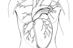
Heart Structure
- The heart has three layers:
- Inner layer = endocardium
- Middle layer = myocardium (formed of cardiac muscle fibers that run in various directions)
- Outer layer = epicardium (formed of simple squamous mesothelium and submesothelial layer of connective tissue containing coronary vessels)
Cardiac Muscle Fibers
- Short, cylindrical cells in the heart wall
- Involuntary
- Cells are joined together by intercalated discs
- Intercaled discs prevent separation of muscle cells during heart contraction
- LM diameter: small (25 µm), branching and anastomosing, show cytoplasmic striation
- Nuclei: oval, central, mono- or bi-nucleated
- Sarcoplasm contains glycogen granules, mitochondria, myofibrils, and lipochrome pigments
Purkinje Fibers
- Modified cardiac muscle fibers
- Form part of conducting system
- Larger diameter than cardiac muscle fibers
Blood Vessels
- Types: arteries, veins, blood capillaries, blood sinusoids, arteriovenous shunts
- Arteries: large elastic (aorta), medium-sized (muscular) arteries, arterioles
- Veins: venules, medium-sized veins, large veins (inferior vena cava)
Blood Capillaries
- Thin blood channels that conduct blood from terminal arterioles to venules (capillary beds)
- Diameter: 8-10 μm
- 1 layer of endothelial cells resting on basement membrane
- Types:
- Continuous (somatic) capillary
- Fenestrated capillary
- Blood sinusoids
Blood Sinusoids
- Dilated, irregular capillaries (5-30 μm)
- Endothelium is fenestrated without diaphragms
- Endothelium rests on reticular connective tissue (no basement membrane)
- Macrophages extend between endothelial cells to engulf any foreign body
- Sites: bone marrow, spleen, liver, endocrine glands
General Structure of Blood Vessels
- Tunica intima: endothelial lining, subendothelial layer, internal elastic lamina
- Tunica media: smooth muscle, elastin, collagenous and reticular fibers
- Tunica adventitia: connective tissue, vasa vasorum (small blood vessels)
Studying That Suits You
Use AI to generate personalized quizzes and flashcards to suit your learning preferences.