Podcast
Questions and Answers
Which of the following is NOT a primary function of the cardiovascular system?
Which of the following is NOT a primary function of the cardiovascular system?
- Transport of oxygen to tissues
- Removal of carbon dioxide from cells
- Production of red blood cells (correct)
- Transport of hormones to organs
The left side of the heart receives blood from the body and pumps it to the lungs.
The left side of the heart receives blood from the body and pumps it to the lungs.
False (B)
What is the approximate volume of blood, in liters, that the heart pumps into the large arteries?
What is the approximate volume of blood, in liters, that the heart pumps into the large arteries?
6 liters
The inner layer of the heart wall is called the ________.
The inner layer of the heart wall is called the ________.
Match the layer of the heart wall with its primary description:
Match the layer of the heart wall with its primary description:
Which of the following is a characteristic of the endocardium?
Which of the following is a characteristic of the endocardium?
The myocardium is thinner in the ventricles compared to the atria.
The myocardium is thinner in the ventricles compared to the atria.
What type of membrane covers the free surface of the epicardium?
What type of membrane covers the free surface of the epicardium?
Valves of the heart are folds of the ________ enclosing a core of collagenous and elastic fibers.
Valves of the heart are folds of the ________ enclosing a core of collagenous and elastic fibers.
Match the heart valve with its location:
Match the heart valve with its location:
Which of the following is NOT a tunic that differs in thickness and composition between blood vessels?
Which of the following is NOT a tunic that differs in thickness and composition between blood vessels?
The tunica intima is the outermost layer of a blood vessel.
The tunica intima is the outermost layer of a blood vessel.
What type of epithelium primarily forms the endothelium of the tunica intima?
What type of epithelium primarily forms the endothelium of the tunica intima?
The ________ is responsible for propelling blood to the end of an artery and controlling the amount of blood entering an organ.
The ________ is responsible for propelling blood to the end of an artery and controlling the amount of blood entering an organ.
Match the tunic with its primary tissue type or component:
Match the tunic with its primary tissue type or component:
Which of the following is found within the tunica adventitia of large vessels?
Which of the following is found within the tunica adventitia of large vessels?
Capillaries and postcapillary venules always have an external elastic lamina within the tunica media.
Capillaries and postcapillary venules always have an external elastic lamina within the tunica media.
What is the function of elastic fibers in the tunica media of arteries during cardiac diastole?
What is the function of elastic fibers in the tunica media of arteries during cardiac diastole?
The vasa vasorum supply the outer part of the tunica media and the ________ with blood and nutrients.
The vasa vasorum supply the outer part of the tunica media and the ________ with blood and nutrients.
Match the artery type with its primary characteristic:
Match the artery type with its primary characteristic:
Which structural feature is characteristic of large arteries (elastic arteries)?
Which structural feature is characteristic of large arteries (elastic arteries)?
The internal elastic lamina is always clearly apparent in the intima of the aorta.
The internal elastic lamina is always clearly apparent in the intima of the aorta.
What cells are joined by gap junctions in the endothelium of the aorta?
What cells are joined by gap junctions in the endothelium of the aorta?
Medium-sized arteries are also referred to as ________ arteries, as they supply blood to organs.
Medium-sized arteries are also referred to as ________ arteries, as they supply blood to organs.
Match the artery type with its function:
Match the artery type with its function:
Which of the following is a key characteristic of arterioles?
Which of the following is a key characteristic of arterioles?
Terminal arterioles have multiple layers of smooth muscle fiber.
Terminal arterioles have multiple layers of smooth muscle fiber.
What is the name of the band of circular smooth muscle fibers at the junction of a metarteriole with a capillary?
What is the name of the band of circular smooth muscle fibers at the junction of a metarteriole with a capillary?
Veins carry ________ blood from the tissues.
Veins carry ________ blood from the tissues.
Match the vein type with its characteristic:
Match the vein type with its characteristic:
What feature distinguishes small post-capillary venules?
What feature distinguishes small post-capillary venules?
Large veins lack smooth muscle bundles in the tunica adventitia.
Large veins lack smooth muscle bundles in the tunica adventitia.
In large veins, what structural adaptation assists in counteracting the effects of gravity?
In large veins, what structural adaptation assists in counteracting the effects of gravity?
Unoxygenated blood is collected from venules toward the heart by ________ veins.
Unoxygenated blood is collected from venules toward the heart by ________ veins.
Match the connection between arteries and veins with its description:
Match the connection between arteries and veins with its description:
Which of the following is NOT a characteristic of blood capillaries?
Which of the following is NOT a characteristic of blood capillaries?
Continuous capillaries have diaphragms spanning their pores.
Continuous capillaries have diaphragms spanning their pores.
What type of capillaries are present in endocrine glands to transport hormones to the blood?
What type of capillaries are present in endocrine glands to transport hormones to the blood?
Blood sinusoids have wider lumens and are lined with simple ________ cells.
Blood sinusoids have wider lumens and are lined with simple ________ cells.
Match the type of blood vessel with its corresponding location:
Match the type of blood vessel with its corresponding location:
Flashcards
What is the heart?
What is the heart?
A central muscular pump that drives the cardiovascular system.
What are blood vessels?
What are blood vessels?
Vessels that carry blood, including arteries, veins, and capillaries.
What are the two systems of circulation?
What are the two systems of circulation?
Two systems: pulmonary (lungs) and systemic (body).
Why are large arteries thick?
Why are large arteries thick?
Signup and view all the flashcards
Why are capillaries very thin?
Why are capillaries very thin?
Signup and view all the flashcards
What is the function of the cardiovascular system?
What is the function of the cardiovascular system?
Signup and view all the flashcards
What does the right side of the heart do?
What does the right side of the heart do?
Signup and view all the flashcards
What does the left side of the heart do?
What does the left side of the heart do?
Signup and view all the flashcards
What are the two chambers of each side of the heart?
What are the two chambers of each side of the heart?
Signup and view all the flashcards
What are the three layers of the heart wall?
What are the three layers of the heart wall?
Signup and view all the flashcards
What is the endocardium?
What is the endocardium?
Signup and view all the flashcards
What is the myocardium?
What is the myocardium?
Signup and view all the flashcards
What is the epicardium?
What is the epicardium?
Signup and view all the flashcards
Name the four valves of the heart.
Name the four valves of the heart.
Signup and view all the flashcards
Structure of heart valves
Structure of heart valves
Signup and view all the flashcards
What are the three layers/tunics of blood vessels?
What are the three layers/tunics of blood vessels?
Signup and view all the flashcards
What is the tunica intima?
What is the tunica intima?
Signup and view all the flashcards
What is the tunica media?
What is the tunica media?
Signup and view all the flashcards
What is the tunica adventitia?
What is the tunica adventitia?
Signup and view all the flashcards
Characteristics of Large Arteries
Characteristics of Large Arteries
Signup and view all the flashcards
Examples of elastic arteries in the body.
Examples of elastic arteries in the body.
Signup and view all the flashcards
Histophysiology of Aorta
Histophysiology of Aorta
Signup and view all the flashcards
Medium sized arteries
Medium sized arteries
Signup and view all the flashcards
Smooth muscle role.
Smooth muscle role.
Signup and view all the flashcards
Regulate blood flow to regions.
Regulate blood flow to regions.
Signup and view all the flashcards
Describe the tunica intima of arterioles.
Describe the tunica intima of arterioles.
Signup and view all the flashcards
What is the function of arterioles?
What is the function of arterioles?
Signup and view all the flashcards
What are Metarterioles?
What are Metarterioles?
Signup and view all the flashcards
What is Function of post capillary venules
What is Function of post capillary venules
Signup and view all the flashcards
What is the function of the structure of large veins.
What is the function of the structure of large veins.
Signup and view all the flashcards
What are Blood capillaries
What are Blood capillaries
Signup and view all the flashcards
Continuous (somatic) capillaries Function
Continuous (somatic) capillaries Function
Signup and view all the flashcards
3- Fenestrated capillaries without diaphragm
3- Fenestrated capillaries without diaphragm
Signup and view all the flashcards
What Size are Blood sinusoids
What Size are Blood sinusoids
Signup and view all the flashcards
what is the Definition of Blood sinusoids:-
what is the Definition of Blood sinusoids:-
Signup and view all the flashcards
3- Arteriovenous anastomosis Definition
3- Arteriovenous anastomosis Definition
Signup and view all the flashcards
N.B Glomus Speciality
N.B Glomus Speciality
Signup and view all the flashcards
Study Notes
- Histology is the study of the cardiovascular system
Cardiovascular System Composition
- Consists of a central muscular pump, the heart
- Includes blood vessels: arteries, veins, and capillaries
- Has two circulation systems: pulmonary and systemic (or peripheral)
Circulation System Details
- Pulmonary systems carry blood to other tissues
- Large arteries are thick to withstand heart's pumping force
- Capillaries are thin for substance exchange with tissues
- Vein thickness varies with gravity; veins against gravity, like the inferior vena cava, have thick walls
Cardiovascular System Function
- Transports oxygen, hormones, nutrients, and macromolecules to organs and tissues
- Removes carbon dioxide and waste from cells
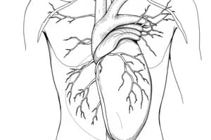
Heart Structure
- The heart has a right side, which receives blood from the body and pumps it to the lungs
- The heart also has a left side, which receives blood from the lungs and pumps it to the body
- The heart pumps 6 liters of blood in large arteries
- Each heart side has two chambers: an atrium for blood collection and a ventricle for ejection
Heart Wall Layers
- Inner endocardium
- Middle myocardium
- Outer epicardium
Endocardium Details
- Lines heart chambers and valves
- Made of:
- Endothelium (simple squamous cells)
- Thin basal lamina layer
- Subendothelial layer of loose connective tissue, elastic fibers, blood vessels, nerves, and Purkinje fibers
Myocardium Details
- Forms the heart's main substance and thickest layer
- Made of cardiac muscle groups joined end-to-end at intercalated discs
- Myocardium is thicker in the ventricles compared to the atria
Epicardium Details
- The free surface is covered by visceral pericardium’s mesothelial membrane
- Serous membrane of simple squamous epithelium and connective tissue, containing fat cells and coronary vessel branches
Heart Valves
- There are four heart valves
- Tricuspid: between right atrium and right ventricle
- Pulmonary (semilunar): between right ventricle and pulmonary artery
- Mitral: between left atrium and left ventricle
- Aortic (semilunar): between left ventricle and aorta
- Valves are endocardium folds with a core of dense collagenous and elastic fibers, covered by endothelium
Blood Vessel Structure
- Walls have 3 coats/tunics with varying thickness and composition
- Tunica intima
- Tunica media
- Tunica adventitia
Tunica Intima Details
- Innermost layer, close to the lumen
- Made of:
- Endothelium (lamina endothelialis): simple squamous epithelium on a thin basal lamina, providing a smooth surface for blood passage
- Subendothelial layer (lamina subendothelialis): loose connective tissue
- Internal elastic lamina (lamina elastica enterna)
Tunica Media Details
- Muscular coat of blood vessels
- Structure is related to the vessel's function
- Made of circular smooth muscle fibers (in distributed arteries)
- Function: produce elastin and collagen and propel blood, controlling blood flow to organs
- Elastic and collagenic fibers between muscle layers (large arteries)
- Function: maintain forward blood movement during diastole, prevent closure during artery muscle contraction, and allow expansion during systole.
- External elastic lamina may be absent in capillary and postcapillary venule
Tunica Adventitia Details
- Outermost coat of blood vessels
- Loose connective tissue containing fibroblasts with few elastic and collagenic fibers
- Contains tiny blood vessels of the vessels called vasa vasorum
- Vasa vasorum are present in large vessels because the thickness of the vessel prevents the cells from being nourished by diffusion from the lumen.
Artery Types
- Large Artery
- Medium Sized Artery
- Small Artery & Arteriole
Large Arteries
- Also called elastic or conducting arteries because they have a thick wall and wide lumen
- Their walls maintain blood pressure during ventricular relaxation
- Their media are rich in elastic fibers
- Examples include the Aorta, aortic arch, common iliac, pulmonary, and subclavian arteries
Aorta Details
- It has a thick wall and wide lumen
- Tunica intima is thick and rich in elastic fibers
- Not apparent internal elastic lamina
- Made oF:
- Endothelium that contains simple squamous epithelium with Endothelial cells joined by gap junctions
- Subendothelium with loose connective tissue containing fibroblasts, collagen (type I, III and IV), and elastic fibers
- Non-clear internal elastic lamina
- The tunica media is a very thick layer made of 40-70 laminae of fenestrated elastic membrane enclosing smooth muscles, collagenic and reticular fibers
- Tunica Adventitia is thin and narrow containing collagen, elastic fibers, nerves, and vasa vasorum, provides blood and nutrients to media and adventitia outer parts
- Elastic fibers allow expansion during systole; elastic recoil maintains blood flow and diastolic pressure during diastole
- They major elastic conducting arteries contain many elastic fibers and can stretch, taking the heart's ejected blood during ventricular conduction
Medium Sized Arteries
- Also called muscular arteries
- Are the most common arteries in the body, supplying blood to organs
Tunica Intima of Medium Sized Arteries
- Innermost layer
- Made oF:
  - Simple squamous epithelium
  - Subendothelial layer of connective tissue
  - Internal elastic lamina
Tunica Media of Medium Sized Arteries
- Includes circular smooth muscle fibers
- May have few scattered elastic fibers
- Includes fine collagenic fibers
Tunica Adventitia of Medium Sized Arteries
- Outermost layer composed of mostly collagenic fibers
- Contains some elastic fibers and connective tissue cells
Function Of Medium Sized Arteries
- Walls of fibers distribute blood to different body parts
- Regulate blood flow through contraction/relaxation
Arterioles/Small Arteries
- Regulate blood flow into capillary beds
- They have a small diameter (0.1 mm) with thick walls related to the lumen.
- Tunica Intima has endothelium; thin, wavy internal elastic lamina; no subendothelial tissue
- Tunica Media has 2-5 smooth muscle layers and scattered elastic fibers without external elastic lamina Tunica Adventitia is thin and contains collagen and elastic fibers
- They decrease pressure in blood entering capillaries
- They have impermeable walls, and aren't involved in interchange between blood and tissue fluid
Terminal Arteriole
- Contians a single layer of smooth muscle fiber
Metarterioles
- They are vessels intermediate between arterioles and capillaries
- They have thin walls with endothelial cells on basal lamina, plus interrupted smooth muscle cells layer
- A band of circular smooth muscle fibers called precapillary sphincters are located at the junction of metarteriole with capillary
- precapillary sphincters control blood flow into the capillary
Veins
- Include small venules, medium sized veins, and large sized veins
- They carry venous blood from tissues
- Similar to arterioles
- Start at the capillary bed as small post-capillary venules, which join to create larger veins
Small Post Capillary Venule
- Have a diameter of 0.2-1mm
- Smooth muscle fibers appear in the media as diameter increases
- Lined by a simple squamous cells layer
- Outside of the squamous endothelial cells there is differentiate in to smooth muscle
- Thin adventitia while large ones have thin media; small venules do not have media
- Function similar to capillaries; sites for gas exchange from blood to tissue
Large Muscular Venules
- They are lined by simple squamous epithelium with rich actin filaments
- Presence of basement membrane outside endothelial lining
- A formed media with a thin layer of smooth muscles
- Contains a thin adventitia formed of areolar CT
Medium Sized/Muscular Veins
- Are made of 3 tunics/layers
- Unoxygenated blood is collected from venules towards the heart by muscular veins, blood only goes towards the heart
Large Veins
- Examples of large veins are the superior vena cava, inferior vena cava, and common iliac vein
- Thicker Tunica intema compared to medium veins
- Tunica media consists of circular smooth muscle fibers with collagen and a few or no elastic fibers
- Tunica adventitia is thick the thickest coat in large bein. contains bundles of smooth muscle and collagen. Vasa vasorum is present
- Wide lumen with many valves in the IVC and I.V.C. is thick
- Smooth muscle bundles help to strengthen the vessel wall against excessive extension
Connections Between Arteries And Veins
- The blood capillaries
- Sinusoids
- Arteriovenous anastomosis
Blood Capillaries
- They connect arteries and veins, found throughout the body
- Thin-walled vessels with 8-10um luminal diameter
Blood Capillary Size
- Diameter ranges from 7-9um and 0.25-1mm in length
- Have a complete regular lumen about 8 microns in diameter
Blood Capillary Function
- Conduct the blood from arteriole branches to venule branches
- Made of a single-layered wall with endothelial cells resting on basal lamina
- Endothelium is formed of simple squamous epithelium
- Pericytes are found around the endothelium
Types of Capillaries
- Continuous (somatic) capillaries
- Fenestrated capillaries with diaphragm
- Fenestrated capillaries without diaphragm
Continuous (Somatic) Capillaries
- Present all over the body; found in CT, skeletal muscles, brain, bone, lung and exocrine glands
- Found in the brain
- Share in formation of blood brain barrier which prevent the passage of some substances as antibiotic, chemical and bacterial toxin from the blood to the nerve tissue
Fenestrated Capillaries with Diaphragm
- Present in endocrine glands, intestine and ciliary body of eye
- The endothelial cells contain pores (fenestrae) and rest on the basement membrane. Through these pours tissue fluid can pass
Fenestrated Capillaries Without Diaphragm
- Present in the glomeriolar blood capillaries of kidney
- Speed up the process of exchange
Blood Sinusoids
- Thin walled capillary like vessel
- A connection between arterieis and veins
- Irregular wide lumens from 5-30 microns in diameter
- They are lined with simple squamous endothelial cells
- Macrophages are present either in or along the outside of the sinusoidal wall
- Walls contain pores not covered by diaphragms
Locations
- Bone marrow allows for formed blood cells to carry
- Found in spleen to store blood and in their wall to filter the blood from any foreign body by phagocytic cells
- Liver allows contact with blood by liver cells and easy passage of protein from phagocytes into the blood stream
- Endocrine glands and endocrine glands to carry the secreted hormones and to increase the blood supply to the endocrine secretory cells
Arteriovenous Anastomosis
- Definition: Arteriovenous anastomoses arise as side branches from arteries and arterioles and pass as straight or tortuous vessels to be connected with the veins or venules
Arteriovenous Anastomosis Type
- Direct connection between an arteriole and a venule by a side branch, which is found in the placenta
- Indirect (or glomus) a complicated side branch, which is a specialized organ present in skin
- Present in exposed parts of the body such as fingers, toes, the external ear, the nose, the lip, tongue, eyelids, as well as liver, and stomach internal organs, intestine
Glomus Details
- On some sites (nail beds/auricle of ear), A-V anastomosis are branched and tortuous an form a specialized organ
- The arterial branch is coiled or branched
- The internal elastic lamina disappears
- The elastic tissue is reduced
- The smooth muscle of the media is replaced by myoepithelial cells (epitheloid like cells)
- The adventitia becomes thicker
Studying That Suits You
Use AI to generate personalized quizzes and flashcards to suit your learning preferences.