Podcast
Questions and Answers
What are the key functions of the foot?
What are the key functions of the foot?
The key functions of the foot include: - Key for gait, propulsion, and balance - Transfer of force within the lower kinetic chain - Shock absorption
How many bones are in the foot?
How many bones are in the foot?
26
How many tarsals are there in the foot? Choose the correct answer:
How many tarsals are there in the foot? Choose the correct answer:
- 7 (correct)
- 5
- 11
- 9
Name the seven tarsal bones.
Name the seven tarsal bones.
How many metatarsals are in the foot?
How many metatarsals are in the foot?
How many phalanges are in the foot?
How many phalanges are in the foot?
What is the hallux?
What is the hallux?
Select the three joints in the foot that drive movement:
Select the three joints in the foot that drive movement:
Choose the four joints in the foot that do not drive movement:
Choose the four joints in the foot that do not drive movement:
What is the talocrural joint?
What is the talocrural joint?
Which of the following are lateral ligaments in the foot? (Select all that apply)
Which of the following are lateral ligaments in the foot? (Select all that apply)
Name the three arches of the foot.
Name the three arches of the foot.
What is the plantar fascia?
What is the plantar fascia?
List the six types of movement that occur at the foot and ankle.
List the six types of movement that occur at the foot and ankle.
What is the gastrocnemius muscle?
What is the gastrocnemius muscle?
Where is the soleus muscle located?
Where is the soleus muscle located?
Which of the following muscles are plantarflexors? (Select all that apply)
Which of the following muscles are plantarflexors? (Select all that apply)
Which of the following muscles are dorsiflexors? (Select all that apply)
Which of the following muscles are dorsiflexors? (Select all that apply)
Which of the following muscles are invertors? (Select all that apply)
Which of the following muscles are invertors? (Select all that apply)
What is the most common ankle sprain among active individuals?
What is the most common ankle sprain among active individuals?
What is tibial stress syndrome?
What is tibial stress syndrome?
Flashcards
What are the key functions of the foot?
What are the key functions of the foot?
The foot is crucial for walking, pushing off, and maintaining balance. It transfers force throughout the lower limb and absorbs shock during movement.
How many bones are in the foot?
How many bones are in the foot?
The foot contains 26 bones, contributing to its complex structure and functionality.
How many tarsals are there? And name them.
How many tarsals are there? And name them.
There are 7 tarsals, which are the bones in the ankle and midfoot. They include the calcaneus (heel bone), talus, navicular, cuboid, and three cuneiforms.
How many metatarsals are there?
How many metatarsals are there?
Signup and view all the flashcards
How many phalanges are there?
How many phalanges are there?
Signup and view all the flashcards
What is the hallux?
What is the hallux?
Signup and view all the flashcards
What are the 3 joints in the foot that drive movement?
What are the 3 joints in the foot that drive movement?
Signup and view all the flashcards
What are the 4 joints that don't drive movement?
What are the 4 joints that don't drive movement?
Signup and view all the flashcards
What is the talocrural joint?
What is the talocrural joint?
Signup and view all the flashcards
What are the 4 lateral ligaments in the foot?
What are the 4 lateral ligaments in the foot?
Signup and view all the flashcards
What are the 5 medial ligaments in the foot?
What are the 5 medial ligaments in the foot?
Signup and view all the flashcards
What are the 3 arches?
What are the 3 arches?
Signup and view all the flashcards
What is the plantar fascia?
What is the plantar fascia?
Signup and view all the flashcards
What are the 6 types of movement at foot/ankle?
What are the 6 types of movement at foot/ankle?
Signup and view all the flashcards
What is the gastrocnemius?
What is the gastrocnemius?
Signup and view all the flashcards
Where is the soleus?
Where is the soleus?
Signup and view all the flashcards
Which muscles are the plantarflexors?
Which muscles are the plantarflexors?
Signup and view all the flashcards
Which muscles are the dorsiflexors?
Which muscles are the dorsiflexors?
Signup and view all the flashcards
Which muscles are the invertors?
Which muscles are the invertors?
Signup and view all the flashcards
Which muscles are the evertors?
Which muscles are the evertors?
Signup and view all the flashcards
What is the most common ankle sprain with active people?
What is the most common ankle sprain with active people?
Signup and view all the flashcards
What is tibial stress syndrome?
What is tibial stress syndrome?
Signup and view all the flashcards
What is the subtalar joint?
What is the subtalar joint?
Signup and view all the flashcards
What is the transverse tarsal joint?
What is the transverse tarsal joint?
Signup and view all the flashcards
What is the intertarsal joint?
What is the intertarsal joint?
Signup and view all the flashcards
What is the tarsometatarsal joint?
What is the tarsometatarsal joint?
Signup and view all the flashcards
What is the metatarsophalangeal joint?
What is the metatarsophalangeal joint?
Signup and view all the flashcards
What is the interphalangeal joint?
What is the interphalangeal joint?
Signup and view all the flashcards
Study Notes
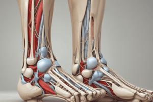
Foot Anatomy and Function
- The foot is crucial for gait, propulsion, and balance, transferring force through the lower body and absorbing shock.
- There are 26 bones in the foot.
- The foot comprises 7 tarsal bones: calcaneus, talus, navicular, cuboid, and three cuneiform bones.
- There are 5 metatarsal bones.
- There are 14 phalanges (toe bones).
- The hallux is the big toe.
Foot Joints
- The foot has key joints driving movement:
- Talocrural (ankle) joint: Connects talus, tibia (medial malleolus), and fibula (lateral malleolus). The lateral malleolus is slightly lower, predisposing it to sprains.
- Subtalar joint: Talus and calcaneus, allowing gliding movement.
- Transverse tarsal joints: Gliding joints for multi-directional foot movement.
- Other joints (non-primary movers):
- Intertarsal joints
- Tarsometatarsal joints
- Metatarsophalangeal joints
- Interphalangeal joints
Foot Ligaments
- Lateral ligaments:
- Anterior talofibular ligament (ATFL)
- Posterior talofibular ligament
- Calcaneofibular ligament
- Medial ligaments (deltoid ligament):
- Posterior tibiotalar ligament
- Tibiocalcaneal ligament
- Anterior tibiotalar ligament
- Tibionavicular joint
- Plantarcalcaneonavicular ligament (important for propulsion).
- The Achilles tendon (gastrocnemius to the calcaneus) connects calf muscles to heel bone, contributing to ankle function.
Foot Arches
- The foot has three arches: medial longitudinal, lateral longitudinal, and transverse arches.
- The plantar fascia is a ligamentous structure that runs along the bottom of the foot and supports the arches. It connects the calcaneus to the metatarsals and phalanges.
Foot Muscle Groups
- Plantarflexors: Muscles that point the foot downward (plantar flexion). Includes gastrocnemius, soleus, tibialis posterior, FDL, FHL, peroneus longus, and brevis.
- Dorsiflexors: Muscles that lift the foot upwards (dorsiflexion). Includes tibialis anterior, EDL, EHL, and peroneus tertius.
- Invertors: Muscles that turn the sole of the foot inward (inversion). Includes tibialis anterior, tibialis posterior, FDL, and FHL.
- Evertors: Muscles that turn the sole of the foot outward (eversion). Includes peroneus longus, brevis, and tertius, and EDL.
Common Foot Conditions & Injuries
- Inversion Ankle Sprain: Most common ankle sprain in active people, often involving the ATFL.
- Tibial Stress Syndrome (Shin Splints): Common overuse injury, usually due to periostitis (inflammation of the periosteum).
Important Muscles
- Gastrocnemius: Calf muscle, origin on femur, inserts on calcaneus via Achilles tendon.
- Soleus: Located beneath the gastrocnemius.
Studying That Suits You
Use AI to generate personalized quizzes and flashcards to suit your learning preferences.