Podcast
Questions and Answers
What is the initial event in the process of neurulation?
What is the initial event in the process of neurulation?
- Differentiation of pre-nodal surface ectoderm into neuroectoderm. (correct)
- Expansion of the primitive node.
- Closure of the cranial neuropore.
- Formation of the neural folds.
The neural tube develops two open ends in the middle of the 4th week. What are these openings called?
The neural tube develops two open ends in the middle of the 4th week. What are these openings called?
- Neural crests
- Neural folds
- Neuropores (correct)
- Neural plates
What does an elevated level of Alpha-fetoprotein in the amniotic fluid after the 5th week of IUL suggest?
What does an elevated level of Alpha-fetoprotein in the amniotic fluid after the 5th week of IUL suggest?
- Proper formation of cranial and caudal neuropores.
- Malformation of the cranial or caudal neuropore. (correct)
- Typical expansion of the brain vesicles.
- Normal development of the neural tube.
Which primary brain vesicle gives rise to the future cerebral hemispheres and basal ganglia?
Which primary brain vesicle gives rise to the future cerebral hemispheres and basal ganglia?
Which of the following is derived from the rhombencephalon?
Which of the following is derived from the rhombencephalon?
Lens placodes develop into which structures?
Lens placodes develop into which structures?
What type of cells are responsible for the pseudostratified appearance of the neuroepithelium?
What type of cells are responsible for the pseudostratified appearance of the neuroepithelium?
Which layer of the neural tube contains the cell bodies of newly formed neurons and neuroglia?
Which layer of the neural tube contains the cell bodies of newly formed neurons and neuroglia?
What structures do alar plates develop into?
What structures do alar plates develop into?
What cell types are derived from neural crest cells?
What cell types are derived from neural crest cells?
From what structure does the medulla oblongata originate?
From what structure does the medulla oblongata originate?
What anatomical feature marks the boundary between the alar and basal plates in the developing neural tube?
What anatomical feature marks the boundary between the alar and basal plates in the developing neural tube?
The somatic efferent group, which forms the nuclei of the abducens nerve, is found in which part of the brain?
The somatic efferent group, which forms the nuclei of the abducens nerve, is found in which part of the brain?
The pons connects the cerebral and cerebellar cortices to the spinal cord. From which of the following does it originate?
The pons connects the cerebral and cerebellar cortices to the spinal cord. From which of the following does it originate?
What part of the developing brain is the lamina terminalis associated with?
What part of the developing brain is the lamina terminalis associated with?
What term describes the smooth surface of the cerebral vesicles before the formation of gyri and sulci?
What term describes the smooth surface of the cerebral vesicles before the formation of gyri and sulci?
The cerebral hemispheres are recognizable as two lateral bulges from which part of the developing brain?
The cerebral hemispheres are recognizable as two lateral bulges from which part of the developing brain?
What is the developmental origin of the caudate nucleus?
What is the developmental origin of the caudate nucleus?
Which components are present in the diencephalon?
Which components are present in the diencephalon?
What shallow groove divides each alar plate to form the thalamus and hypothalamus?
What shallow groove divides each alar plate to form the thalamus and hypothalamus?
During what week of development does the anterior commissure arise?
During what week of development does the anterior commissure arise?
The corpus callosum connects the neocortex on both sides and develops during which months of gestation?
The corpus callosum connects the neocortex on both sides and develops during which months of gestation?
From which structure does the anterior pituitary gland originate?
From which structure does the anterior pituitary gland originate?
In what week of development does the Rathke's pouch appear?
In what week of development does the Rathke's pouch appear?
Sensory (dorsal) horns of the spinal cord are formed by
Sensory (dorsal) horns of the spinal cord are formed by
Flashcards
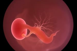
Neurulation
Neurulation
The process leading to the formation of the neural plate, neural folds, and the neural tube which begins in the 3rd week of IUL and is completed by the end of the 4th week.
Appearance of the neural plate
Appearance of the neural plate
The differentiation of pre-nodal surface ectoderm into neuroectoderm and formation of the neural plate under the influence of the notochord.
Formation of the neural tube
Formation of the neural tube
Fusion of neural folds begins in the future cervical region and moves in both directions. The neural tube becomes like a straw with two open ends.
Neuropores
Neuropores
Signup and view all the flashcards
Primary brain vesicles
Primary brain vesicles
Signup and view all the flashcards
Myelencephalon
Myelencephalon
Signup and view all the flashcards
Placodes
Placodes
Signup and view all the flashcards
Neuroepithelium
Neuroepithelium
Signup and view all the flashcards
Mantle layer
Mantle layer
Signup and view all the flashcards
Neural crest cells
Neural crest cells
Signup and view all the flashcards
Telencephalon
Telencephalon
Signup and view all the flashcards
Lissencephalic
Lissencephalic
Signup and view all the flashcards
Diencephalon
Diencephalon
Signup and view all the flashcards
Anterior commissure
Anterior commissure
Signup and view all the flashcards
Rathke's pouch
Rathke's pouch
Signup and view all the flashcards
Expansion of the neural plate
Expansion of the neural plate
Signup and view all the flashcards
Neural folds and groove formation
Neural folds and groove formation
Signup and view all the flashcards
Alpha-fetoprotein
Alpha-fetoprotein
Signup and view all the flashcards
Cephalic/Midbrain flexure
Cephalic/Midbrain flexure
Signup and view all the flashcards
Somatic efferent group
Somatic efferent group
Signup and view all the flashcards
Corpus callosum
Corpus callosum
Signup and view all the flashcards
Infundibulum
Infundibulum
Signup and view all the flashcards
Study Notes
- Neurulation leads to the formation of the neural plate, neural folds, and the neural tube.
- It begins in the 3rd week of IUL and is completed by the end of the 4th week.
Appearance of Neural Plate
- Differentiation of pre-nodal surface ectoderm into Neuroectoderm.
- Formation of the 'neural plate’ occurs under the influence of the notochord.
- Represents the initial event in neurulation.
Expansion of Neural Plate
- Occurs in a cranio-caudal direction between the prechordal plate and primitive node.
Appearance of Neural Folds and Groove
- Around day 20, lateral edges of the neural plate become elevated due to rapid proliferation of neuroectodermal cells, forming neural folds.
- The depressed mid-region forms the neural groove.
- Neural folds are the first sign of brain development.
Formation of Neural Tube & Closure of Neuropores
- Fusion of neural folds begins in the future cervical region and moves in both directions.
- By the middle of the 4th week, the neural tube resembles a straw with two open ends called neuropores.
- The cranial and caudal neuropores stay open for a period, communicating with the amniotic cavity.
- Cranial neuropore closes around day 25 of IUL.
- Caudal neuropore closes around day 27/28 of IUL.
- The cranial end of the neural tube expands to give rise to the brain.
- The caudal end of the neural tube elongates and becomes the spinal cord.
- By the end of the 4th week, there should be no communication between the neural tube cavity and the amniotic cavity.
- High levels of Alpha-fetoprotein in the amniotic fluid after the 5th week of IUL are a key indicator of malfusion of the cranial/caudal neuropore.
Formation of Primitive Brain Vesicles and Primary Flexures
- Immediately after the closure of the cranial neuropore, the cephalic end of the neural tube expands rapidly, forming three primary/primitive brain vesicles.
- The three vesicles include:
- Prosencephalon (forebrain)
- Mesencephalon (midbrain)
- Rhombencephalon (hindbrain)
- The cranial end of the tube shows two dorsal primary flexures.
- Cephalic/Midbrain flexure, where the forebrain meets the midbrain.
- Cervical/Hindbrain flexure, where the hindbrain meets the spinal cord.
Structural & Function Differentiation of Brain Vesicles & Formation of Secondary Flexure
- By the 5th week of intrauterine life:
- The Prosencephalon differentiates into the Telencephalon (future cerebral hemispheres and basal ganglia) and Diencephalon (future thalamus, hypo, epi, and sub thalami).
- The Rhombencephalon differentiates into the Metencephalon (future cerebellum & pons) and Myelencephalon (future medulla oblongata).
- The upper half of the Mesencephalon is developmentally part of the Diencephalon, and the lower half is associated with the metencephalon.
- A secondary ventral flexure appears in the region of the hindbrain, known as the pontine flexure.
- By the end of the 5th week, the embryonic brain exhibits all major components of the adult-form brain.
Formation of Primitive Special Sensory Organs
- Regional thickenings of the surface ectoderm around the developing brain vesicles are called placodes.
- Each pair of placodes are influenced by the underlying neural epithelium, developing into sensory epithelia of special sense organs (vision, smell, hearing, etc.).
- Olfactory placodes, induced by the neuroepithelium of the prosencephalon, lead to the formation of the olfactory mucosa. The olfactory mucosa comprises special bipolar olfactory neurons and olfactory bulbs.
- Lens placodes, induced by the optic vesicle (neuroepithelium of the diencephalon), develop into the retina and lens of the eye.
- Auditory placodes, induced by the neuroepithelium of the rhombencephalon, form the special sensory organs of balance and hearing of the internal ear.
Initial Histogenesis in the Neural Tube & Formation of Three Zones
- The wall of the newly closed neural tube consists of cells extending over the entire thickness.
- Overcrowded alignment of elongated neuroepithelial cells gives a pseudostratified appearance to the epithelium of the neural tube called the 'neuroepithelium'.
- The neuroepithelium lines the central cavity of the neural tube.
- After the complete closure of the neural tube, the neuroepithelium produces different cell types: neuroblasts, glioblasts, and ependymal cells.
- A cut section through the neural tube shows a central fluid-filled cavity surrounded by cells, forming three zones:
- Ventricular zone
- Intermediate zone
- Marginal zone
Basal, Alar, Roof, and Floor Plates of the Neural Tube
- The mantle layer consists of the cell bodies of newly formed neurons and neuroglia.
- The marginal layer consists of the cell processes of the neurons.
- Rapid proliferation of cells in the mantle layer forms ventral and dorsal thickenings.
- Ventral thickenings or Basal plates are composed of motor neuron cell bodies.
- Dorsal thickenings or Alar plates are composed of sensory neuron cell bodies.
- A longitudinal groove, called the Sulcus limitans, marks the boundary between the alar and basal plates.
- The floor plate is the ventral midline portion.
- The roof plate is the dorsal midline portion.
- The floor and roof plates do not directly contribute to neurogenesis.
Neural Crest Cells & Their Neural Derivatives
- While the neural folds are fusing, neural crest cells (NCC) disengage from the crests and initially lie between the surface ectoderm and mesoderm.
- Some NCC lie parallel to the developing spinal cord and give rise to Leptomeninges (arachnoid and pia), sensory (DRG), and sympathetic ganglia.
- In the lumbar region, some NCC lose processes and migrate laterally to invade the developing adrenal gland, forming chromaffin cells of the adrenal medulla (adrenaline, noradrenaline producing cells).
- Some NCC on either side of the tube dive into the underlying mesoderm undergoing an epithelial-to-mesenchymal transformation.
- These modified cells give rise to melanocytes, Schwann cells, parasympathetic ganglia, pharyngeal arches’ cartilage & muscles, etc.
Development of the Spinal Cord
- Closure of the caudal neuropore on day 27 or 28 triggers the elongation of the neural tube towards the caudal end of the embryo.
- Blocks of paraxial mesoderm (somites) flank the developing spinal cord, giving rise to the vertebral column.
- The mantle and marginal layers of the developing spinal cord form the gray and white matter of the adult-form spinal cord.
- Basal plates will form the motor/ventral horns.
- Alar plates will form the sensory/dorsal horns.
Development and Differentiation of the Rhombencephalon/Hindbrain
- Distinct basal & alar plates, representing motor and sensory areas, are present on each side of the midline.
- The sulcus limitans dividing the sensory & motor areas is visible in the rhombencephalon and mesencephalon.
- The Myelencephalon represents the most caudal part of the rhombencephalon, giving rise to the Medulla oblongata.
- The caudal portion of the medulla is simply the upward continuation of the spinal cord.
- The dorsolateral walls of the upper/rostral part of the Medulla move laterally around an imaginary longitudinal axis.
- Resulting in two alar plates coming to lie lateral to the two basal plates, stretching the roof of the rostral myelencephalon.
- Basal plates give rise to three groups of efferent nuclei
- Somatic efferent group
- Special visceral efferent group
- General visceral efferent group
- Alar plates give rise to three groups of afferent nuclei
- General visceral afferent
- Special visceral afferent
- Somatic afferent.
Metencephalon
- Characterized by basal and alar plates.
- The metencephalon gives rise to two separate structures:
- Pons (ventral)
- Cerebellum (dorsal)
- Each basal plate contains three groups of motor nuclei forming several different nuclei
- Somatic efferent group that forms the nuclei of the Abducens nerve.
- SVE group forming the Trigeminal & Facial nerves' motor nuclei.
- GVE group (superior salivatory nucleus) whose axons supply the submandibular & sublingual salivary glands.
- The zone of the basal plates in the metencephalon expands, including transversely running bundles of myelinated axons coming from the cerebrum & cerebellum.
- This is also known as the Pons (=bridge) which connects the cerebral & cerebellar cortices with the spinal cord.
- Clusters of neurons (Pontine nuclei) are also found in the pons.
- The roof plate is stretched and expands over the 4th ventricle.
Development of the Telencephalon
- The most rostral section of the Prosencephalon
- The two lateral outpocketings are called the cerebral hemispheres.
- It also has a median portion called the lamina terminalis.
- Interventricular foramina of Monro connect the cavities of the hemispheres.
Shape Change and Lobar differentiation of Cerebral Hemispheres
- The cerebral vesicles acquire a horseshoe curvature through dorso-caudal and ventro-rostral expansion.
- Insula, the curvature's central region, doesn't develop and is covered by the hemisphere's fast-growing regions.
- The lateral sulcus develops above the insular lobe from a deep furrow.
- Initially the vesicles exhibit a smooth surface (lissencephalic)
- The cortex of each hemisphere gets a rush of neuroblasts and the formation of gyri and sulci becomes evident on the surface by the 18th week.
- By the sixth month of IUL the four cerebral lobes are distinctly recognizable.
Cortex and Deep Nuclei development in Telencephalon
- the cerebral hemispheres are easily recognized from 33 days IUL.
- The cortex is the result of migrating neuroblasts from the the ventricle in a cerebral direction.
Basal Nuclei and Internal Capsule development
- The hemispheres expand and starts to cover much of the sides.
- Axons in the nuclear mass that produce striatum divides in two
- Dorsomedial portion produces caudate nucleus
- Ventrolateral region generates lentiform nucleus.
Development of the Diencephalon
- Arises from the 'median section of prosencephalon
- Mostly contains roof and alar plates, but does not have floor or basal plates.
Formation & Differentiation of Thalamus, Subthalamus, & Hypothalamus
- Formed by two Alar plates on lateral diencephalon sides
- Each Alar plate gets divided by, the hypothalamic sulcus;
- Dorsal parts turns into Thalamus
- Ventral parts develop into Subthalamus and hypothalamus.
- A number of side-to-side Thalamic cells reduces ventricular cavity size.
- Alar Plates on lower parts of form hypothalamus that help to process digestion and feelings.
Formation of Major White Matter Commissures
- Paleo and limbic Cortex get joined to the hemispheres at 7th week by the anterior commissure
- The olfactory bulb lies between tw hemispheres.
- 10th week sees the formation of diencephalon commissures.
- 3 to 6 months : corpus callosum gets built to help join neocortex from sides
Development of the Pituitary Gland
- An ectodermal section of the upward of stomodeum creates Rathke's pouch at what will bucopharyngeal membrane.
- The pituitary Rathke's pouch creates extension for the oral cavity.
- The gland losses connection and creates anterior portion.
Studying That Suits You
Use AI to generate personalized quizzes and flashcards to suit your learning preferences.
Related Documents
Description
Neurulation is the process of forming the neural plate, neural folds, and neural tube. It starts in the 3rd week and concludes by the end of the 4th week of intrauterine life. The neural plate forms from the differentiation of the pre-nodal surface ectoderm into neuroectoderm under the notochord's influence.