Podcast
Questions and Answers
What type of joint is the elbow classified as?
What type of joint is the elbow classified as?
- Saddle joint
- Hinge type synovial (correct)
- Ball and socket
- Pivot joint
Which muscle is primarily responsible for flexion at the elbow?
Which muscle is primarily responsible for flexion at the elbow?
- Biceps brachii (correct)
- Anconeus
- Brachioradialis
- Triceps brachii
Which structure provides the strongest support to the elbow joint?
Which structure provides the strongest support to the elbow joint?
- Annular ligament
- Fibrous capsule
- Ulnar collateral ligament (correct)
- Radial collateral ligament
What angle does the ulna make with the humerus at full extension?
What angle does the ulna make with the humerus at full extension?
What is the primary role of the synovial membrane in the elbow joint?
What is the primary role of the synovial membrane in the elbow joint?
Which nerve passes posterior to the medial epicondyle?
Which nerve passes posterior to the medial epicondyle?
What type of joint is the proximal radioulnar joint classified as?
What type of joint is the proximal radioulnar joint classified as?
Which structure is primarily responsible for ensuring the integrity of the distal radioulnar joint?
Which structure is primarily responsible for ensuring the integrity of the distal radioulnar joint?
Which movement occurs at both the elbow and forearm joints during pronation?
Which movement occurs at both the elbow and forearm joints during pronation?
What is the main role of the interosseous membrane?
What is the main role of the interosseous membrane?
What defines a subluxation?
What defines a subluxation?
Which muscle is not involved in pronation?
Which muscle is not involved in pronation?
What is the primary function of the annular ligament?
What is the primary function of the annular ligament?
Flashcards
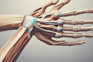
Elbow Joint
Elbow Joint
This is the joint formed between the humerus, ulna and radius.
Elbow Joint Capsule
Elbow Joint Capsule
This is the strong fibrous membrane that surrounds the elbow joint.
Collateral Ligaments
Collateral Ligaments
These are strong, fibrous bands that connect bones and help to stabilize the elbow joint.
Carrying Angle
Carrying Angle
Signup and view all the flashcards
Bursae
Bursae
Signup and view all the flashcards
Subtendinous olecranon bursa
Subtendinous olecranon bursa
Signup and view all the flashcards
Subcutaneous olecranon bursa
Subcutaneous olecranon bursa
Signup and view all the flashcards
Hilton's law
Hilton's law
Signup and view all the flashcards
Subluxation
Subluxation
Signup and view all the flashcards
Dislocation
Dislocation
Signup and view all the flashcards
Interosseous membrane
Interosseous membrane
Signup and view all the flashcards
Annular ligament
Annular ligament
Signup and view all the flashcards
Proximal radioulnar joint
Proximal radioulnar joint
Signup and view all the flashcards
Study Notes
Elbow and Forearm Anatomy
- The elbow joint is a hinge type synovial joint
- At full extension, the ulna makes a 170° angle with the humerus (long axis)
- In females, the forearm is angled further away from the trunk (carrying angle)
Joint Considerations
- Articulations: The bones forming the joint (humerus, radius, ulna)
- Capsule: Connective tissue sac surrounding the joint
- Ligaments: Strong bands of tissue reinforcing the joint
- Movements: Actions possible at the joint (flexion, extension, etc.)
- Muscles: Muscles produce the movements
- Neurovascular supply: Nerves and blood vessels supplying the joint
- Bursae: Small fluid-filled sacs reducing friction
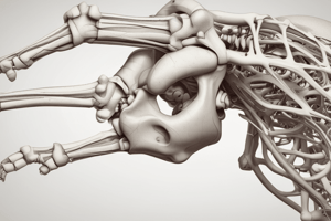
Elbow Articulations (Osteology)
-
Humerus:
- Trochlea
- Capitulum
- Olecranon fossa
- Coronoid fossa
- Radial fossa
-
Ulna:
- Coronoid process
- Trochlear notch
- Olecranon process
-
Radius:
- Head
Elbow Movements
- Flexion: Bending the elbow
- Extension: Straightening the elbow
- Carrying angle: Forearm angled further away from the trunk in females
Muscles
- Biceps brachii: Flexor of the elbow
- Triceps brachii: Extensor of the elbow
- Branchioradialis: Flexor, innervated by the radial nerve
Elbow Capsule
- Weak anteriorly and posteriorly
- Strengthened by collateral ligaments (medially and laterally)
- Elbow and proximal radioulnar joints share a capsule
- Synovial membrane lines the fibrous capsule and humerus
Elbow Ligaments (Ulnar Collateral Ligament)
- Three bands:
- Anterior (strongest)
- Posterior
- Oblique (deepens the socket for the trochlea)
- Radial collateral ligament: Fan-like, blends with annular ligament
Elbow Bursae
- Many bursa (fluid-filled sacs)
- Problems are rare except some:
- Subcutaneous olecranon bursa
- Subtendinous olecranon bursa
Nerves
- Radial nerve passes anterior to the lateral epicondyle
- Ulnar nerve passes posterior to the medial epicondyle
Neurovascular Supply
- Hilton's law: nerves and vessels supplying a joint also innervate the muscles moving the joint.
- Anastomoses formed by collateral arteries (and recurrent branches) of ulnar, radial, and interosseous arteries
Radioulnar Joints
- Proximal: Pivot joint, osteology of the radius head and ulna's radial notch
- Interosseous membrane: Fibrous joint, allows force distribution to the ulna
- Distal: Osteology of the ulna head, rounded head of radius, ulnar notch on medial border, articular disk.
- The articular disk is important for maintaining joint integrity and separating the distal radioulnar joint cavity from the wrist cavity.
Radioulnar Movements
- Pronation/Supination: Rotation of the forearm
- The head of the radius pivots on the capitulum of the humerus
Proximal Radioulnar Joint
- Pivot joint
- Osteology: radius head, ulna's radial notch
Annular Ligament
- Ligamentous collar and attached to the ulna, anterior and posterior to its radial notch
Pulled Elbow (Children)
- Dislocation of the radial head from the annular ligament
Dislocation/Subluxation
- Dislocation: Complete loss of joint contact of joint surfaces.
- Subluxation: Partial dislocation (misalignment) but bone ends remain in contact
Interosseous Membrane
- Fibrous joint
- Fibers run inferomedially
- Allows force distribution from radius to ulna
Distal Radioulnar Joint
- Osteology: rounded head of radius, ulnar notch on medial border
- Articular disk
- Maintains joint integrity, separating distal radioulnar joint cavity from wrist cavity
Distal Radioulnar Joint in Pronation/Supination
- Articular disk ensures maintenance of joint integrity
- Sacciform recess is a superior expansion of the synovial capsule that allows twisting of the capsule
Movements of Radioulnar Joints
- Supinator: Muscle assisting in supination
- Biceps brachii: Muscle assisting in supination
- Pronator teres: Muscle assisting in pronation
- Pronator quadratus: Muscle assisting in pronation
Wrist
- Radiocarpal joint: Articulation of distal radius and articular disk with proximal carpal bones
- Osteology: Radius (distal), carpus (proximal row excluding pisiform)
- Ligaments: Collateral, palmar radiocarpal, and dorsal radiocarpal ligaments
- Movements: Flexion/extension, abduction/adduction (radial/ulnar deviation), limited by the radial styloid process
Wrist Fractures
- Colles fracture: Posterior displacement of the distal radius fragment
- Scaphoid fracture: Fall onto outstretched hand causes tenderness over the anatomical snuffbox. Complications can include avascular necrosis.
Self-Study
- Review upper limb osteology, joint classifications (synovial and fibrous joints), and clinically relevant sections from anatomy texts.
Studying That Suits You
Use AI to generate personalized quizzes and flashcards to suit your learning preferences.