Podcast
Questions and Answers
The elbow joint is comprised of which two main components?
The elbow joint is comprised of which two main components?
- Humero-ulnar and humero-radial joints (correct)
- Humero-ulnar and radio-carpal joints
- Radio-ulnar and humero-radial joints
- Humero-radial and ulno-carpal joints
Which movements primarily occur at the humero-radial and humero-ulnar joints of the elbow?
Which movements primarily occur at the humero-radial and humero-ulnar joints of the elbow?
- Pronation and supination
- Circumduction and rotation
- Flexion and extension (correct)
- Abduction and adduction
A patient presents with pain located posterolaterally during elbow flexion and extension but not along the lateral epicondyle. Which condition is most likely?
A patient presents with pain located posterolaterally during elbow flexion and extension but not along the lateral epicondyle. Which condition is most likely?
- Elbow synovial fold syndrome (correct)
- Medial epicondylitis
- Ulnar nerve entrapment
- Lateral epicondylitis
The lateral collateral ligament complex of the elbow primarily stabilizes against which type of force?
The lateral collateral ligament complex of the elbow primarily stabilizes against which type of force?
What is the primary function of the annular ligament at the elbow joint?
What is the primary function of the annular ligament at the elbow joint?
A patient has sustained an injury resulting in an inward angulation of the distal bone relative to the joint. This type of force is best described as:
A patient has sustained an injury resulting in an inward angulation of the distal bone relative to the joint. This type of force is best described as:
Damage to which of the following may lead to elbow instability, according to the concept of the ring of columns?
Damage to which of the following may lead to elbow instability, according to the concept of the ring of columns?
What anatomical characteristic predisposes children (usually below 5 years) to proximal radioulnar dislocations (pulled elbow)?
What anatomical characteristic predisposes children (usually below 5 years) to proximal radioulnar dislocations (pulled elbow)?
In a posterior dislocation of the humero-radio-ulnar joint, what is the typical direction of displacement of the forearm bones?
In a posterior dislocation of the humero-radio-ulnar joint, what is the typical direction of displacement of the forearm bones?
What combination of injuries constitutes the 'terrible triad' of the elbow?
What combination of injuries constitutes the 'terrible triad' of the elbow?
A Monteggia fracture-dislocation involves which combination of injuries?
A Monteggia fracture-dislocation involves which combination of injuries?
Which nerve(s) and artery are most at risk in the context of neurovascular complications following an elbow dislocation or associated fractures?
Which nerve(s) and artery are most at risk in the context of neurovascular complications following an elbow dislocation or associated fractures?
What is the primary mechanism by which supracondylar humeral fractures can lead to Volkmann ischemic contracture?
What is the primary mechanism by which supracondylar humeral fractures can lead to Volkmann ischemic contracture?
A patient presents with pallor, pulselessness, pain, paraesthesia, and paralysis in their forearm following a supracondylar fracture. Which condition is most likely indicated by these 5 'P's'?
A patient presents with pallor, pulselessness, pain, paraesthesia, and paralysis in their forearm following a supracondylar fracture. Which condition is most likely indicated by these 5 'P's'?
In the context of bone ossification, when do primary ossification centers typically appear?
In the context of bone ossification, when do primary ossification centers typically appear?
In pediatric elbow radiographs, what might translucent gaps between ossified areas indicate?
In pediatric elbow radiographs, what might translucent gaps between ossified areas indicate?
What is the approximate order of appearance (in years) of the 2ry ossification centers around the elbow, starting from the earliest?
What is the approximate order of appearance (in years) of the 2ry ossification centers around the elbow, starting from the earliest?
In a lateral radiograph of the elbow, what does the presence of a posterior fat pad typically indicate?
In a lateral radiograph of the elbow, what does the presence of a posterior fat pad typically indicate?
Which of the following statements accurately describes the Anterior Humeral Line (AHL) in the context of elbow injuries?
Which of the following statements accurately describes the Anterior Humeral Line (AHL) in the context of elbow injuries?
A radiocapitellar line (RCL) that does not intersect the capitellum on any projection (AP or Lateral) suggests which type of injury?
A radiocapitellar line (RCL) that does not intersect the capitellum on any projection (AP or Lateral) suggests which type of injury?
Which of the following muscles does NOT act on the digits (Digitorum)?
Which of the following muscles does NOT act on the digits (Digitorum)?
Which nerve is responsible for innervating almost all of the anterior compartment muscles of the forearm?
Which nerve is responsible for innervating almost all of the anterior compartment muscles of the forearm?
What condition is characterized by inflammation and pain at the medial epicondyle of the elbow due to overuse of flexors?
What condition is characterized by inflammation and pain at the medial epicondyle of the elbow due to overuse of flexors?
Which muscle is located between the flexor and extensor compartments of the wrist and elbow?
Which muscle is located between the flexor and extensor compartments of the wrist and elbow?
Which nerve provides innervation to the brachioradialis muscle?
Which nerve provides innervation to the brachioradialis muscle?
What nerve innervates the Extensor Carpi Radialis Longus (ECRL) muscle?
What nerve innervates the Extensor Carpi Radialis Longus (ECRL) muscle?
Which of the following structures is found in the floor of the anatomical snuffbox:
Which of the following structures is found in the floor of the anatomical snuffbox:
What is the medial border of the anatomical snuff box?
What is the medial border of the anatomical snuff box?
What structure passes between the heads of the flexor carpi ulnaris?
What structure passes between the heads of the flexor carpi ulnaris?
The anterior interosseous branch innervates?
The anterior interosseous branch innervates?
Which artery travels inside Anatomical Snuff Box:
Which artery travels inside Anatomical Snuff Box:
What happens to the deep radial nerve after it passes through the supinator:
What happens to the deep radial nerve after it passes through the supinator:
Where are both Median vs Ulnar nerves Located in relation to the FDP (Flexor Digitorum Profundus):
Where are both Median vs Ulnar nerves Located in relation to the FDP (Flexor Digitorum Profundus):
Which is most closely associated with the Mallet finger?
Which is most closely associated with the Mallet finger?
Flashcards
Elbow Joint
Elbow Joint
Complex synovial joint with two components: humero-ulnar and humero-radial joints inside a common synovial cavity.
Lateral Collateral Ligament (LCL)
Lateral Collateral Ligament (LCL)
Ligament complex providing lateral elbow stability, attaching from the humerus to the radius and ulna.
Medial Collateral Ligament (MCL)
Medial Collateral Ligament (MCL)
Ligament complex providing medial elbow stability, connecting humerus to ulna.
Annular Ligament
Annular Ligament
Signup and view all the flashcards
Varus Force
Varus Force
Signup and view all the flashcards
Valgus Force
Valgus Force
Signup and view all the flashcards
Elbow Stability Factors
Elbow Stability Factors
Signup and view all the flashcards
Pulled Elbow (nursemaid's elbow)
Pulled Elbow (nursemaid's elbow)
Signup and view all the flashcards
Humero-radio-ulnar joint dislocation
Humero-radio-ulnar joint dislocation
Signup and view all the flashcards
Monteggia Fracture
Monteggia Fracture
Signup and view all the flashcards
Neurovascular relations of Elbow
Neurovascular relations of Elbow
Signup and view all the flashcards
Volkmann Ischemic Contracture
Volkmann Ischemic Contracture
Signup and view all the flashcards
Primary ossification centres
Primary ossification centres
Signup and view all the flashcards
Secondary ossification centres
Secondary ossification centres
Signup and view all the flashcards
2ry ossification centers
2ry ossification centers
Signup and view all the flashcards
Supracondylar Fracture (SC # ) of Humerus
Supracondylar Fracture (SC # ) of Humerus
Signup and view all the flashcards
Fat Pad sign
Fat Pad sign
Signup and view all the flashcards
Anterior Humeral Line
Anterior Humeral Line
Signup and view all the flashcards
Pronators
Pronators
Signup and view all the flashcards
Wrist Flexors
Wrist Flexors
Signup and view all the flashcards
Muscles that act of the digits
Muscles that act of the digits
Signup and view all the flashcards
Brachioradialis
Brachioradialis
Signup and view all the flashcards
Posterior compartment muscles
Posterior compartment muscles
Signup and view all the flashcards
Anatomical Snuff Box
Anatomical Snuff Box
Signup and view all the flashcards
Extensor Expansion
Extensor Expansion
Signup and view all the flashcards
Ulnar nerve in the forearm
Ulnar nerve in the forearm
Signup and view all the flashcards
Radial artery in the forearm
Radial artery in the forearm
Signup and view all the flashcards
Study Notes
Clinical Anatomy of Upper Limb 2: Elbow and Forearm
- Covers the clinical anatomy of the upper limb, focusing on the elbow and forearm.
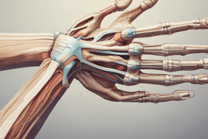
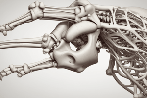
Elbow Joint: Relevant Bony Anatomy
- The elbow joint involves bony landmarks in the supracondylar region.
- Key bony features include the epicondyles (lateral and medial), condyles, capitulum, and trochlea.
Elbow Joint Bony Features & Radiology
- Anterior view shows the relationship between the capitulum, trochlea, medial and lateral epicondyles.
- Posterior view presents the olecranon fossa, trochlea.
- Lateral view radiographs highlight the coronoid fossa, capitellum, radial head, olecranon fossa and process, plus the trochlea.
Elbow Joint
- It's a complex synovial joint with two main components.
- The humero-ulnar joint connects the trochlea of the humerus with the trochlear notch of the ulna.
- Humero-radial joints link the capitellum of the humerus to the radial head, facilitating flexion and extension.
- Proximal radio-ulnar joint is between the radial head and radial notch of ulna.
- Both joints share a common synovial cavity allowing supination and pronation.
Elbow Synovial Fold Syndrome
- A 20-year-old female presents with snapping pain and elbow locking during elbow flexion and extension.
- During examination, pain is usually located posterolaterally.
- Elbow synovial fold syndrome involves a thickened and inflamed plica, plus chondral fraying of the radial head and capitellum.
- MR arthrography images reveal the thickened posterolateral fold indicative of the syndrome.
Ligaments of the Elbow Joint
- Lateral collateral ligament complex's proximal attachment is at lateral epicondyle of humerus, and its distal attachment is annular ligament and proximal ulna.
- Medial collateral ligament complex's proximal attachment is at medial epicondyle of humerus, and its distal attachment is at proximal ulna.
- Annular ligament holds the head of the radius against radial notch of ulna allowing rotatory movement.
Valgus and Varus Forces
- Varus force refers to an inward force on the distal bone, and valgus force refers to an outward force on the distal bone.
- The lateral collateral ligament (LCL) protects against varus forces.
- The medial collateral ligament (MCL) protects against valgus forces.
Stability of the Elbow Joint
- Elbow joint stability relies on bony factors, ligaments, and muscles.
- Damage to these columns may lead to elbow instability.
- The stabilizing factors are described as part of a ring of 4 columns.
- Isolated large coronoid or medial epicondylar fractures may cause instability.
Dislocation of Elbow 1: Proximal Radioulnar (Pulled Elbow/Nurse Maid’s Elbow)
- Typically occurs in children under 5 years old due to not well developed radial head.
- Mechanism involves sudden, longitudinal traction applied to the forearm with elbow extension.
- Presentation includes pain with reluctance and the elbow in extension with the forearm in pronation.
Dislocation of Elbow 2: Humero-radio-ulnar Joint
- Second most common joint dislocation, generally posterior.
- Posterior movement of forearm bones in relation to the humerus typically from a FOOSH injury.
- Both the radius and ulna dislocate posteriorly.
- Coronoid process resists posterior displacement of ulna.
- Can result in joint instability and recurrent dislocations.
- The "terrible triad" includes elbow dislocation + radial head fracture (Axial compression) + coronoid fracture.
- Damage to neurovascular structures is a risk and associated fractures increase this risk.
- Ulnar nerve, median nerve, and the brachial artery may be damaged.
Dislocation of Elbow 3: Monteggia Fracture Dislocation
- Anterior dislocation of radial head with a fracture of the proximal one third of the ulna.
Neurovascular Relations of Elbow
- Elbow injuries such as supracondylar fractures pose a risk to neurovascular structures.
- Median, radial, and ulnar nerves are vulnerable.
- Brachial artery and veins can also be affected.
Volkmann Ischemic Contracture
- Supracondylar humeral fractures may damage the brachial artery causing contraction.
- Acute symptoms are the 5 "P"s: Pallor, Pulselessness, Pain, Paraesthesia, and Paralysis.
- Ischemia of forearm muscles results in muscle necrosis, fibrosis, contractures and ultimately deformity.
2ry Ossification Centers
- Long bones ossify using primary and secondary ossification centers.
- Primary centers appear during prenatal development in the diaphysis (shaft).
- Secondary centers appear postnatally in the epiphysis region, with multiple centers present.
- Cartilages may appear as translucent gaps on radiographs.
2ry Ossification Centers - Clinical Scenario
- There are six 2ry ossification centers in relation to elbow.
- Times of appearance of 2ry ossification centres in elbow:
- Capitellum: 1 year
- Radial head: 3 years
- Int. epicondyle: 5 years
- Trochlear: 7 years
- Olecranon: 9 years
- Ext. epicondyle: 11 years
- A 7 year-old boy presenting with swollen elbow after a fall on the elbow.
Supracondylar Fracture (SC #) of Humerus
- Note the thin bone above the condyles.
- Hyperextension causes fracture at the narrow SC region.
- Can cause bleeding into the joint space.
Anterior & Posterior Fat Pad Sign
- The posterior fat pad resides inside the olecranon fossa in normal conditions.
- The anterior fat pad slightly projects outside the coronoid fossa, visible on lateral radiographs.
- Elevating fat pads indicates intraarticular fractures and the presence of hemarthrosis.
- The fat pad signs aid in detecting subtle elbow joint effusion or haemarthrosis.
- If positive for fat pad signs after trauma you should look carefully for fractures.
Clinical Scenario 2
- A 12 year old boy is presenting with swollen elbow after a FOOSH.
- Anterior humeral line (AHL) and radiocapitellar line (RCL) helps identify/differentiate subtle supracondylar fractures and radial head dislocation.
- AHL should intersect the middle 1/3 of the capitellum on an AP view – if disrupted in SC.
- RCL should intersect the middle of the capitellum regardless projection - if disrupted when there is radial head dislocation.
Radial Head Dislocation
- Normal radiocapitellar line intersect the mid-capitellum.
- Abnormal radiocapitellar line does not intersect the capitellum indicating radial head dislocation.
Clinical Scenarios 3 – 4
- Triangle is maintained in Supra condylar.
- Triangle is disrupted in elbow dislocation.
Anterior Compartment Muscles of the Forearm
- Muscles act on both the proximal and distal radio-ulnar joints.
- Pronators include the pronator teres and pronator quadratus.
- Flexors include the flexor carpi radialis and ulnaris.
- Flexors act on the digits (digitorum) and thumb (pollicis).
- Are arranged in three groups: superficial, intermediate, and deep.
Anterior Compartment Muscles of the Forearm Details
- Innervation: all muscles are supplied by the median nerve, deep layer by anterior interosseous branch.
- All muscles are supplied by Median N – Deep layer by Anterior interosseous Br. Of Median N, EXCEPT 2 medial (FCU and Medial 2 heads of FDP)
- Medial Epicondylitis (Golfer's elbow): Overuse of flexors inflammation and pain @medial epicondyle.
Brachioradialis
- Positioned between the flexor and extensor compartments of the wrist and elbow.
- Strong flexor of the semi-pronated forearm.
- Supinates the pronated forearm and pronates the supinated forearm to mid-position.
- Innervated by the radial nerve.
Posterior Compartment Muscles of the Forearm
- Muscles include extensor digitorum, extensor digiti minimi, extensor carpi ulnaris.
- Others include the extensor pollicis brevis and longus.
- Also, the abductor pollicis longus, extensor indicis, and anconeus.
- Common extensor origin occurs at the lateral epicondyle.
- ECRL is innervated by the radial nerve; all others are innervated by the deep radial or posterior interosseous nerve.
- Lateral Epicondylitis (Tennis elbow) Overuse of extensors Inflammation & Pain @ lateral epicondyle.
Anatomical Snuff Box
- Triangular depression is bounded by the tendons of the extensor pollicis longus, and extensor pollicis brevis laterally.
- The floor is the scaphoid and trapezium.
- In floor Radial artery and Superficial branch of the radial nerve runs inside.
- Tender when scaphoid # or rarely trapezium #.
Extensor Expansion
- Includes lateral bands, medial band, and extensor expansion.
- Mallet finger is the result of the rupture of extensor tendon at the DIP joint.
Nerves in the Forearm
- Ulnar nerve passes between the ulnar nerve and the flexor carpi ulnaris.
- Median nerve passes between the 2 heads of the pronator teres.
- Anterior interosseous Br. innervates the Flexor pollicis longus, FDP lateral ½ and Pronator quadratus.
- Deep Radial nerve passes between 2 heads of the supinator.
- Superficial branch continues under the brachioradialis.
- Deep radial branch passes into the posterior compartment as the posterior interosseous N.
- Both Ulnar & Median nerves stay anterior and run on the FDP.
Arteries in the Forearm
- The principal arteries are the brachial, radial, and ulnar arteries.
- The common interosseous artery and anterior/posterior interosseous arteries are also present.
- The deep and superficial palmer arches are located in the hand.
Studying That Suits You
Use AI to generate personalized quizzes and flashcards to suit your learning preferences.