Podcast
Questions and Answers
Which part of the ear is MOSTLY involved in both hearing and balance?
Which part of the ear is MOSTLY involved in both hearing and balance?
- Outer ear
- Middle ear
- External ear
- Inner ear (correct)
The pinna primarily amplifies sound waves before they enter the ear canal.
The pinna primarily amplifies sound waves before they enter the ear canal.
False (B)
What are the names of the three ossicles found in the middle ear?
What are the names of the three ossicles found in the middle ear?
malleus, incus, stapes
The __________ is the main organ of hearing, converting sound waves into neural signals.
The __________ is the main organ of hearing, converting sound waves into neural signals.
Match the following ear structures with their primary function:
Match the following ear structures with their primary function:
Which fluid is found in the Scala media and is characterized by a high concentration of potassium ions (K+)?
Which fluid is found in the Scala media and is characterized by a high concentration of potassium ions (K+)?
The endocochlear potential refers to the electrically negative charge of the Scala media relative to the Scala vestibuli and Scala tympani.
The endocochlear potential refers to the electrically negative charge of the Scala media relative to the Scala vestibuli and Scala tympani.
What structure separates the middle ear from the inner ear and allows sound waves to enter the cochlea?
What structure separates the middle ear from the inner ear and allows sound waves to enter the cochlea?
Bending of hair cells in the cochlea triggers the release of the neurotransmitter __________.
Bending of hair cells in the cochlea triggers the release of the neurotransmitter __________.
Match the cochlear structure with its primary function:
Match the cochlear structure with its primary function:
What is the approximate resting membrane potential of hair cells in the organ of Corti?
What is the approximate resting membrane potential of hair cells in the organ of Corti?
Kinocilia are hairlike protrusions on hair cells that are arranged in rows of progressively decreasing height.
Kinocilia are hairlike protrusions on hair cells that are arranged in rows of progressively decreasing height.
What is the function of tip links in hair cells?
What is the function of tip links in hair cells?
Deflection of stereocilia toward the kinocilium opens __________ channels, depolarizing the hair cell.
Deflection of stereocilia toward the kinocilium opens __________ channels, depolarizing the hair cell.
Match the following terms with their role in hair cell function:
Match the following terms with their role in hair cell function:
Approximately how many inner hair cells are found in the organ of Corti?
Approximately how many inner hair cells are found in the organ of Corti?
Outer hair cells primarily transmit sensory input to the auditory nerve fibers.
Outer hair cells primarily transmit sensory input to the auditory nerve fibers.
What percentage of sensory neurons innervate the inner hair cells?
What percentage of sensory neurons innervate the inner hair cells?
Outer hair cells __________ sound signals before they are transmitted to the auditory nerve fibers.
Outer hair cells __________ sound signals before they are transmitted to the auditory nerve fibers.
Match the type of hair cell with its primary function:
Match the type of hair cell with its primary function:
What type of neurons are spiral ganglion neurons?
What type of neurons are spiral ganglion neurons?
Spiral ganglion do not form synapses with hair cells.
Spiral ganglion do not form synapses with hair cells.
Where are the cell bodies of sensory neurons that innervate the hair cells located?
Where are the cell bodies of sensory neurons that innervate the hair cells located?
Spiral ganglion neurons transmit electrical signals to the __________ for interpretation as sound.
Spiral ganglion neurons transmit electrical signals to the __________ for interpretation as sound.
Match the following structures with their function in regards to the spiral ganglion:
Match the following structures with their function in regards to the spiral ganglion:
Where on the basilar membrane do high-frequency sounds cause maximum displacement?
Where on the basilar membrane do high-frequency sounds cause maximum displacement?
The basilar membrane is more flexible at the base of the cochlea compared to the apex.
The basilar membrane is more flexible at the base of the cochlea compared to the apex.
What is the role of the basilar membrane in hearing?
What is the role of the basilar membrane in hearing?
Hair cells at the __________ of the cochlea respond to low-frequency sounds.
Hair cells at the __________ of the cochlea respond to low-frequency sounds.
Match the location on the basilar membrane with the sound frequency it responds to:
Match the location on the basilar membrane with the sound frequency it responds to:
According to the Traveling Wave Theory of Von Bekesy, in what direction does sound travel along the basilar membrane?
According to the Traveling Wave Theory of Von Bekesy, in what direction does sound travel along the basilar membrane?
The traveling wave peaks at the same location on the basilar membrane regardless of the sound frequency.
The traveling wave peaks at the same location on the basilar membrane regardless of the sound frequency.
How do outer hair cells contribute to the traveling wave theory of hearing?
How do outer hair cells contribute to the traveling wave theory of hearing?
In the Traveling Wave Theory, __________ frequencies cause maximum displacement near the base of the cochlea.
In the Traveling Wave Theory, __________ frequencies cause maximum displacement near the base of the cochlea.
Match the following components of the Traveling Wave Theory with their description:
Match the following components of the Traveling Wave Theory with their description:
What is the main function of prestin?
What is the main function of prestin?
Opening potassium channels inhibits signal transmittion by afferent neurons
Opening potassium channels inhibits signal transmittion by afferent neurons
What type of stimulation to the hair cells causes potassion channels to close?
What type of stimulation to the hair cells causes potassion channels to close?
The hair cells are located in the __________ within the cochlea
The hair cells are located in the __________ within the cochlea
Match the following structures to their description:
Match the following structures to their description:
Flashcards
Pinna
Pinna
The outer part of the ear that collects and directs sound waves.
External Auditory Canal
External Auditory Canal
A tube that amplifies and carries sound waves to the eardrum.
Ossicles
Ossicles
Three small bones in the middle ear that amplify sound: malleus, incus, and stapes.
Cochlea
Cochlea
Signup and view all the flashcards
Vestibule and Semicircular Canals
Vestibule and Semicircular Canals
Signup and view all the flashcards
Perilymph
Perilymph
Signup and view all the flashcards
Endolymph
Endolymph
Signup and view all the flashcards
Endocochlear Potential
Endocochlear Potential
Signup and view all the flashcards
Oval Window
Oval Window
Signup and view all the flashcards
Basilar Membrane
Basilar Membrane
Signup and view all the flashcards
Hair Cells
Hair Cells
Signup and view all the flashcards
Tectorial Membrane
Tectorial Membrane
Signup and view all the flashcards
Glutamate
Glutamate
Signup and view all the flashcards
Cochlear Amplifier
Cochlear Amplifier
Signup and view all the flashcards
Organ of Corti
Organ of Corti
Signup and view all the flashcards
Prestin
Prestin
Signup and view all the flashcards
Stereocilia
Stereocilia
Signup and view all the flashcards
Tip Links
Tip Links
Signup and view all the flashcards
Cation Channels
Cation Channels
Signup and view all the flashcards
Stereocilia Deflection
Stereocilia Deflection
Signup and view all the flashcards
Afferent Neurons
Afferent Neurons
Signup and view all the flashcards
Outer Hair Cells
Outer Hair Cells
Signup and view all the flashcards
Inner Hair Cells
Inner Hair Cells
Signup and view all the flashcards
Spiral Ganglion
Spiral Ganglion
Signup and view all the flashcards
Spiral Ganglion Neurons
Spiral Ganglion Neurons
Signup and view all the flashcards
Basilar Membrane Role
Basilar Membrane Role
Signup and view all the flashcards
Frequency Displacement
Frequency Displacement
Signup and view all the flashcards
High-Frequency Displacement
High-Frequency Displacement
Signup and view all the flashcards
Low-Frequency Displacement
Low-Frequency Displacement
Signup and view all the flashcards
Traveling Wave Theory
Traveling Wave Theory
Signup and view all the flashcards
Wave Peak Location
Wave Peak Location
Signup and view all the flashcards
Study Notes
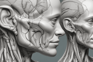
Ear Anatomy
- The ear has three regions: external (outer), middle, and internal (inner).
- The external and middle ear are involved in hearing.
- The internal ear is involved in both hearing and equilibrium.
Outer Ear
- The outer ear consists of the pinna and the external auditory canal.
- The pinna collects sound waves and directs them into the external auditory canal.
- The pinna aids in sound localization.
- The external auditory canal amplifies sound waves.
Middle Ear
- The middle ear is an air-filled space behind the eardrum.
- It contains three small bones called ossicles: the malleus, incus, and stapes.
- The ossicles amplify sound waves and transmit them from the eardrum to the inner ear.
Inner Ear
- The inner ear is located deep within the temporal bone.
- It comprises the cochlea, vestibule, and semicircular canals.
- The cochlea is the main organ of hearing, converting sound waves into neural signals.
- The vestibule and semicircular canals are responsible for balance.
Cochlea Physiology
- The cochlea is 35 mm long with 2 and 3/4 turns.
- The Scala vestibuli and Scala tympani contain perilymph, which is rich in sodium ions (Na+).
- The Scala media is the middle cochlear chamber, containing endolymph secreted by the stria vascularis.
- Endolymph is rich in potassium ions (K+).
- The Scala media is electrically positive by 85 mV, known as endocochlear potential, relative to the Scala vestibuli and Scala tympani.
- The oval window separates the middle ear from the inner ear, allowing sound waves to enter the cochlea.
- Sound waves traveling through the cochlea cause the basilar membrane to vibrate.
- The basilar membrane is a thin, flexible structure running the length of the cochlea.
- Thousands of hair cells are embedded in the basilar membrane.
- Hair cells convert sound waves into electrical impulses.
- Hair cells are overlaid by the tectorial membrane, a gel-like structure.
- Vibration of the basilar membrane causes hair cells to move back and forth.
- Bending of hair cells triggers the release of glutamate.
- Glutamate stimulates auditory nerve fibers.
- Auditory nerve fibers transmit electrical signals to the brainstem.
- Different frequencies of sound waves stimulate different parts of the basilar membrane.
- The location of stimulated hair cells corresponds to the frequency of the sound waves.
- The cochlear amplifier involves outer hair cells that contract and expand in response to signals from the brainstem.
- Contraction and expansion of outer hair cells amplify basilar membrane vibrations.
- The cochlear amplifier enhances the sensitivity and selectivity of the cochlea to different sound frequencies.
Organ of Corti
- The organ of Corti contains the hair cells, which are the receptors for hearing.
- The resting membrane potential of hair cells is about -60 mV.
- Hair cells have motor protein called prestin.
- Hair cells have stereocilia and kinocilia.
- Stereocilia are hairlike protrusions.
- Kinocilia are specialized single long cilium originally but during mammalian development it disappears in maturation.
- Stereocilia are arranged in rows of progressively decreasing height.
- Tip links connect stereocilia to each other.
- Tip links contain mechanically sensitive cation channels.
- Cation channels allow positively charged ions (potassium and calcium) to flow into the cell.
- When sound waves cause stereocilia to bend, tip links stretch and open cation channels.
- Deflection of stereocilia toward the kinocilium opens potassium channels, depolarizing the hair cell.
- Influx of calcium stimulates the release of glutamate.
- Glutamate stimulates afferent neurons to transmit neural impulses to the auditory cortex.
- At rest, potassium channels are partially open.
- Deflection of stereocilia away from the kinocilium closes potassium channels.
- Closing potassium channels inhibits signal transmission by afferent neurons.
Outer and Inner Hair Cells
- Hair cells in the organ of Corti are arranged in four rows: three rows of outer hair cells and one row of inner hair cells.
- There are approximately 20,000 outer hair cells.
- There are approximately 3,500 inner hair cells.
- Inner hair cells transmit most of the sensory input to the auditory nerve fibers.
- Outer hair cells amplify sound signals before they're transmitted to the auditory nerve fibers.
- 90-95% of sensory neurons innervate the inner hair cells.
- 5-10% of sensory neurons innervate the outer hair cells.
Spiral Ganglion
- The spiral ganglion contains the cell bodies of sensory neurons that innervate the hair cells.
- Sensory neurons are called spiral ganglion neurons.
- Spiral ganglion neurons form synapses with hair cells.
- They transmit electrical signals to the brain.
- Spiral ganglion neurons are bipolar neurons.
- One dendrite synapses with hair cells, and one axon extends to the brainstem.
- Spiral ganglion neurons are essential for transmitting sensory information to the brain for interpretation as sound.
Role of Basilar Membrane in Hearing
- The basilar membrane is critical for frequency analysis of sound waves.
- It separates the fluid-filled chambers of the cochlea.
- Sound waves cause the basilar membrane to vibrate.
- The basilar membrane is stiffer at the base of the cochlea and more flexible towards the apex.
- Different frequencies cause maximum displacement at different points along the membrane.
- Higher frequencies cause displacement near the base.
- Lower frequencies cause displacement near the apex.
- The frequency analysis allows the auditory system to distinguish between different frequencies and identify pitch.
- Hair cells on the basilar membrane respond to mechanical vibrations by generating electrical signals.
- Hair cells at the base of the cochlea respond to high-frequency sounds.
- Hair cells at the apex respond to low-frequency sounds.
Traveling Wave Theory of Von Bekesy
- Sound waves create a traveling wave along the basilar membrane.
- The wave moves from the base to the apex of the cochlea.
- The wave peaks at a specific location corresponding to the frequency of the sound wave.
- Higher frequencies cause maximum displacement near the base; lower frequencies near the apex.
- As the traveling wave moves along the basilar membrane, it causes hair cells to bend and generate electrical signals.
- Outer hair cells amplify the traveling wave, enhancing sensitivity and selectivity.
Studying That Suits You
Use AI to generate personalized quizzes and flashcards to suit your learning preferences.