Podcast
Questions and Answers
During the development of the spinal cord, what is the key factor that leads to the formation of the ventral motor horn cells?
During the development of the spinal cord, what is the key factor that leads to the formation of the ventral motor horn cells?
- The presence of the floor plate, which provides pathways for nerve fibers crossing from one side to the other.
- Formation of the sulcus limitans, which separates the basal and alar plates.
- Continuous addition of neuroblasts to the mantle layer, resulting in ventral thickening known as the basal plates. (correct)
- Migration of neuroblasts into the mantle layer and subsequent differentiation into bipolar neuroblasts.
What is the primary role of the sulcus limitans in the development of the spinal cord?
What is the primary role of the sulcus limitans in the development of the spinal cord?
- It plays a critical role in the migration of neuroblasts from the neuroepithelial layer to the mantle layer.
- It marks the boundary between the basal and alar plates, indicating the future locations of motor and sensory areas. (correct)
- It serves as a pathway for nerve fibers to cross from one side of the cord to the other.
- It defines the boundary between the ventral motor horn and dorsal sensory horn.
Which of the following statements accurately describes the relationship between the mantle layer and the white matter of the spinal cord?
Which of the following statements accurately describes the relationship between the mantle layer and the white matter of the spinal cord?
- The mantle layer forms the gray matter, and the white matter develops from the marginal layer surrounding it. (correct)
- The mantle layer and white matter are separate structures with no direct developmental relationship.
- The mantle layer forms the white matter, while the gray matter develops from the neuroepithelial layer.
- The mantle layer differentiates directly into the white matter, forming the myelinated axons of nerve fibers.
Which of the following statements is TRUE about the relationship between the notochord and the neural plate?
Which of the following statements is TRUE about the relationship between the notochord and the neural plate?
Which of the following correctly identifies a key characteristic of the floor plate and roof plate in the spinal cord?
Which of the following correctly identifies a key characteristic of the floor plate and roof plate in the spinal cord?
How does the differentiation of neuroblasts within the mantle layer contribute to the formation of the spinal cord's structure?
How does the differentiation of neuroblasts within the mantle layer contribute to the formation of the spinal cord's structure?
What is the correct sequence of events during neurulation?
What is the correct sequence of events during neurulation?
Which of the following is NOT a correct statement about the neural tube?
Which of the following is NOT a correct statement about the neural tube?
Which of the following INCORRECTLY matches a neural tube defect with its description?
Which of the following INCORRECTLY matches a neural tube defect with its description?
Which of the following is a derivative of the neural crest?
Which of the following is a derivative of the neural crest?
What is the primary function of the notochord during embryonic development?
What is the primary function of the notochord during embryonic development?
Which of the following statements regarding the early development of the brain vesicles is TRUE?
Which of the following statements regarding the early development of the brain vesicles is TRUE?
What is the primary structure formed from the caudal part of the neural tube?
What is the primary structure formed from the caudal part of the neural tube?
On which day does the anterior neuropore close during neurulation?
On which day does the anterior neuropore close during neurulation?
Which neural tube defect is characterized by the closure failure of the anterior neuropore?
Which neural tube defect is characterized by the closure failure of the anterior neuropore?
What condition is a precursor to anencephaly?
What condition is a precursor to anencephaly?
Where do neural crest cells reside?
Where do neural crest cells reside?
What is commonly associated with spina bifida?
What is commonly associated with spina bifida?
Which part of the neural tube is responsible for forming the brain?
Which part of the neural tube is responsible for forming the brain?
When does neurulation start according to developmental timelines?
When does neurulation start according to developmental timelines?
What does microcephaly refer to?
What does microcephaly refer to?
What do neural crest cells give rise to?
What do neural crest cells give rise to?
Which brain vesicle develops into structures such as the cerebral cortex and basal ganglia?
Which brain vesicle develops into structures such as the cerebral cortex and basal ganglia?
What structures are derived from the neural tube?
What structures are derived from the neural tube?
Which of the following structures are classified as part of the ectoderm?
Which of the following structures are classified as part of the ectoderm?
What is the primary function of Schwann cells?
What is the primary function of Schwann cells?
What is the role of the optic vesicles in early development?
What is the role of the optic vesicles in early development?
Which cranial nerve is primarily associated with the optic vesicle?
Which cranial nerve is primarily associated with the optic vesicle?
Which secondary brain vesicle gives rise to the medulla oblongata?
Which secondary brain vesicle gives rise to the medulla oblongata?
What do dorsal root ganglia primarily contain?
What do dorsal root ganglia primarily contain?
Which structure does NOT originate from the neural crest?
Which structure does NOT originate from the neural crest?
Which of the following statements accurately describes the development of the mantle layer in the spinal cord?
Which of the following statements accurately describes the development of the mantle layer in the spinal cord?
What is the primary function of the neuroepithelial layer in spinal cord development?
What is the primary function of the neuroepithelial layer in spinal cord development?
Which of the following accurately reflects the relationship between the marginal layer and the white matter of the spinal cord?
Which of the following accurately reflects the relationship between the marginal layer and the white matter of the spinal cord?
Which of the following best describes the neuroepithelial cells during the early stages of spinal cord development?
Which of the following best describes the neuroepithelial cells during the early stages of spinal cord development?
Based on the provided information, what is the primary function of the neuroblasts during spinal cord development?
Based on the provided information, what is the primary function of the neuroblasts during spinal cord development?
What is the primary difference between the mantle layer and the marginal layer in the developing spinal cord?
What is the primary difference between the mantle layer and the marginal layer in the developing spinal cord?
Which of these accurately describes the development of the optic nerve (CN II)?
Which of these accurately describes the development of the optic nerve (CN II)?
Which of the following statements is TRUE about the neuroepithelial cells?
Which of the following statements is TRUE about the neuroepithelial cells?
After the closure of the neural tube, what happens to the neuroepithelial cells?
After the closure of the neural tube, what happens to the neuroepithelial cells?
What is the fate of the neuroblasts formed from the neuroepithelium?
What is the fate of the neuroblasts formed from the neuroepithelium?
Flashcards
Marginal layer
Marginal layer
The outer region of the developing spinal cord that eventually forms the white matter.
Mantle layer
Mantle layer
The middle layer of the developing spinal cord that gives rise to the gray matter.
Neuroepithelial layer
Neuroepithelial layer
The inner layer of the developing spinal cord, composed of neuroepithelial cells that divide to form neuroblasts.
Basal plate
Basal plate
Signup and view all the flashcards
Alar plate
Alar plate
Signup and view all the flashcards
Neurulation
Neurulation
Signup and view all the flashcards
Neural Canal
Neural Canal
Signup and view all the flashcards
Anencephaly
Anencephaly
Signup and view all the flashcards
Spinal Bifida
Spinal Bifida
Signup and view all the flashcards
Ectoderm
Ectoderm
Signup and view all the flashcards
Notochord
Notochord
Signup and view all the flashcards
Neural Crest
Neural Crest
Signup and view all the flashcards
Anterior Neuropore
Anterior Neuropore
Signup and view all the flashcards
Posterior Neuropore
Posterior Neuropore
Signup and view all the flashcards
Cephalic part of the neural tube
Cephalic part of the neural tube
Signup and view all the flashcards
Caudal part of the neural tube
Caudal part of the neural tube
Signup and view all the flashcards
Microcephaly
Microcephaly
Signup and view all the flashcards
Neural Crest Cells
Neural Crest Cells
Signup and view all the flashcards
Neural Tube
Neural Tube
Signup and view all the flashcards
Neuroectoderm
Neuroectoderm
Signup and view all the flashcards
Surface Ectoderm
Surface Ectoderm
Signup and view all the flashcards
Primary Brain Vesicles
Primary Brain Vesicles
Signup and view all the flashcards
Secondary Brain Vesicles
Secondary Brain Vesicles
Signup and view all the flashcards
Cranial Nerves
Cranial Nerves
Signup and view all the flashcards
Optic Cup
Optic Cup
Signup and view all the flashcards
Neuroblasts
Neuroblasts
Signup and view all the flashcards
Closure of the neural tube
Closure of the neural tube
Signup and view all the flashcards
Formation of the mantle zone
Formation of the mantle zone
Signup and view all the flashcards
Neural Groove Stage
Neural Groove Stage
Signup and view all the flashcards
Neuroepithelial cell division
Neuroepithelial cell division
Signup and view all the flashcards
Grey matter development
Grey matter development
Signup and view all the flashcards
White matter development
White matter development
Signup and view all the flashcards
Study Notes
Development of the Nervous System
-
The nervous system develops from the ectoderm, one of the three germ layers that form during embryonic development.
-
Neurulation is the process by which the neural tube forms. This process begins at day 17 after fertilization.
-
Overlying ectoderm is induced by the notochord to form the neural plate.
-
The neural plate then folds to form the neural groove.
-
The neural folds fuse to form the neural tube.
-
The neural canal is a cavity inside the neural tube, which will later develop into the ventricles, a series of cavities in the brain.
-
The neural tube is separated from the surface ectoderm.
-
The closure of the neural tube disconnects the neural crest from the surface ectoderm.
-
The neural crest cells migrate to numerous locations and give rise to many different structures, including the peripheral nervous system.
-
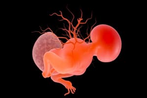
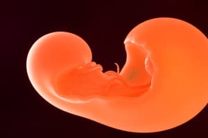
The cephalic part of the neural tube develops into the brain, and the caudal part develops into the spinal cord.
-
The neural canal develops into the ventricular system and the central canal.
-
Neural tube defects, including anencephaly, exencephaly, and microcephaly, can result from incomplete closure of the neural tube.
-
Spina bifida is a neural tube defect that results from incomplete closing of the vertebrae.
-
The three primary brain vesicles are the prosencephalon (forebrain), mesencephalon (midbrain), and rhombencephalon (hindbrain).
-
The five secondary brain vesicles are the telencephalon, diencephalon, mesencephalon, metencephalon, and myelencephalon.
-
The brainstem is a direct continuation of the spinal cord and has a similar organization but shows accentuation of the alar plate and regression of the basal plate in higher centers.
-
12 pairs of cranial nerves arise from the brainstem.
-
The optic nerve (CN II) is an extension of the forebrain.
-
During the neural groove stage and immediately after closure of the neural tube, neuroepithelial cells divide rapidly, forming neuroepithelial layers.
-
The mantle layer forms the grey matter of the spinal cord and contains neuroblasts.
-
The marginal layer develops into the white matter of the spinal cord.
-
The neural tube is divided into three main layers: neuroepithelial layer, mantle layer, and marginal layer.
-
Neuroblasts arise from the division of neuroepithelial cells.
-
Neuroblasts migrate into the mantle layer.
-
Multipolar neuroblasts develop from bipolar ones which develop from apolar ones.
-
Axons of neurones in the basal plate break through the marginal layer and become visible on the ventral aspect of the spinal cord.
-
Axons of neurones in the alar plate penetrate into the marginal layer – ascend to either higher or lower levels to form association neurones (interneurons).
-
Glial cells, also known as glial blasts, are formed from neuroepithelial cells after the neuroblasts cease to form.
-
Glial cells include protoplasmic astrocytes, fibrillar astrocytes, oligodendrocytes, and microglia.
-
Different types of glial cells migrate to different locations, and perform different metabolic functions in the spinal cord and brain.
-
Ependymal cells line the central canal of the spinal cord and ventricles.
-
Glial cells develop into Schwann cells which are responsible for myelination in the peripheral nervous system.
-
Oligodendrocytes are responsible for myelination in the central nervous system.
Studying That Suits You
Use AI to generate personalized quizzes and flashcards to suit your learning preferences.