Podcast
Questions and Answers
What typically accompanies esophageal atresia in newborns?
What typically accompanies esophageal atresia in newborns?
- Duodenal atresia
- Tracheoesophageal fistula (correct)
- Congenital diaphragmatic hernia
- Pneumothorax
Which condition occurs when the esophagus fails to recanalize?
Which condition occurs when the esophagus fails to recanalize?
- Esophageal stenosis (correct)
- Tracheoesophageal fistula
- Congenital hiatal hernia
- Esophageal atresia
What prenatal condition is usually associated with both esophageal atresia and stenosis?
What prenatal condition is usually associated with both esophageal atresia and stenosis?
- Placenta previa
- Polyhydramnios (correct)
- Amniotic band syndrome
- Oligohydramnios
How does a congenital hiatal hernia occur?
How does a congenital hiatal hernia occur?
What happens to the stomach during early development?
What happens to the stomach during early development?
What is primarily formed from endoderm during the development of the primitive gut tube?
What is primarily formed from endoderm during the development of the primitive gut tube?
Which structure primarily derives from mesoderm in the developing gut tube?
Which structure primarily derives from mesoderm in the developing gut tube?
Which arteries supply the foregut derivatives in the abdomen?
Which arteries supply the foregut derivatives in the abdomen?
What does the posterior intestinal portal ultimately form in the embryo's gut development?
What does the posterior intestinal portal ultimately form in the embryo's gut development?
Which of the following organs is NOT derived from the foregut?
Which of the following organs is NOT derived from the foregut?
What type of mesoderm surrounds the gut tube during its development?
What type of mesoderm surrounds the gut tube during its development?
Which structure is part of the midgut derivatives?
Which structure is part of the midgut derivatives?
What role do cranio-caudal and lateral folding play in the development of the gut tube?
What role do cranio-caudal and lateral folding play in the development of the gut tube?
What mechanism is primarily responsible for the concentric layering of the gut tube?
What mechanism is primarily responsible for the concentric layering of the gut tube?
What occurs around the 5th week of development regarding the gut tube?
What occurs around the 5th week of development regarding the gut tube?
Which mesenteric structure is associated with the stomach?
Which mesenteric structure is associated with the stomach?
What happens to the SHH expression later in development?
What happens to the SHH expression later in development?
Which of the following structures is NOT made from mesentery?
Which of the following structures is NOT made from mesentery?
What is the outcome of errors in the occlusion and re-canalization process of the gut tube?
What is the outcome of errors in the occlusion and re-canalization process of the gut tube?
What structures are formed by the tracheoesophageal folds during the development of the foregut?
What structures are formed by the tracheoesophageal folds during the development of the foregut?
What does the term 'secondarily retroperitoneal' refer to in the context of the GI tract?
What does the term 'secondarily retroperitoneal' refer to in the context of the GI tract?
What is the primary consequence of differential growth on the left and right sides of the stomach?
What is the primary consequence of differential growth on the left and right sides of the stomach?
Hypertrophic pyloric stenosis is most commonly characterized by which symptom?
Hypertrophic pyloric stenosis is most commonly characterized by which symptom?
Which factor contributes to the formation of the pyloric sphincter?
Which factor contributes to the formation of the pyloric sphincter?
What is the relationship between the liver and the septum transversum?
What is the relationship between the liver and the septum transversum?
What arises from the caudal end of the foregut during pancreas development?
What arises from the caudal end of the foregut during pancreas development?
Which statement correctly describes the structure of the adult liver?
Which statement correctly describes the structure of the adult liver?
What is a characteristic feature of the endocrine pancreas?
What is a characteristic feature of the endocrine pancreas?
What is typically observed in infants with hypertrophic pyloric stenosis?
What is typically observed in infants with hypertrophic pyloric stenosis?
What condition can result from inadequate mesoderm generation during gastrulation?
What condition can result from inadequate mesoderm generation during gastrulation?
Which neural crest cell migration failure results in an aganglionic segment of the gut?
Which neural crest cell migration failure results in an aganglionic segment of the gut?
What is the primary complication of an aganglionic segment of the colon?
What is the primary complication of an aganglionic segment of the colon?
In which part of the colon is Hirschsprung disease most commonly found?
In which part of the colon is Hirschsprung disease most commonly found?
What is the main function of the submucosal and myenteric plexuses?
What is the main function of the submucosal and myenteric plexuses?
What is the origin of the uncinate process of the pancreas?
What is the origin of the uncinate process of the pancreas?
Which artery serves the proximal duodenum?
Which artery serves the proximal duodenum?
What condition may arise from the failure to recanalize the duodenum during development?
What condition may arise from the failure to recanalize the duodenum during development?
Which part of the GI tract is formed by the cranial loop of the U-shaped loop during development?
Which part of the GI tract is formed by the cranial loop of the U-shaped loop during development?
What happens to the duodenum and pancreas during gut tube rotation?
What happens to the duodenum and pancreas during gut tube rotation?
What is a possible result of failure to obliterate the vitelline duct?
What is a possible result of failure to obliterate the vitelline duct?
From which part of the midgut does the distal duodenum arise?
From which part of the midgut does the distal duodenum arise?
What structure is an elongated remnant of the yolk sac that becomes obliterated during development?
What structure is an elongated remnant of the yolk sac that becomes obliterated during development?
Flashcards
Gut tube formation
Gut tube formation
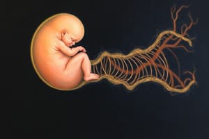
Folding of the embryo's body causes the endoderm-lined yolk sac cavity to form a tube known as the gut tube.
Gut region division: arterial supply
Gut region division: arterial supply
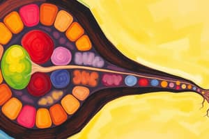
The gut is divided into three regions (foregut, midgut, and hindgut) based on its arterial supply.
Gut region derivatives
Gut region derivatives
The foregut, midgut, and hindgut are each associated with specific organs.
Foregut blood supply
Foregut blood supply
Signup and view all the flashcards
Midgut blood supply
Midgut blood supply
Signup and view all the flashcards
Hindgut blood supply
Hindgut blood supply
Signup and view all the flashcards
Mesoderm contribution to gut wall
Mesoderm contribution to gut wall
Signup and view all the flashcards
Neural crest contribution to gut wall
Neural crest contribution to gut wall
Signup and view all the flashcards
Esophageal Atresia
Esophageal Atresia
Signup and view all the flashcards
Esophageal Stenosis
Esophageal Stenosis
Signup and view all the flashcards
Congenital Hiatal Hernia
Congenital Hiatal Hernia
Signup and view all the flashcards
Esophageal Lengthening
Esophageal Lengthening
Signup and view all the flashcards
Stomach Rotation
Stomach Rotation
Signup and view all the flashcards
Segmental Patterning of the Gut Tube
Segmental Patterning of the Gut Tube
Signup and view all the flashcards
Radial Patterning of the Gut Tube
Radial Patterning of the Gut Tube
Signup and view all the flashcards
Gut Tube Occlusion
Gut Tube Occlusion
Signup and view all the flashcards
Gut Tube Recanalization
Gut Tube Recanalization
Signup and view all the flashcards
Gut Stenosis
Gut Stenosis
Signup and view all the flashcards
Retroperitoneal Organs
Retroperitoneal Organs
Signup and view all the flashcards
Intraperitoneal Organs
Intraperitoneal Organs
Signup and view all the flashcards
Tracheoesophageal Folds
Tracheoesophageal Folds
Signup and view all the flashcards
Anal Atresia
Anal Atresia
Signup and view all the flashcards
Cloacal malformations
Cloacal malformations
Signup and view all the flashcards
Hirschsprung Disease (Congenital Megacolon)
Hirschsprung Disease (Congenital Megacolon)
Signup and view all the flashcards
Aganglionic Segment
Aganglionic Segment
Signup and view all the flashcards
Innervation of the Hindgut
Innervation of the Hindgut
Signup and view all the flashcards
Differential Growth of the Stomach
Differential Growth of the Stomach
Signup and view all the flashcards
Cranio-Caudal Rotation of the Stomach
Cranio-Caudal Rotation of the Stomach
Signup and view all the flashcards
Fate of Mesenteries in the Stomach
Fate of Mesenteries in the Stomach
Signup and view all the flashcards
Formation of the Pyloric Sphincter
Formation of the Pyloric Sphincter
Signup and view all the flashcards
Hypertrophic Pyloric Stenosis
Hypertrophic Pyloric Stenosis
Signup and view all the flashcards
Liver Development
Liver Development
Signup and view all the flashcards
Pancreas Development: Exocrine
Pancreas Development: Exocrine
Signup and view all the flashcards
Pancreas Development: Endocrine
Pancreas Development: Endocrine
Signup and view all the flashcards
Pancreatic bud origins
Pancreatic bud origins
Signup and view all the flashcards
Duodenum development
Duodenum development
Signup and view all the flashcards
Duodenal atresia or stenosis
Duodenal atresia or stenosis
Signup and view all the flashcards
Midgut herniation
Midgut herniation
Signup and view all the flashcards
Appendix development
Appendix development
Signup and view all the flashcards
Vitelline duct abnormalities
Vitelline duct abnormalities
Signup and view all the flashcards
Midgut loop derivatives
Midgut loop derivatives
Signup and view all the flashcards
Midgut loop orientation
Midgut loop orientation
Signup and view all the flashcards
Study Notes
Development of Endodermal Derivatives
- The gut tube forms from endoderm lining the yolk sac, enveloped by developing coelom.
- Cranial and caudal folding apply somatic mesoderm to the body wall (parietal peritoneum) and visceral mesoderm wraps around the gut tube, creating mesenteries.
- Mesoderm contributes to gut tube wall.
- Nerves and neurons originate from neural crest.
- Endoderm forms mucosal epithelium, mucosal glands, submucosal glands of the GI tract.
- Mesoderm forms lamina propria, muscularis mucosae, submucosal connective tissue, blood vessels, muscularis externa, adventitia/serosa.
- Neural crest forms neurons and nerves of the submucosal and myenteric plexuses.
Gut Tube Regions and Refinement
- Cranio-caudal and lateral folding form anterior/cranial and posterior/caudal intestinal portals.
- Foregut and hindgut form from these portals; the midgut remains open to the yolk sac.
- Yolk sac connection to the gut shrinks as the embryo develops.
- Foregut derivatives include trachea, lungs, esophagus, stomach, liver, gallbladder, bile duct, pancreas, upper duodenum.
- Midgut derivatives include lower duodenum, jejunum, ileum, cecum, appendix, ascending colon, proximal 3 of transverse colon.
- Hindgut derivatives include distal transverse colon, descending colon, sigmoid colon, rectum, upper anal canal, urogenital sinus.
- Gut regions are defined by arterial supply: foregut by branches of the celiac artery, midgut by branches of the superior mesenteric artery, and hindgut by branches of the inferior mesenteric artery.
Cranio-caudal Patterning of the Gut Tube
- HOX genes determine regions of the gut tube (lung, esophagus, stomach).
- Sonic Hedgehog (SHH) in endoderm inhibits smooth muscle and neuron differentiation near the endoderm.
- Higher SHH concentration allows differentiation in external layers.
- Later development allows smooth muscle in the muscularis mucosae and neurons in the submucosal plexus.
- Gut tube lumen occlusion (5th week) followed by re-canalization (9th week) throughout the tube.
- Errors in this process may cause stenosis.
Radial Patterning of the Gut Tube
- Concentric layering of the gut tube is due to SHH expression
- SHH inhibits smooth muscle and neuronal differentiation near the endoderm.
- SHH concentration decreases farther away allowing smooth muscle and neuron differentiation.
- Occlusion and recanalization affect the esophageal and anal region.
Clinical Considerations
- Esophageal atresia: Failure of tracheoesophageal ridges to separate esophagus and trachea.
- Symptoms include aspiration pneumonia (gut contents/contaminants entering lungs).
- Typically associated with polyhydramnios (excess amniotic fluid).
- Esophageal stenosis: Failure of recanalization of the esophagus leads to narrowing.
- Symptoms include regurgitation, and potentially no tracheoesophageal fistula.
- Congenital hiatal hernia: Inguinal hernia in diaphragm.
- Incomplete growth of the esophagus and allows the stomach to form into the esophageal hiatus in diaphragm, causing a hiatal hernia.
Stomach
- Stomach rotates 90˚ during development
- Left side moves ventrally; right side moves dorsally.
- Greater and lesser curvatures develop due to differential growth.
- Pyloric sphincter (smooth muscle) forms at the caudal end of the stomach.
Liver
- The liver develops from the ventral foregut endoderm, close to the septum transversum.
- The parenchyma is composed of hepatocytes, and branched bile ducts and is surrounded by vascular sinusoids (mesoderm derived).
Pancreas
- Two outgrowths from the foregut: ventral and dorsal pancreatic buds.
- Exocrine pancreas develops from the endodermal lining.
- Endocrine pancreas (islets of Langerhans) develop from stem cells that form in the areas of the duct branch points.
- Rotation causes the merging of the ventral and dorsal buds into a single pancreas.
Proximal or Upper Duodenum
- Arises from caudal part of foregut
- Served by branches of the superior pancreaticoduodenal artery.
- Becomes secondarily retroperitoneal due to gut rotation.
Distal or Lower Duodenum
- Arises from cranial part of midgut.
- Served by branches of the inferior pancreaticoduodenal artery.
- Becomes secondarily retroperitoneal due to gut rotation.
Jejunum, Ileum, Cecum, Appendix, Ascending Colon, Proximal 2/3 of Transverse Colon
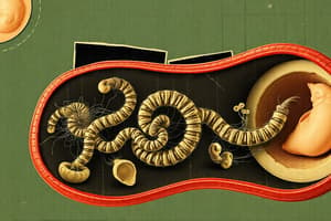
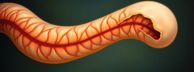
- Rapid elongation of the midgut forms a U-shaped loop that herniates into the umbilicus.
- The loop contains the future jejunum, ileum, cecum, appendix, ascending colon and proximal two-thirds of transverse colon.
- The appendix usually points caudally.
- The loop rotates 90 degrees counterclockwise.
- Vitelline duct connects to the yolk sac; obliteration is normal; failure leads to complications (Meckel's diverticulum, vitelline cyst, or vitelline fistula).
Derivatives of the Hindgut
- Includes distal 1/3 of transverse colon, descending colon, sigmoid colon, rectum, and upper anal canal.
- Hindgut ends in the cloaca (a common chamber with the urogenital tract).
- Septa divide the cloaca into the urogenital sinus and the rectoanal canal.
- Failure of the cloacal membrane to close or the failure of the cloaca to divide leads to anatomical abnormalities such as imperforate anus.
- Hindgut is innervated by vagal and sacral neural crest cells.
Clinical Considerations
- Hypertrophic pyloric stenosis: Overdeveloped pyloric sphincter.
- Hirschsprung disease: Absence of ganglion cells in a region of the distal colon and rectum causes obstruction.
Studying That Suits You
Use AI to generate personalized quizzes and flashcards to suit your learning preferences.