Podcast
Questions and Answers
Match the embryonic layers with their respective roles:
Match the embryonic layers with their respective roles:
Ectoderm = Forms the neural tube Endoderm = Forms the gut tube Mesoderm = Holds the two tubes together Lateral plate mesoderm = Splits into visceral and somatic layers
Match the types of embryonic folding with their descriptions:
Match the types of embryonic folding with their descriptions:
Lateral folding = Closes the ventral body wall Cephalocaudal folding = Forms the foregut and hindgut Neurulation = Forms the brain and spinal cord Mesoderm splitting = Creates the primitive body cavity
Match the terms with their definitions:
Match the terms with their definitions:
Primitive gut = Created during the 3rd and 4th week of embryo development Coelomic cavity = Space between visceral and somatic layers Peritoneal cavity = Part of the embryological development Mesenteries = Connectsome abdominal organs to the wall
Match the week of development with the corresponding embryonic event:
Match the week of development with the corresponding embryonic event:
Match the structures with their fate in embryonic development:
Match the structures with their fate in embryonic development:
Match the following terms with their functions:
Match the following terms with their functions:
Match the organ with its developmental origin:
Match the organ with its developmental origin:
Match the outcome of embryonic folding with its explanation:
Match the outcome of embryonic folding with its explanation:
Match the following anatomical structures with their description:
Match the following anatomical structures with their description:
Match the following ligaments with their related organs:
Match the following ligaments with their related organs:
Match the following stages with their significance in development:
Match the following stages with their significance in development:
Match the following blood supplies with their respective regions:
Match the following blood supplies with their respective regions:
Match the following conditions to their characteristics:
Match the following conditions to their characteristics:
Match the following systems with their development source:
Match the following systems with their development source:
Match the esophageal abnormalities with their descriptions:
Match the esophageal abnormalities with their descriptions:
Match the following developmental weeks with their events:
Match the following developmental weeks with their events:
Match the stomach features with their details:
Match the stomach features with their details:
Match the following descriptions with the corresponding abnormality:
Match the following descriptions with the corresponding abnormality:
Match the following organs with their developmental origins:
Match the following organs with their developmental origins:
Match the parts of the stomach with their movements during rotation:
Match the parts of the stomach with their movements during rotation:
Match the mesenteries with their corresponding positions after stomach rotation:
Match the mesenteries with their corresponding positions after stomach rotation:
Match the functions of the vagus nerve with their roles:
Match the functions of the vagus nerve with their roles:
Match the statements about esophagus structure:
Match the statements about esophagus structure:
Match the terms with their definitions:
Match the terms with their definitions:
Match the following types of cells with their primary functions:
Match the following types of cells with their primary functions:
Match the following structures formed by the septum transversum to their anatomical locations:
Match the following structures formed by the septum transversum to their anatomical locations:
Match the following pathologies with their descriptions:
Match the following pathologies with their descriptions:
Match the following terms with their definitions:
Match the following terms with their definitions:
Match the following embryological structures with their origins:
Match the following embryological structures with their origins:
Match the following parts of the embryonic gut with their descriptions:
Match the following parts of the embryonic gut with their descriptions:
Match the following types of organs to their characteristics:
Match the following types of organs to their characteristics:
Match the following mesentery types with their locations:
Match the following mesentery types with their locations:
Match the following structures with their associated regions:
Match the following structures with their associated regions:
Match the following processes with their corresponding types of folding:
Match the following processes with their corresponding types of folding:
Match the following embryonic phases with their advancements:
Match the following embryonic phases with their advancements:
Match the following terms to their definitions:
Match the following terms to their definitions:
Match the following structures to their roles in gut development:
Match the following structures to their roles in gut development:
Flashcards are hidden until you start studying
Study Notes
Learning Outcomes
- Understand embryonic folding processes that create the primitive gut and abdominal wall during the 3rd and 4th week.
- Describe the development of pregut derivatives.
- Explain the development of the coelomic and peritoneal cavities.
- Discuss the fates of embryonic dorsal and ventral mesenteries.
- Recognize why some abdominal organs have mesenteries while others are retroperitoneal.
- Understand the positioning of the viscera and developmental causes of congenital gastrointestinal defects.
Embryonic Folding
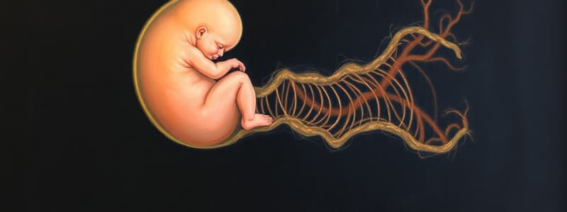
- During the 3rd and 4th weeks, ectoderm forms the neural plate, which rolls into the neural tube via neurulation.
- The endoderm rolls down to form the gut tube, resulting in a tube-on-a-tube structure: neural tube dorsally and gut tube ventrally.
- Mesoderm holds the two tubes together and splits into visceral and somatic layers, forming the lateral body wall.
Gut Tube Formation
- Cephalocaudal and lateral folding create the primitive gut tube.
- The primitive gut divides into foregut (cephalic), midgut (connected to yolk sac), and hindgut (caudal).
Mesenteries and Organ Suspension
- Mesenteries (peritoneum) suspend some gut portions from the dorsal and ventral body wall.
- Intraperitoneal organs are enclosed by double layers of peritoneum, connecting them to the body wall.
- Retroperitoneal organs, such as kidneys, lie against the posterior wall with peritoneum only on the anterior surface.
Dorsal and Ventral Mesentery Development
- The dorsal mesentery extends from the lower end of the esophagus to the cloacal region.
- Dorsal mesogastrium forms the greater omentum; dorsal mesoduodenum and dorsal mesocolon also develop from the dorsal mesentery.
- Ventral mesentery originates from the septum transversum and gives rise to the lesser omentum and falciform ligament.
Foregut Development
- The respiratory diverticulum appears at the foregut's ventral wall, with the tracheoesophageal septum separating it from the dorsal foregut.
- The esophagus rapidly lengthens, with varying muscular innervation: striated upper 2/3 by the vagus and smooth lower third by the splanchnic plexus.
Clinical Correlates: Esophageal Abnormalities
- Esophageal atresia/tracheoesophageal fistula develops from anomalies during septum formation.
- Esophageal stenosis primarily occurs in the lower third, linked to incomplete recanalization or vascular abnormalities.
- Congenital hiatal hernia results from insufficient esophageal lengthening, pulling the stomach into the diaphragm.
Stomach Development
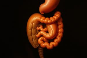
- The stomach appears as a fusiform expansion of the foregut by the 4th week.
- It undergoes 90° clockwise rotation around its longitudinal axis, altering the positions of its anterior and posterior walls.
- Stomach rotation creates the omental bursa and affects mesogastrium position; the dorsal mesogastrium lengthens while the ventral moves right.
Stomach Abnormalities
- Pyloric stenosis arises from hypertrophy of the pyloric musculature, causing severe food passage obstruction in infants.
- Other rare stomach malformations include duplications and prepyloric septums.
Duodenum Development
- Formed from the terminal foregut and cephalic midgut, the duodenum rotates into a C-shape during stomach development, becoming retroperitoneal.
Liver Development
- The liver primordium appears in the 3rd week as an outgrowth from the endoderm at the foregut's distal end.
- Liver cords differentiate into functional liver cells and structures for the biliary system, mixing with vitelline and umbilical veins.
Septum Transversum Contributions
- Mesoderm of the septum transversum forms hepatocytes, Kupffer cells, connective tissues, peritoneum of the liver, falciform ligament, lesser omentum, and central tendon of the diaphragm.
Bare Area of the Liver
- The liver's cranial surface remains uncovered by peritoneum, termed the bare area, while the rest is enveloped in visceral peritoneum.
Clinical Correlates: Biliary Atresia
- Extrahepatic biliary atresia has low correctable defect rates, with most untreated cases leading to mortality without transplants.
- Intrahepatic biliary duct atresia is a rare condition possibly linked to fetal infections, occurring in about 1 in 100,000 live births.
Studying That Suits You
Use AI to generate personalized quizzes and flashcards to suit your learning preferences.


