Podcast
Questions and Answers
What do the pleuroperitoneal membranes fuse with during diaphragm development?
What do the pleuroperitoneal membranes fuse with during diaphragm development?
- Pectoral muscles
- Esophageal muscles
- Mesentery of the esophagus (correct)
- Phrenic nerves
From which cervical segments do the muscle cells of the diaphragm originate?
From which cervical segments do the muscle cells of the diaphragm originate?
- C2—C4
- C4—C6
- C3—C5 (correct)
- C1—C3
What is the primary function of the phrenic nerves in relation to the diaphragm?
What is the primary function of the phrenic nerves in relation to the diaphragm?
- Control digestion
- Support spinal column stability
- Provide blood supply
- Innervate diaphragm for motor and sensory functions (correct)
What is the main consequence of differential growth during the sixth week of embryo development?
What is the main consequence of differential growth during the sixth week of embryo development?
Which structure is NOT directly involved in the formation of the diaphragm?
Which structure is NOT directly involved in the formation of the diaphragm?
What is the primary origin of the septum transversum?
What is the primary origin of the septum transversum?
What do the pericardioperitoneal canals become as lung buds grow?
What do the pericardioperitoneal canals become as lung buds grow?
Which structures fuse to form the fibrous pericardium in adults?
Which structures fuse to form the fibrous pericardium in adults?
How does the thoracic cavity divide into definitive compartments?
How does the thoracic cavity divide into definitive compartments?
What is the role of the pleuroperitoneal folds during development?
What is the role of the pleuroperitoneal folds during development?
What tissue gives rise to the definitive wall of the thorax?
What tissue gives rise to the definitive wall of the thorax?
What embryological change causes the enlargement of the pleuropericardial folds?
What embryological change causes the enlargement of the pleuropericardial folds?
What do the pleuropericardial membranes contain?
What do the pleuropericardial membranes contain?
What does the ectoderm layer form during the third and fourth weeks of embryonic development?
What does the ectoderm layer form during the third and fourth weeks of embryonic development?
Which layer of the embryonic disc is primarily responsible for forming the gut tube?
Which layer of the embryonic disc is primarily responsible for forming the gut tube?
What is the term for the space between the visceral and parietal layers of lateral plate mesoderm?
What is the term for the space between the visceral and parietal layers of lateral plate mesoderm?
At the end of the third week of embryonic development, what does the lateral plate mesoderm differentiate into?
At the end of the third week of embryonic development, what does the lateral plate mesoderm differentiate into?
Which structure forms the somatopleure?
Which structure forms the somatopleure?
What occurs to the lateral plate mesoderm during the formation of the body cavity?
What occurs to the lateral plate mesoderm during the formation of the body cavity?
Which layer is intimately connected to the gut tube?
Which layer is intimately connected to the gut tube?
What is the role of the intermediate mesoderm during embryonic development?
What is the role of the intermediate mesoderm during embryonic development?
What structures begin to form the lateral body wall folds in the embryo during the fourth week?
What structures begin to form the lateral body wall folds in the embryo during the fourth week?
Which region does not undergo closure of the ventral body wall?
Which region does not undergo closure of the ventral body wall?
What structure is formed from the endoderm layer folding ventrally during the fourth week?
What structure is formed from the endoderm layer folding ventrally during the fourth week?
What happens to the Vitelline duct between the second and third months of gestation?
What happens to the Vitelline duct between the second and third months of gestation?
What do the cells of the parietal layer of lateral plate mesoderm develop into?
What do the cells of the parietal layer of lateral plate mesoderm develop into?
Where is the dorsal mesentery located in relation to the gut tube?
Where is the dorsal mesentery located in relation to the gut tube?
What is the role of the ventral mesentery?
What is the role of the ventral mesentery?
Which of the following statements about visceral and parietal layers of mesoderm is NOT true?
Which of the following statements about visceral and parietal layers of mesoderm is NOT true?
Flashcards
Pleuroperitoneal membranes
Pleuroperitoneal membranes
A pair of thin membranes that develop from the folds in the embryo's body wall; these membranes grow inward and fuse with the mesentery of the esophagus, forming the crucial structures of the diaphragm.
Central tendon of the diaphragm
Central tendon of the diaphragm
The central, tendon-like part of the diaphragm; it's formed by the fusion of the pleuroperitoneal membranes.
Crura of the diaphragm
Crura of the diaphragm
The two muscular pillars that extend from the diaphragm to the lumbar vertebrae; they contribute to the diaphragm's shape and function.
Where do diaphragm muscle cells originate?
Where do diaphragm muscle cells originate?
Signup and view all the flashcards
Phrenic nerves
Phrenic nerves
Signup and view all the flashcards
Neurulation
Neurulation
Signup and view all the flashcards
Gut tube
Gut tube
Signup and view all the flashcards
Mesoderm
Mesoderm
Signup and view all the flashcards
Ectoderm
Ectoderm
Signup and view all the flashcards
Endoderm
Endoderm
Signup and view all the flashcards
Primitive body cavity
Primitive body cavity
Signup and view all the flashcards
Parietal (somatic) layer
Parietal (somatic) layer
Signup and view all the flashcards
Visceral (splanchnic) layer
Visceral (splanchnic) layer
Signup and view all the flashcards
Septum transversum
Septum transversum
Signup and view all the flashcards
Pericardioperitoneal canals
Pericardioperitoneal canals
Signup and view all the flashcards
Pleuropericardial membranes
Pleuropericardial membranes
Signup and view all the flashcards
Pleuropericardial membrane fusion
Pleuropericardial membrane fusion
Signup and view all the flashcards
Fibrous pericardium
Fibrous pericardium
Signup and view all the flashcards
Pleuroperitoneal folds
Pleuroperitoneal folds
Signup and view all the flashcards
Pleuroperitoneal fold closure
Pleuroperitoneal fold closure
Signup and view all the flashcards
Diaphragm
Diaphragm
Signup and view all the flashcards
Ventral Body Wall Closure
Ventral Body Wall Closure
Signup and view all the flashcards
Components of Lateral Body Wall Folds
Components of Lateral Body Wall Folds
Signup and view all the flashcards
Gut Tube Formation
Gut Tube Formation
Signup and view all the flashcards
Incomplete Ventral Body Wall Closure
Incomplete Ventral Body Wall Closure
Signup and view all the flashcards
Vitelline Duct
Vitelline Duct
Signup and view all the flashcards
Parietal Layer of Serous Membranes
Parietal Layer of Serous Membranes
Signup and view all the flashcards
Visceral Layer of Serous Membranes
Visceral Layer of Serous Membranes
Signup and view all the flashcards
Dorsal Mesentery
Dorsal Mesentery
Signup and view all the flashcards
Study Notes
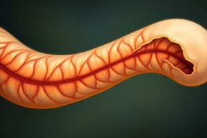
Embryology L7: The Gut Tube and Body Cavities
- The gut tube forms during the third and fourth weeks of development.
- The neural tube forms simultaneously, creating a tube-on-a-tube structure for the embryo.
- The mesoderm layer holds the neural tube and gut tube together with its visceral (splanchnic) and parietal (somatic) components.
- The visceral layer is connected to the gut tube.
- The parietal layer forms the lateral body wall folds.
- The space between the visceral and parietal layers is the primitive body cavity, which is initially continuous.
- The primitive body cavity eventually develops into the pericardial, pleural, and abdomino-pelvic regions.
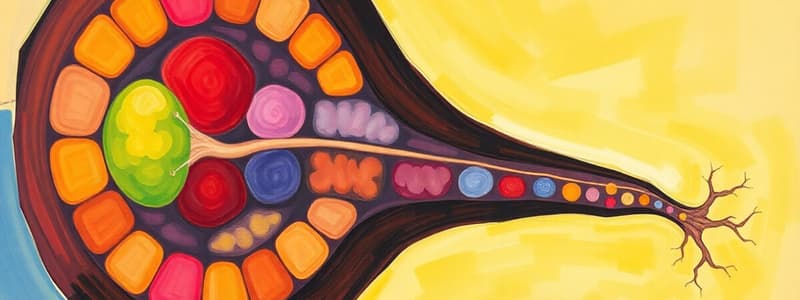
Formation of the Body Cavity
- At the end of the third week, intraembryonic mesoderm differentiates into paraxial, intermediate, and lateral plate mesoderm.
- Paraxial mesoderm forms somitomeres and somites crucial for the skull and vertebrae.
- Intermediate mesoderm contributes to the urogenital system.
- Lateral plate mesoderm forms the body cavity.
- Initially, the lateral plate mesoderm is a solid sheet that splits into two layers; parietal and visceral.
- The parietal layer, adjacent to the surface ectoderm, is continuous with the extraembryonic parietal layer.
- The visceral layer is adjacent to the endoderm forming the gut tube.
- Together, the parietal and visceral layers form the somatopleure and splanchnopleure, respectively.
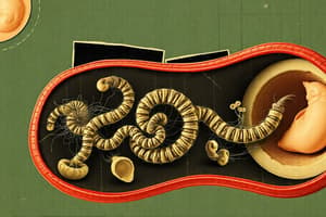
Serous Membranes
- Some parietal layer cells form the parietal layer of the serous membranes (peritoneal, pleural, pericardial).
- Visceral layer cells form the visceral layer of the serous membranes.
- Visceral and parietal layers connect as the dorsal mesentery, suspending the gut tube from the posterior body wall.
- The dorsal mesentery extends continuously from the foregut to the hindgut.
- The ventral mesentery connects the caudal foregut to the duodenum.
- It results from the thinning of the mesoderm of the septum transversum, a structure important for liver and diaphragm formation.
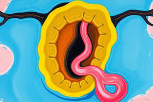
Diaphragm and Thoracic Cavity
- The septum transversum is a mesodermal plate between the thoracic and abdominal cavities.
- It is initially continuous with the visceral mesoderm surrounding the heart.
- As the lungs expand, the pericardioperitoneal canals become smaller.
- Pleuropericardial folds grow, fusing with each other and forming the pleuroperitoneal membranes.
- Mesodermal folds migrate to fuse, forming the pleuroperitoneal membranes and fully closing the thoracic cavity.
- The pleuropericardial membranes create the fibrous pericardium in the adult.
Formation of the Diaphragm
- The diaphragm forms from pleuroperitoneal membranes, the muscular components from cervical segments three to five, and the mesentery of the esophagus.
- Phrenic nerves innervate the diaphragm, originating in the ventral primary rami of C3 to C5.
- The cervical origin is due to early development of the diaphragm in the fourth week.
- The diaphragm's descent and final location occur due to differential growth in the embryo.
Clinical Correlates: Ventral Body Wall Defects
- Defects in the ventral body wall can lead to the protrusion of abdominal organs outside the body, such as hernias.
Studying That Suits You
Use AI to generate personalized quizzes and flashcards to suit your learning preferences.