Podcast
Questions and Answers
What is the primary action described regarding the palatine process of the maxillary bone?
What is the primary action described regarding the palatine process of the maxillary bone?
- To create the nasal septum.
- To separate from the palatine process of the palatine bone. (correct)
- To attach to the temporal bone.
- To fuse with the mandible.
Which action is described to occur with the teeth?
Which action is described to occur with the teeth?
- To be fused into a solid mass.
- To be extracted completely from the skull.
- To be separated from each other. (correct)
- To be joined with the palatine bone.
The described separation process involves which two bony structures?
The described separation process involves which two bony structures?
- Zygomatic and lacrimal bones.
- Mandible and hyoid bones.
- Maxillary and temporal bones.
- Palatine processes of the maxillary and palatine bones. (correct)
What is the relationship of the processes mentioned?
What is the relationship of the processes mentioned?
In addition to the bony separation, what other physical alteration is mentioned?
In addition to the bony separation, what other physical alteration is mentioned?
Which muscle is NOT considered an extraocular muscle?
Which muscle is NOT considered an extraocular muscle?
Which of the following is classified as a masticatory muscle?
Which of the following is classified as a masticatory muscle?
What is the primary function of the levator palpebrae superioris?
What is the primary function of the levator palpebrae superioris?
Which muscle is considered an accessory muscle of mastication?
Which muscle is considered an accessory muscle of mastication?
How many distinct facial expressions is it estimated that a human can make?
How many distinct facial expressions is it estimated that a human can make?
What is a primary function of the sinuses?
What is a primary function of the sinuses?
What potential issue should be considered during a maxillary molar extraction or root canal due to the sinuses?
What potential issue should be considered during a maxillary molar extraction or root canal due to the sinuses?
Approximately how many muscles are there in the face?
Approximately how many muscles are there in the face?
Why is it important to identify facial muscles when viewing an image of them?
Why is it important to identify facial muscles when viewing an image of them?
Which of the following is NOT directly related to the content provided?
Which of the following is NOT directly related to the content provided?
Which cranial nerve provides motor innervation to the majority of the tongue's muscles?
Which cranial nerve provides motor innervation to the majority of the tongue's muscles?
The muscle of the tongue that is NOT innervated by the hypoglossal nerve (CN XII) is the:
The muscle of the tongue that is NOT innervated by the hypoglossal nerve (CN XII) is the:
What is the primary function of the hypoglossal nerve (CN XII)?
What is the primary function of the hypoglossal nerve (CN XII)?
Which cranial nerve provides motor innervation to the palatoglossus muscle?
Which cranial nerve provides motor innervation to the palatoglossus muscle?
What is the significance of the palatoglossus muscle's innervation being different from other tongue muscles?
What is the significance of the palatoglossus muscle's innervation being different from other tongue muscles?
Which cranial nerve provides motor innervation to the tensor veli palatini muscle?
Which cranial nerve provides motor innervation to the tensor veli palatini muscle?
The sensory innervation of the soft palate is primarily provided by which nerve?
The sensory innervation of the soft palate is primarily provided by which nerve?
Which of the following arteries does NOT contribute directly to the rich blood supply of the face?
Which of the following arteries does NOT contribute directly to the rich blood supply of the face?
The facial and superficial temporal arteries form a/an:
The facial and superficial temporal arteries form a/an:
The lesser palatine nerve is a branch of which cranial nerve?
The lesser palatine nerve is a branch of which cranial nerve?
Which cranial nerve provides sensory innervation to the face?
Which cranial nerve provides sensory innervation to the face?
Which branch of the trigeminal nerve is solely dedicated to sensory function?
Which branch of the trigeminal nerve is solely dedicated to sensory function?
Which nerve primarily controls the muscles of mastication?
Which nerve primarily controls the muscles of mastication?
Besides sensation, the mandibular branch of the trigeminal nerve has which additional function?
Besides sensation, the mandibular branch of the trigeminal nerve has which additional function?
Which nerve is solely responsible for the motor control of facial muscles?
Which nerve is solely responsible for the motor control of facial muscles?
Flashcards
Palatine Process
Palatine Process
A bony structure that forms part of the roof of the mouth (hard palate).
Maxillary Palatine Process
Maxillary Palatine Process
The palatine process of the maxillary bone is a bony projection that extends towards the midline of the skull.
Palatine Palatine Process
Palatine Palatine Process
The palatine process of the palatine bone is also a bony projection that extends towards the midline, connecting with the maxillary process.
Palatal Fusion
Palatal Fusion
Signup and view all the flashcards
Tooth Separation
Tooth Separation
Signup and view all the flashcards
What is the maxillary sinus?
What is the maxillary sinus?
Signup and view all the flashcards
How do maxillary molars change with age?
How do maxillary molars change with age?
Signup and view all the flashcards
How can the maxillary sinus impact dental procedures?
How can the maxillary sinus impact dental procedures?
Signup and view all the flashcards
Extraocular Muscles
Extraocular Muscles
Signup and view all the flashcards
How many facial muscles are there?
How many facial muscles are there?
Signup and view all the flashcards
Why is it important to be able to identify facial muscles in images?
Why is it important to be able to identify facial muscles in images?
Signup and view all the flashcards
Rectus Muscles (Superior, Interior, Medial, Lateral)
Rectus Muscles (Superior, Interior, Medial, Lateral)
Signup and view all the flashcards
Oblique Muscles (Superior, Inferior)
Oblique Muscles (Superior, Inferior)
Signup and view all the flashcards
Levator Palpebrae Superioris
Levator Palpebrae Superioris
Signup and view all the flashcards
Masticatory Muscles
Masticatory Muscles
Signup and view all the flashcards
Tensor Veli Palatini Muscle Innervation
Tensor Veli Palatini Muscle Innervation
Signup and view all the flashcards
Soft Palate Sensory Innervation
Soft Palate Sensory Innervation
Signup and view all the flashcards
Facial Blood Supply
Facial Blood Supply
Signup and view all the flashcards
External Carotid Artery
External Carotid Artery
Signup and view all the flashcards
What nerve controls tongue movement?
What nerve controls tongue movement?
Signup and view all the flashcards
Which muscle is the exception to the Hypoglossal nerve's rule?
Which muscle is the exception to the Hypoglossal nerve's rule?
Signup and view all the flashcards
What nerve controls the Palatoglossus muscle?
What nerve controls the Palatoglossus muscle?
Signup and view all the flashcards
What is the primary role of the Hypoglossal nerve?
What is the primary role of the Hypoglossal nerve?
Signup and view all the flashcards
Why is the Palatoglossus muscle's innervation unique?
Why is the Palatoglossus muscle's innervation unique?
Signup and view all the flashcards
What is the trigeminal nerve?
What is the trigeminal nerve?
Signup and view all the flashcards
How many branches does the trigeminal nerve have?
How many branches does the trigeminal nerve have?
Signup and view all the flashcards
What is the function of the ophthalmic branch of the trigeminal nerve?
What is the function of the ophthalmic branch of the trigeminal nerve?
Signup and view all the flashcards
What is the function of the maxillary branch of the trigeminal nerve?
What is the function of the maxillary branch of the trigeminal nerve?
Signup and view all the flashcards
What is the function of the facial nerve?
What is the function of the facial nerve?
Signup and view all the flashcards
Study Notes
Dental Anatomy
- Sheet number: 14
- Document date: 2024
- Authors: Ahmad Al-Nsour, Nadeen Ramadan
- Editor: Ahmad Al-Nsour, Nadeen Ramadan
- Doctor: Aseel sharaireh
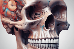
Anatomy of the Head (Revision)
- The face structure is similar to the scalp, composed of 5 layers:
- Skin (outermost layer covering the face)
- Subcutaneous layer (made of fat and fascia)
- Muscular-aponeurotic layer (innervation of nerves and muscles, creating facial expressions)
- Loose areolar tissue (spaces, ligaments, some muscles)
- Fixed periosteum and deep fascia
- Musculoaponeurotic layer (important):
- Innervation of blood vessels
- Sensory nerve supply to the face
- Trigeminal ganglion (within the skull) and its branches
The Skull
- Bone of the neurocranium (skull bones that cover the brain):
- Frontal
- Parietal (2)
- Occipital
- Temporal (2)
- Sphenoid
- Ethmoid
Bone of the Viscerocranium (Facial Bones)
- Vomer
- Inferior nasal concha (2)
- Nasal (2)
- Zygomatic (2)
- Maxilla (2)
- Mandible
Classifications of Joints
- Fibrous (held together by dense connective tissue)
- Cartilaginous (held together by cartilage)
- Synovial (separated by a fluid-filled cavity)
Gomphosis
- Specific to the dental part, attaching teeth to the bone through the periodontal ligament.
Sutures of the Skull
- Sagittal suture (midline between parietal bones)
- Coronal suture (between frontal and parietal bones)
- Lambdoidal suture (between parietal and occipital bones)
- Squamosal suture (between temporal and parietal bones)
Additional Sutures
- Metopic suture (between frontal bones)
- Sphenosquamous suture (between sphenoid and anterior temporal)
- Sphenofrontal suture (between sphenoid and frontal)
- Parietomastoid suture (between parietal and mastoid temporal)
- Sphenoparietal suture (between sphenoid and parietal)
- Occipitomastoid suture (between occipital and mastoid temporal)
Bone of the Viscerocranium (Facial Bones)
- Comprises the facial bones
- Includes various numbered bones like Vomer, Inferior nasal concha, Nasal, Zygomatic, Maxilla
Maxillary Bone
- Continuous with the zygomatic bone.
- Contains various processes (e.g., zygomatic, frontal) and surfaces (e.g., orbital, infraorbital).
- Contains alveolus cavities for teeth.
- Parts vital for creating the framework
Mandible Bone
- Contains various processes and surfaces, including alveolar for teeth.
- Attaches with the TMJ to the Cranium for jaw movement.
Cavities of the Skull
- Cranial fossae (middle & posterior)
- Orbital cavity
- Nasal cavity
- Oral cavity
- Paranasal sinuses (frontal, maxillary, sphenoidal, and ethmoidal sinuses)
Paranasal Sinuses
- Air-filled spaces with mucus lining moisturizing the air.
- All openings into the nasal cavity.
- Reduces the skull's weight.
Muscles of the Face
- Total number of major facial muscles: 42
- Ability to create over 10,000 facial expressions
- Extraocular muscle groups (oblique and rectus, and Levator palpebrae superioris).
Masticatory Muscles
- Masseter
- Temporalis
- Pterygoids (lateral & medial)
- Buccinator (accessory muscle)
Facial Muscles
- Ear: Auricular (anterior, superior, posterior)
- Temporoparietalis muscle
- Scalp/Eyelid: Occipitofrontalis
- Nose: Procerus, Nasalis, Dilator naris, Depressor septi nasi
- Mouth: Levator Labii Superioris and others
- Descriptions of muscles and their placements
Soft Palate
- Muscles (e.g., Veli palatini, Musculus uvulae).
Tongue
- Extrinsic tongue muscles
- Intrinsic tongue muscles
Vasovagal Syncope
- Cause of fainting in dental patients
- Origin from emotional distress or stimulating the vagus nerve
- Overstimulation of vagus nerve may result in changes to heart rate, leading to fainting.
Blood Supply of the Face
- Rich blood supply from mainly two arteries: Facial and Superficial temporal.
- Anastomoses (delicate connections) between these two arteries.
- Internal carotid arteries (supra-orbital and supratrochlear arteries) are part of the system.
- Dangerous area due to anastomoses.
Nervous Innervation of the Face
- Trigeminal nerve (Ophthalmic, Maxillary, Mandibular) branches - sensory/motor function
- Facial nerve (motor) for mastication muscles.
Studying That Suits You
Use AI to generate personalized quizzes and flashcards to suit your learning preferences.
Related Documents
Description
This quiz covers the essential aspects of dental anatomy and the structural organization of the skull. Students will explore the layers of the face, the composition of the neurocranium, and key facial bones. Enhance your understanding of these critical topics in head anatomy through this comprehensive quiz.