Podcast
Questions and Answers
Which proteins are responsible for cross-linking actin into parallel bundles within microvilli?
Which proteins are responsible for cross-linking actin into parallel bundles within microvilli?
Spectrins provide flexible stiffness to most vertebrate cells, but not red blood cells.
Spectrins provide flexible stiffness to most vertebrate cells, but not red blood cells.
False (B)
What is the primary structural component found at the core of microvilli?
What is the primary structural component found at the core of microvilli?
Parallel actin filaments
Some types of cancer cells, especially malignant melanomas, lack ______ and cannot crawl properly.
Some types of cancer cells, especially malignant melanomas, lack ______ and cannot crawl properly.
Signup and view all the answers
Match the protein with its primary function:
Match the protein with its primary function:
Signup and view all the answers
Which protein is typically found in the axon of a neuron?
Which protein is typically found in the axon of a neuron?
Signup and view all the answers
Microtubules in dendrites have a smaller space between them compared to microtubules in axons.
Microtubules in dendrites have a smaller space between them compared to microtubules in axons.
Signup and view all the answers
What protein stabilizes actin filaments by binding to 7 adjacent actin subunits?
What protein stabilizes actin filaments by binding to 7 adjacent actin subunits?
Signup and view all the answers
______ destabilizes actin filaments by forcing the filament to twist more tightly.
______ destabilizes actin filaments by forcing the filament to twist more tightly.
Signup and view all the answers
Which protein protects actin filaments from the destabilizing effects of cofilin?
Which protein protects actin filaments from the destabilizing effects of cofilin?
Signup and view all the answers
Proteins that bind to the ends of filaments need to be present in high stoichiometric ratios to have dramatic effects.
Proteins that bind to the ends of filaments need to be present in high stoichiometric ratios to have dramatic effects.
Signup and view all the answers
Match the following proteins with their function:
Match the following proteins with their function:
Signup and view all the answers
What disease can result from mutations in the gene for plectin?
What disease can result from mutations in the gene for plectin?
Signup and view all the answers
Which of the following proteins is NOT primarily associated with microfilaments?
Which of the following proteins is NOT primarily associated with microfilaments?
Signup and view all the answers
Filamins form rigid, low-angle links between actin filaments.
Filamins form rigid, low-angle links between actin filaments.
Signup and view all the answers
What is a key similarity between the γ-tubulin ring complex and the ARP complex?
What is a key similarity between the γ-tubulin ring complex and the ARP complex?
Signup and view all the answers
The concentration of actin in cells is ______ times greater than the critical concentration observed for pure actin in a test tube.
The concentration of actin in cells is ______ times greater than the critical concentration observed for pure actin in a test tube.
Signup and view all the answers
Match the following proteins with their primary cytoskeletal association:
Match the following proteins with their primary cytoskeletal association:
Signup and view all the answers
What is the primary function of the centrosome (MTOC)?
What is the primary function of the centrosome (MTOC)?
Signup and view all the answers
Microtubules always grow from the MTOC.
Microtubules always grow from the MTOC.
Signup and view all the answers
What is the MT aster formation?
What is the MT aster formation?
Signup and view all the answers
Centrioles are arranged at ______ angles to each other.
Centrioles are arranged at ______ angles to each other.
Signup and view all the answers
What is a key component of the centrosome matrix that is capable of polymerizing MTs?
What is a key component of the centrosome matrix that is capable of polymerizing MTs?
Signup and view all the answers
The plus end of the microtubule is relatively stable compared to the minus end.
The plus end of the microtubule is relatively stable compared to the minus end.
Signup and view all the answers
What does the astral configuration help the cell to achieve?
What does the astral configuration help the cell to achieve?
Signup and view all the answers
Match the component with its description:
Match the component with its description:
Signup and view all the answers
What is the primary function of formin in actin polymerization?
What is the primary function of formin in actin polymerization?
Signup and view all the answers
Profilin competes with stathmin to bind actin monomers.
Profilin competes with stathmin to bind actin monomers.
Signup and view all the answers
What cellular structure is profilin associated with, awaiting activation?
What cellular structure is profilin associated with, awaiting activation?
Signup and view all the answers
Unpolymerized tubulin subunits are sequestered by the protein ______.
Unpolymerized tubulin subunits are sequestered by the protein ______.
Signup and view all the answers
What effect does stathmin phosphorylation have on tubulin?
What effect does stathmin phosphorylation have on tubulin?
Signup and view all the answers
Overexpression of stathmins in cancer cells leads to a decreased rate of microtubule turnover.
Overexpression of stathmins in cancer cells leads to a decreased rate of microtubule turnover.
Signup and view all the answers
How does severing of existing actin filaments accelerate the assembly of new filaments?
How does severing of existing actin filaments accelerate the assembly of new filaments?
Signup and view all the answers
Match the following proteins with their primary function:
Match the following proteins with their primary function:
Signup and view all the answers
In a typical cell, where is the centrosome usually located?
In a typical cell, where is the centrosome usually located?
Signup and view all the answers
In differentiated epithelial cells, microtubules always originate from the MTOC.
In differentiated epithelial cells, microtubules always originate from the MTOC.
Signup and view all the answers
What is the primary function of the orientation of microtubules in polarized cells?
What is the primary function of the orientation of microtubules in polarized cells?
Signup and view all the answers
Actin filaments are often nucleated at the ______.
Actin filaments are often nucleated at the ______.
Signup and view all the answers
Which end of the microtubule is oriented towards the apical side of a polarized epithelial cell?
Which end of the microtubule is oriented towards the apical side of a polarized epithelial cell?
Signup and view all the answers
Which of the following is NOT a structure formed by actin filaments at the cell surface?
Which of the following is NOT a structure formed by actin filaments at the cell surface?
Signup and view all the answers
Match the cell structure with its location or characteristic:
Match the cell structure with its location or characteristic:
Signup and view all the answers
The orientation of microtubules is not important for directionality of transport in cells.
The orientation of microtubules is not important for directionality of transport in cells.
Signup and view all the answers
Flashcards
What is the centrosome?
What is the centrosome?
A structure found in the center of animal cells. It acts as a microtubule organizing center (MTOC) and is crucial for the organization and formation of microtubule networks. The minus end of microtubules is anchored to the centrosome.
What is the 'plus end' of a microtubule?
What is the 'plus end' of a microtubule?
The plus end of a microtubule is the end that grows outwards from the centrosome. It is the more dynamic end, meaning it can grow or shrink faster than the minus end.
What is the 'minus end' of a microtubule?
What is the 'minus end' of a microtubule?
The minus end of the microtubule is anchored to the centrosome. It is more stable compared to the plus end.
What are centrioles?
What are centrioles?
Signup and view all the flashcards
What is mitosis?
What is mitosis?
Signup and view all the flashcards
What is the gamma tubulin ring complex?
What is the gamma tubulin ring complex?
Signup and view all the flashcards
How are microtubules dynamic?
How are microtubules dynamic?
Signup and view all the flashcards
What is the 'astral configuration'?
What is the 'astral configuration'?
Signup and view all the flashcards
Microtubule Organization in Dividing Cells
Microtubule Organization in Dividing Cells
Signup and view all the flashcards
Microtubule Organization in Differentiated Cells
Microtubule Organization in Differentiated Cells
Signup and view all the flashcards
Microtubule Orientation in Polarized Cells
Microtubule Orientation in Polarized Cells
Signup and view all the flashcards
Actin Nucleation at the Plasma Membrane
Actin Nucleation at the Plasma Membrane
Signup and view all the flashcards
Actin-Based Cell Surface Structures
Actin-Based Cell Surface Structures
Signup and view all the flashcards
MAP2 function
MAP2 function
Signup and view all the flashcards
Tau function
Tau function
Signup and view all the flashcards
Tropomyosin function
Tropomyosin function
Signup and view all the flashcards
Cofilin function
Cofilin function
Signup and view all the flashcards
CapZ function
CapZ function
Signup and view all the flashcards
ARP complex function
ARP complex function
Signup and view all the flashcards
Filaggrin function
Filaggrin function
Signup and view all the flashcards
Plectin function
Plectin function
Signup and view all the flashcards
What is formin's role in actin polymerization?
What is formin's role in actin polymerization?
Signup and view all the flashcards
How does profilin influence actin polymerization?
How does profilin influence actin polymerization?
Signup and view all the flashcards
How is profilin activated?
How is profilin activated?
Signup and view all the flashcards
What is the role of stathmins in microtubule dynamics?
What is the role of stathmins in microtubule dynamics?
Signup and view all the flashcards
How does phosphorylation affect stathmins?
How does phosphorylation affect stathmins?
Signup and view all the flashcards
How do severing proteins influence actin dynamics?
How do severing proteins influence actin dynamics?
Signup and view all the flashcards
How are stathmins related to cancer cells?
How are stathmins related to cancer cells?
Signup and view all the flashcards
What is the significance of the plasma membrane in actin polymerization?
What is the significance of the plasma membrane in actin polymerization?
Signup and view all the flashcards
What are microvilli and how are they formed?
What are microvilli and how are they formed?
Signup and view all the flashcards
What is the difference between actin bundles and actin webs?
What is the difference between actin bundles and actin webs?
Signup and view all the flashcards
Why is filamin important for cancer cell invasion?
Why is filamin important for cancer cell invasion?
Signup and view all the flashcards
How do spectrins contribute to red blood cell function?
How do spectrins contribute to red blood cell function?
Signup and view all the flashcards
Why are actin filaments considered dynamic?
Why are actin filaments considered dynamic?
Signup and view all the flashcards
What is γ-tubulin?
What is γ-tubulin?
Signup and view all the flashcards
What is the ARP complex?
What is the ARP complex?
Signup and view all the flashcards
How does cofilin recognize old actin filaments?
How does cofilin recognize old actin filaments?
Signup and view all the flashcards
Why is the concentration of actin in cells much higher than its critical concentration?
Why is the concentration of actin in cells much higher than its critical concentration?
Signup and view all the flashcards
How does the centrosome locate the cell’s center?
How does the centrosome locate the cell’s center?
Signup and view all the flashcards
Study Notes
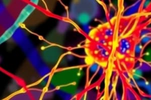
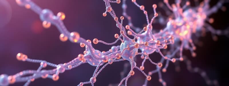
Cytoskeletal Filaments: Regulation and Structure
- Microtubules (MTs):
- Nucleated from the microtubule-organizing center (MTOC), typically near the nucleus.
- Minus ends are anchored, plus ends grow outward.
- 𝛄-tubulin nucleates MTs, and the structure allows for quicker addition of tubulin dimers
- γ-tubulin:
- Anchored to accessory proteins that create the MTOC.
- Allows for the initial formation of the structure.
- Mutating or removing this can cause slower and less organized MTOC formation.
Cytoskeletal Filaments: Dynamics and Structure
- Accessory Proteins:
- Modify MT dynamics and structure.
- Y-tubulin nucleates MTs from the MTOC.
- MTs are assembled from alpha and beta tubulin dimers.
- Plus ends grow outward from the MTOC, minus ends remain at the MTOC or are anchored.
- The structural orientation of the MT (alpha and beta) is important because they are always oriented toward the plus or minus ends.
- Centrosome:
- A well-defined MTOC in most animal cells.
- Contains a fibrous matrix with 50 copies of γ-tubulin.
- Centrioles are rod-like structures within the centrosome, arranged at right angles to each other.
- Astral Configuration:
- Plus ends of MTs outward from the MTOC towards the outer cell regions,
- The centrosome is positioned at the cell center.
- This arrangement and positioning of MTs within the cell are important for proper distribution of organelles and maintaining cellular shape.
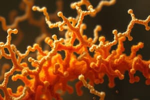
Actin Filaments
- Nucleation at the Plasma Membrane:
- Actin nucleation occurs frequently at, or near, the cell membrane.
- Different types of cell surface projections form from actin: microvilli, filopodia, lamellipodia, and phagocytic cups.
Actin Filament Organization
- Polymerization:
- Actin filaments are formed from the polymerization of actin subunits.
- External stimuli frequently regulate the nucleation of actin filaments.
- ARP complex and formins catalyze this nucleation.
- ARP Complex:
- Nucleates new actin filaments at a 70-degree angle from pre-existing filaments.
- Responsible for forming branched actin networks.
Actin Filament Organization: Formins
- Formins:
- Stabilize growing plus ends of actin structures.
- Formins move along the plus end as polymerization occurs.
- Interact with minus end, creating linear polymerization, not creating branched networks as ARP does.
Proteins Binding to Filament Ends
- Proteins that Interact with Filament Ends:
- Dynamically change filament properties.
- Proteins that bind along sides of filaments can stabilize or destabilize those filaments.
- Proteins with high ratio for binding can induce dramatic effects at low concentrations.
Intermediate Filaments
- Cross-Linked and Bundled:
- Filaggrin bundles keratin filaments to make the outer skin tougher.
- Plectin links intermediate filaments to microtubules and actin bundles.
- Plectin mutations can cause a devastating disease combining epidermis bullosa, muscular dystrophy, and neurodegeneration.
Actin Parallel Filaments vs Actin Webs
- Parallel Bundles:
- Villin and fimbrin cross-link actin into parallel bundles.
- Web-Forming/Stabilizing Proteins:
- Filamins and spectrins regulate actin stability.
- Critically important in maintaining cell shape and providing flexibility.
Proteins for Microtubules and Microfilaments
- Microtubule:
- γ-tubulin
- Stathmins
- Katanin
- Gelsolin
- MAPS (MAP2)
- Tau
- Microfilaments:
- Tropomyosin
- Cofilin
- Villin
- Fimbrin
- Filamins
- Spectrins
- CAPZ
- ARP
- Formins
- Thymosin
- Profilin
- Plectin
- Filaggrin
Studying That Suits You
Use AI to generate personalized quizzes and flashcards to suit your learning preferences.
Related Documents
Description
Explore the regulation and structure of cytoskeletal filaments, particularly focusing on microtubules, γ-tubulin, and accessory proteins. This quiz will test your understanding of the nucleation process, dynamics, and the key components involved in microtubule organization and function.