Podcast
Questions and Answers
What is the main function of the cardiovascular system?
What is the main function of the cardiovascular system?
Circulates blood to supply oxygen and nutrients to tissues, and remove carbon dioxide and waste products
Which system provides blood flow to support the heart's work in pumping oxygenated blood to the body?
Which system provides blood flow to support the heart's work in pumping oxygenated blood to the body?
- Systemic system
- Pulmonary system
- Coronary system (correct)
- Vascular system
The left ventricle works harder than any other heart chamber.
The left ventricle works harder than any other heart chamber.
True (A)
The ________ valves are located between the right ventricle and the trunk of the pulmonary arteries.
The ________ valves are located between the right ventricle and the trunk of the pulmonary arteries.
Match the heart sound with its description:
Match the heart sound with its description:
What are the two possible rates at which the heart may beat abnormally?
What are the two possible rates at which the heart may beat abnormally?
What is the primary cause of over 90% of essential or primary hypertension cases?
What is the primary cause of over 90% of essential or primary hypertension cases?
Which element of the ECG recording represents the repolarization (relaxation) of the ventricles?
Which element of the ECG recording represents the repolarization (relaxation) of the ventricles?
Match the following diagnostic tests with their descriptions:
Match the following diagnostic tests with their descriptions:
What is the term for a slow and progressive circulation disorder due to narrowing, blockage, or spasms in a blood vessel in the periphery?
What is the term for a slow and progressive circulation disorder due to narrowing, blockage, or spasms in a blood vessel in the periphery?
Which of the following conditions is associated with peripheral vascular disease?
Which of the following conditions is associated with peripheral vascular disease?
Is claudication known as ischaemic pain in legs and hands?
Is claudication known as ischaemic pain in legs and hands?
Peripheral vascular disease leads to ______ in the peripheral tissues, potentially resulting in tissue necrosis.
Peripheral vascular disease leads to ______ in the peripheral tissues, potentially resulting in tissue necrosis.
What is the name of the inflammation of the innermost lining of the heart and the valves?
What is the name of the inflammation of the innermost lining of the heart and the valves?
What is the most common cause of inflammation of the muscular layer of the heart?
What is the most common cause of inflammation of the muscular layer of the heart?
What is the term for inflammation of the pericardium?
What is the term for inflammation of the pericardium?
What are the three main types of cardiomyopathies?
What are the three main types of cardiomyopathies?
What is the treatment for inflammation of the pericardium?
What is the treatment for inflammation of the pericardium?
Septal defects can lead to compromised cardiac output and heart failure.
Septal defects can lead to compromised cardiac output and heart failure.
Define anatomy and physiology.
Define anatomy and physiology.
Which of the following is NOT one of the four basic types of tissues in the body?
Which of the following is NOT one of the four basic types of tissues in the body?
A cell is the basic structural and functional unit of __________.
A cell is the basic structural and functional unit of __________.
Apoptosis is a form of programmed cell death.
Apoptosis is a form of programmed cell death.
Match the following terms with their definitions:
Match the following terms with their definitions:
Define incidence in epidemiology.
Define incidence in epidemiology.
Explain what prevalence refers to in epidemiology.
Explain what prevalence refers to in epidemiology.
Which of the following are the levels of prevention discussed in the content?
Which of the following are the levels of prevention discussed in the content?
What is evidence-based practice in healthcare?
What is evidence-based practice in healthcare?
What is the process of transporting blood to and from the body tissues called?
What is the process of transporting blood to and from the body tissues called?
What components make up the cellular components of blood? (Select all that apply)
What components make up the cellular components of blood? (Select all that apply)
Normal haematocrit levels are lower in females than in males.
Normal haematocrit levels are lower in females than in males.
____ is the liquid portion of blood that carries various nutrients, gases, and electrolytes.
____ is the liquid portion of blood that carries various nutrients, gases, and electrolytes.
Match the white blood cell type with their function:
Match the white blood cell type with their function:
What are the two main catecholamines secreted by the Adrenal Medulla?
What are the two main catecholamines secreted by the Adrenal Medulla?
What are the common symptoms of Phaeochromocytoma?
What are the common symptoms of Phaeochromocytoma?
Thyroxin (T4) accounts for less than 90% of all thyroid hormones secreted.
Thyroxin (T4) accounts for less than 90% of all thyroid hormones secreted.
Goitre is classified as non-toxic or toxic, and ___________ or nodular.
Goitre is classified as non-toxic or toxic, and ___________ or nodular.
Match the following between Hypothyroidism and Hyperthyroidism:
Match the following between Hypothyroidism and Hyperthyroidism:
What is the most common cause of primary hyperparathyroidism?
What is the most common cause of primary hyperparathyroidism?
What are the two regulatory systems of the body?
What are the two regulatory systems of the body?
What are the chemical messengers called in the Endocrine system?
What are the chemical messengers called in the Endocrine system?
______ glands are often referred to as ductless glands.
______ glands are often referred to as ductless glands.
The hypothalamic-pituitary axis controls the regulation of the endocrine system.
The hypothalamic-pituitary axis controls the regulation of the endocrine system.
Match the type of endocrine disease with its description:
Match the type of endocrine disease with its description:
What are the common conditions associated with the Adrenal gland?
What are the common conditions associated with the Adrenal gland?
What are the two types of Diabetes Insipidus?
What are the two types of Diabetes Insipidus?
What is the major regulator of metabolism and organ function in Diabetes Mellitus?
What is the major regulator of metabolism and organ function in Diabetes Mellitus?
Which cells in the alveoli are responsible for reducing surface tension?
Which cells in the alveoli are responsible for reducing surface tension?
Acute cough typically lasts longer than 3 weeks.
Acute cough typically lasts longer than 3 weeks.
Coughing enables the airways to be cleared of secretions and ____________.
Coughing enables the airways to be cleared of secretions and ____________.
Match the respiratory symptom with its description:
Match the respiratory symptom with its description:
What are the common presentations of inflammation and swelling of the mucous membrane of the nose?
What are the common presentations of inflammation and swelling of the mucous membrane of the nose?
Acute sinusitis is mostly viral, which is common after a common cold.
Acute sinusitis is mostly viral, which is common after a common cold.
What is the recommended management for acute inflammation of bronchi?
What is the recommended management for acute inflammation of bronchi?
_______ is one of the most common COPDs.
_______ is one of the most common COPDs.
Match the following lung diseases with their primary cause:
Match the following lung diseases with their primary cause:
What is the gold standard for diagnosing pulmonary embolism?
What is the gold standard for diagnosing pulmonary embolism?
What is considered a high probability of pulmonary embolism according to the simplified Wells score?
What is considered a high probability of pulmonary embolism according to the simplified Wells score?
Primary lung cancer originates outside of the lung.
Primary lung cancer originates outside of the lung.
____ is a major risk factor for the 5th leading cause of death in Australia in 2022.
____ is a major risk factor for the 5th leading cause of death in Australia in 2022.
Match the lung cancer type with its percentage of occurrence:
Match the lung cancer type with its percentage of occurrence:
What are some of the common invaders the immune system protects the body against?
What are some of the common invaders the immune system protects the body against?
Name five components of the lymphatic system that work with the immune system.
Name five components of the lymphatic system that work with the immune system.
Neutrophils play an important role in bacterial infection through a process called ____________.
Neutrophils play an important role in bacterial infection through a process called ____________.
Secondary immunodeficiency affects the immune function as a consequence of an issue elsewhere in the body.
Secondary immunodeficiency affects the immune function as a consequence of an issue elsewhere in the body.
Match the following antibodies with their functions:
Match the following antibodies with their functions:
What are the phases of digestion?
What are the phases of digestion?
What nutrients are considered macronutrients?
What nutrients are considered macronutrients?
Excessive caloric intake can lead to malnutrition.
Excessive caloric intake can lead to malnutrition.
A FOBT is a test that detects tiny amounts of __________ from bowel cancers or their precursors into the bowel lumen.
A FOBT is a test that detects tiny amounts of __________ from bowel cancers or their precursors into the bowel lumen.
Match the following blood test with its use:
Match the following blood test with its use:
What is another name for Coeliac Disease?
What is another name for Coeliac Disease?
What are consistent symptoms associated with malabsorption in Coeliac Disease?
What are consistent symptoms associated with malabsorption in Coeliac Disease?
Obesity is a highly complex and multifactorial condition.
Obesity is a highly complex and multifactorial condition.
Match the following obesity BMI categories with their respective ranges:
Match the following obesity BMI categories with their respective ranges:
What does GORD stand for?
What does GORD stand for?
What can be a complication of untreated GORD?
What can be a complication of untreated GORD?
Peptic Ulcer Disease can be caused by H.pylori infection.
Peptic Ulcer Disease can be caused by H.pylori infection.
What are the functions of the kidneys? Select all that apply.
What are the functions of the kidneys? Select all that apply.
Name the microscopic functional units of the kidneys.
Name the microscopic functional units of the kidneys.
Glomerular filtration primarily occurs in the loop of Henle.
Glomerular filtration primarily occurs in the loop of Henle.
Glomerular filtration barrier helps in the first stage of urine formation, where the fluid part of blood is forced from the glomerulus into Bowman's ______.
Glomerular filtration barrier helps in the first stage of urine formation, where the fluid part of blood is forced from the glomerulus into Bowman's ______.
What are common clinical features of UTI? (Select all that apply)
What are common clinical features of UTI? (Select all that apply)
Antimicrobial agents are the primary means of therapy for UTIs.
Antimicrobial agents are the primary means of therapy for UTIs.
What is the primary goal of UTI treatment?
What is the primary goal of UTI treatment?
The most common bacteria causing Chlamydial infection is Chlamydia ________.
The most common bacteria causing Chlamydial infection is Chlamydia ________.
Match the STIs with their respective causes:
Match the STIs with their respective causes:
What are the functions of the skin?
What are the functions of the skin?
List some of the structures that are part of the integumentary system.
List some of the structures that are part of the integumentary system.
Malignant Melanoma is a type of skin cancer that spreads slowly.
Malignant Melanoma is a type of skin cancer that spreads slowly.
_____ is needed for calcium absorption.
_____ is needed for calcium absorption.
Match the following skin lesions with their descriptions:
Match the following skin lesions with their descriptions:
Which of the following is true about Glucosuria?
Which of the following is true about Glucosuria?
What does Bilirubinuria indicate?
What does Bilirubinuria indicate?
Ketones in the urine are considered normal.
Ketones in the urine are considered normal.
A high Specific gravity in a dipstick test may indicate ____________.
A high Specific gravity in a dipstick test may indicate ____________.
Match the dipstick test result with its potential cause:
Match the dipstick test result with its potential cause:
What are the general functions of the liver and the digestive system?
What are the general functions of the liver and the digestive system?
Which phase of digestion slows down gastric emptying to allow absorption in the small intestine?
Which phase of digestion slows down gastric emptying to allow absorption in the small intestine?
Deficiencies, excesses, or imbalances in a person's intake of energy and nutrients can cause weight loss or gain.
Deficiencies, excesses, or imbalances in a person's intake of energy and nutrients can cause weight loss or gain.
______ is a common symptom of malnutrition and can be caused by various conditions.
______ is a common symptom of malnutrition and can be caused by various conditions.
Match the abdominal region with the common disorders associated with it:
Match the abdominal region with the common disorders associated with it:
What are some skin manifestations of GI disorders?
What are some skin manifestations of GI disorders?
What stimulates saliva secretion in the cephalic phase of digestion?
What stimulates saliva secretion in the cephalic phase of digestion?
What nerve stimulates acid secretion in the stomach during the cephalic phase?
What nerve stimulates acid secretion in the stomach during the cephalic phase?
What are the effects of the gastric phase on the stomach?
What are the effects of the gastric phase on the stomach?
What is the purpose of the intestinal phase of digestion?
What is the purpose of the intestinal phase of digestion?
What effect does the intestinal phase have on acid secretion?
What effect does the intestinal phase have on acid secretion?
Which condition is associated with Terry's nail?
Which condition is associated with Terry's nail?
Which of the following can cause palmar erythema?
Which of the following can cause palmar erythema?
What is a possible cause of pallor on palmar creases?
What is a possible cause of pallor on palmar creases?
Quae conditio clara eicere Hyperbilirubinaemiam facit?
Quae conditio clara eicere Hyperbilirubinaemiam facit?
Quae caussa est periorbital purpura?
Quae caussa est periorbital purpura?
Xanthelasmata connexa sunt cum ______________.
Xanthelasmata connexa sunt cum ______________.
What are some common causes of generalised abdominal distension?
What are some common causes of generalised abdominal distension?
What condition might be indicated by prominent abdominal veins resembling Caput Medusae?
What condition might be indicated by prominent abdominal veins resembling Caput Medusae?
What might a pulsation in the abdomen indicate?
What might a pulsation in the abdomen indicate?
What are some generalised symptoms of acute liver disease?
What are some generalised symptoms of acute liver disease?
What symptom typically appears as acute hepatitis progresses?
What symptom typically appears as acute hepatitis progresses?
What are some symptoms of chronic liver disease?
What are some symptoms of chronic liver disease?
What are the characteristics of pre-hepatic jaundice?
What are the characteristics of pre-hepatic jaundice?
What are the characteristics of hepatic jaundice?
What are the characteristics of hepatic jaundice?
What are the characteristics of post-hepatic (cholestatic) jaundice?
What are the characteristics of post-hepatic (cholestatic) jaundice?
What are some investigations used for COPD diagnosis?
What are some investigations used for COPD diagnosis?
Which of the following are included in COPD management? (Select all that apply)
Which of the following are included in COPD management? (Select all that apply)
COPD management includes bronchodilators such as short- or long-acting β2-______.
COPD management includes bronchodilators such as short- or long-acting β2-______.
Match the following medications with their use in COPD management:
Match the following medications with their use in COPD management:
What color is typically associated with reliever medications for asthma?
What color is typically associated with reliever medications for asthma?
Which color is usually linked to preventer medications for asthma?
Which color is usually linked to preventer medications for asthma?
Which color represents symptom controller medications for asthma?
Which color represents symptom controller medications for asthma?
What color is associated with combination medications for asthma?
What color is associated with combination medications for asthma?
What is pneumonia?
What is pneumonia?
What are common causes of pneumonia?
What are common causes of pneumonia?
What are some common symptoms of pneumonia?
What are some common symptoms of pneumonia?
How is pneumonia classified?
How is pneumonia classified?
What is the main characteristic of pneumothorax?
What is the main characteristic of pneumothorax?
What is one common risk factor for primary pneumothorax?
What is one common risk factor for primary pneumothorax?
Secondary pneumothorax occurs in the absence of any lung pathology.
Secondary pneumothorax occurs in the absence of any lung pathology.
Iatrogenic pneumothorax can occur due to procedures like pleural or lung ____________.
Iatrogenic pneumothorax can occur due to procedures like pleural or lung ____________.
What is pleural effusion?
What is pleural effusion?
Which conditions can cause transudative pleural effusion? Select all that apply.
Which conditions can cause transudative pleural effusion? Select all that apply.
What is empyema?
What is empyema?
What causes blood effusion?
What causes blood effusion?
What is a pulmonary embolism?
What is a pulmonary embolism?
What are some thrombotic causes of pulmonary embolism?
What are some thrombotic causes of pulmonary embolism?
Fat embolism is a non-thrombotic cause of pulmonary embolism.
Fat embolism is a non-thrombotic cause of pulmonary embolism.
What are some common causes of gastritis?
What are some common causes of gastritis?
What are some common symptoms of gastritis?
What are some common symptoms of gastritis?
How can gastritis be managed?
How can gastritis be managed?
What is the purpose of Upper GI Endoscopy & biopsy in gord management?
What is the purpose of Upper GI Endoscopy & biopsy in gord management?
What are some lifestyle changes recommended for patients with GERD?
What are some lifestyle changes recommended for patients with GERD?
What medications are commonly used in the treatment of GERD?
What medications are commonly used in the treatment of GERD?
When is H. pylori eradication treatment indicated in the management of GERD?
When is H. pylori eradication treatment indicated in the management of GERD?
What are some complications of long-lasting GORD?
What are some complications of long-lasting GORD?
What are the two main types of peptic ulcers?
What are the two main types of peptic ulcers?
Which type of peptic ulcer is more common, gastric ulcer (GU) or duodenal ulcer (DU)?
Which type of peptic ulcer is more common, gastric ulcer (GU) or duodenal ulcer (DU)?
Peptic ulcers are typically associated with chest pain.
Peptic ulcers are typically associated with chest pain.
Peptic ulcers improve with ________.
Peptic ulcers improve with ________.
What type of pain is associated with peptic ulcers and tends to worsen with food but can be relieved by food intake?
What type of pain is associated with peptic ulcers and tends to worsen with food but can be relieved by food intake?
What are some investigations done for peptic ulcer disease?
What are some investigations done for peptic ulcer disease?
What is the primary management approach for peptic ulcer disease?
What is the primary management approach for peptic ulcer disease?
What are the risk factors associated with peptic ulcer disease?
What are the risk factors associated with peptic ulcer disease?
What are the complications of peptic ulcer disease?
What are the complications of peptic ulcer disease?
What does IBD stand for?
What does IBD stand for?
Name two factors that contribute to the development of IBD.
Name two factors that contribute to the development of IBD.
IBD is a curable disease.
IBD is a curable disease.
Qual es alcun scintillas de IBS?
Qual es alcun scintillas de IBS?
Which of the following symptoms are common in Irritable Bowel Syndrome (IBS)? (Select all that apply)
Which of the following symptoms are common in Irritable Bowel Syndrome (IBS)? (Select all that apply)
What is a common gastrointestinal symptom in IBS besides abdominal pain?
What is a common gastrointestinal symptom in IBS besides abdominal pain?
Is the presence of fever a common symptom of Irritable Bowel Syndrome (IBS)?
Is the presence of fever a common symptom of Irritable Bowel Syndrome (IBS)?
What are two major causes of hepatitis?
What are two major causes of hepatitis?
What are some common symptoms of hepatitis? (Select all that apply)
What are some common symptoms of hepatitis? (Select all that apply)
Is viral hepatitis a major health problem worldwide?
Is viral hepatitis a major health problem worldwide?
Complications of hepatitis can include the existence of a chronic ____ state.
Complications of hepatitis can include the existence of a chronic ____ state.
Quin test acciona l'ACTH per avaluar la funció adrenal?
Quin test acciona l'ACTH per avaluar la funció adrenal?
Quins són els dos hormones principals avaluts en els tests tiroïdals?
Quins són els dos hormones principals avaluts en els tests tiroïdals?
Quin test avalua els nivells de calci en sang?
Quin test avalua els nivells de calci en sang?
Relaciona els tests paratiroïdals amb els components correctes:
Relaciona els tests paratiroïdals amb els components correctes:
What does a glucose tolerance test measure?
What does a glucose tolerance test measure?
What does HbA1C test for?
What does HbA1C test for?
What is the purpose of testing serum electrolytes?
What is the purpose of testing serum electrolytes?
What does a urine ketones test detect?
What does a urine ketones test detect?
What does a urine/serum creatinine test measure?
What does a urine/serum creatinine test measure?
Quid est neurohypophysis?
Quid est neurohypophysis?
Quid est diabetes insipidus?
Quid est diabetes insipidus?
Quid est dysfunctio pancreatis endocrini?
Quid est dysfunctio pancreatis endocrini?
Quid est diabetes mellitus (T1DM, T2DM)?
Quid est diabetes mellitus (T1DM, T2DM)?
What is another term for Hyperaldosteronism?
What is another term for Hyperaldosteronism?
Which condition is characterized by excessive production of cortisol?
Which condition is characterized by excessive production of cortisol?
What is the primary hormone affected in Addison's disease?
What is the primary hormone affected in Addison's disease?
What is the most common type of thyroid dysfunction where the thyroid gland is underactive?
What is the most common type of thyroid dysfunction where the thyroid gland is underactive?
Which condition results from the overproduction of parathyroid hormone?
Which condition results from the overproduction of parathyroid hormone?
What are the main features of Diabetes Insipidus (DI)?
What are the main features of Diabetes Insipidus (DI)?
What are the two types of Diabetes Insipidus (DI) and their causes?
What are the two types of Diabetes Insipidus (DI) and their causes?
What is the body's inability to regulate blood glucose level called?
What is the body's inability to regulate blood glucose level called?
Which type of Diabetes Mellitus is characterized by severe or absolute insulin deficiency?
Which type of Diabetes Mellitus is characterized by severe or absolute insulin deficiency?
What can cause Chronic hyperglycemia to affect every system in the body?
What can cause Chronic hyperglycemia to affect every system in the body?
What are some symptoms of hyperglycaemia?
What are some symptoms of hyperglycaemia?
Name a complication of diabetes related to blood vessels.
Name a complication of diabetes related to blood vessels.
Name a microvascular complication of diabetes affecting the eyes.
Name a microvascular complication of diabetes affecting the eyes.
Which complication of diabetes affects the kidneys?
Which complication of diabetes affects the kidneys?
What type of complication of diabetes is neuropathy?
What type of complication of diabetes is neuropathy?
Name a complication of diabetes related to the nervous system.
Name a complication of diabetes related to the nervous system.
What are the common investigations used to confirm Cushing's syndrome?
What are the common investigations used to confirm Cushing's syndrome?
What are the common investigations used to differentiate the cause of Cushing's syndrome?
What are the common investigations used to differentiate the cause of Cushing's syndrome?
What are the causes of secondary hyperaldosteronism?
What are the causes of secondary hyperaldosteronism?
Which condition is associated with primary hyperaldosteronism?
Which condition is associated with primary hyperaldosteronism?
What is another name for Addison’s disease?
What is another name for Addison’s disease?
Which of the following hormones production is severely reduced in Addison’s disease? (Select all that apply)
Which of the following hormones production is severely reduced in Addison’s disease? (Select all that apply)
Addison’s disease should be considered in patients with unexplained fatigue plus hypernatremia or hypertension.
Addison’s disease should be considered in patients with unexplained fatigue plus hypernatremia or hypertension.
Addison’s disease must be ruled out in any patient with unexplained fatigue plus ________ or hypotension.
Addison’s disease must be ruled out in any patient with unexplained fatigue plus ________ or hypotension.
Match the common causes of Addison’s disease with the correct description:
Match the common causes of Addison’s disease with the correct description:
What are some of the investigations done for Addison's disease?
What are some of the investigations done for Addison's disease?
What is recommended for management of Addison's disease?
What is recommended for management of Addison's disease?
What are the two main catecholamines produced and secreted by the Medulla in response to Sympathetic NS stimulation?
What are the two main catecholamines produced and secreted by the Medulla in response to Sympathetic NS stimulation?
What is Phaeochromocytoma?
What is Phaeochromocytoma?
Where can Phaeochromocytoma arise in the body?
Where can Phaeochromocytoma arise in the body?
What does Phaeochromocytoma secrete?
What does Phaeochromocytoma secrete?
What percentage of Phaeochromocytoma cases are malignant?
What percentage of Phaeochromocytoma cases are malignant?
Which of the following are clinical manifestations of Phaeochromocytoma? (Select all that apply)
Which of the following are clinical manifestations of Phaeochromocytoma? (Select all that apply)
Which symptom is NOT typically associated with Phaeochromocytoma?
Which symptom is NOT typically associated with Phaeochromocytoma?
What hormone constitutes more than 90% of all secreted hormones by the thyroid?
What hormone constitutes more than 90% of all secreted hormones by the thyroid?
Which hormone is more potent but less durable compared to T4?
Which hormone is more potent but less durable compared to T4?
Which hormone is responsible for regulating calcium levels in the blood?
Which hormone is responsible for regulating calcium levels in the blood?
What do Thyroid function tests (TFTs) measure in the blood?
What do Thyroid function tests (TFTs) measure in the blood?
What is goitre?
What is goitre?
Goitre can be associated with which of the following thyroid conditions?
Goitre can be associated with which of the following thyroid conditions?
Goitre may enlarge as a compensatory mechanism in __________.
Goitre may enlarge as a compensatory mechanism in __________.
In hyperthyroidism, goitre enlarges as part of the primary __________ process.
In hyperthyroidism, goitre enlarges as part of the primary __________ process.
What is the main characteristic of non-toxic goitre?
What is the main characteristic of non-toxic goitre?
Which type of goitre is associated with thyroid dysfunction?
Which type of goitre is associated with thyroid dysfunction?
What does a diffuse goitre refer to?
What does a diffuse goitre refer to?
What characterizes a nodular goitre?
What characterizes a nodular goitre?
What is hypothyroidism characterized by?
What is hypothyroidism characterized by?
What is the primary cause of hypothyroidism?
What is the primary cause of hypothyroidism?
What type of hypothyroidism is characterized by underproduction of TSH by the pituitary gland?
What type of hypothyroidism is characterized by underproduction of TSH by the pituitary gland?
What is the most common cause of Hashimoto's thyroiditis in the West?
What is the most common cause of Hashimoto's thyroiditis in the West?
Which group is 5-10 times more likely to develop Hashimoto's thyroiditis?
Which group is 5-10 times more likely to develop Hashimoto's thyroiditis?
Which of the following are clinical manifestations of hypothyroidism? (Select all that apply)
Which of the following are clinical manifestations of hypothyroidism? (Select all that apply)
What are two symptoms of hypothyroidism related to the skin and hair?
What are two symptoms of hypothyroidism related to the skin and hair?
What is the rare and serious medical emergency associated with hypothyroidism?
What is the rare and serious medical emergency associated with hypothyroidism?
Which of the following are potential complications of hypothyroidism? (Select all that apply)
Which of the following are potential complications of hypothyroidism? (Select all that apply)
What is the key component used in the management of hypothyroidism?
What is the key component used in the management of hypothyroidism?
True or False: The prognosis for hypothyroidism is generally excellent with full recovery upon adequate replacement of thyroid hormones.
True or False: The prognosis for hypothyroidism is generally excellent with full recovery upon adequate replacement of thyroid hormones.
What is Graves’ disease?
What is Graves’ disease?
What are the common investigations for hyperthyroidism?
What are the common investigations for hyperthyroidism?
What is the primary cause of primary hyperparathyroidism?
What is the primary cause of primary hyperparathyroidism?
Flashcards are hidden until you start studying
Study Notes
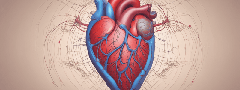
Cardiovascular System: Heart Structure and Function
- The cardiovascular system consists of the heart and vascular system
- The heart is a pump that supplies oxygen and nutrients to the body and removes carbon dioxide and waste products
Heart Chambers and Septum
- The heart has two atria and two ventricles separated by septa
- The interatrial septum separates the right and left atria
- The interventricular septum separates the right and left ventricles
- The atrioventricular septum separates the atria from the ventricles
Heart Valves
- Semilunar valves: pulmonary valve (between right ventricle and pulmonary artery) and aortic valve (between left ventricle and aorta)
- Atrioventricular valves: tricuspid valve (between right atrium and ventricle) and bicuspid (mitral) valve (between left atrium and ventricle)
Cardiac Cycle
- The cardiac cycle consists of systole (contraction) and diastole (relaxation)
- The cardiac cycle can be divided into atrial systole, atrial diastole, ventricular systole, and ventricular diastole
- The cardiac cycle is regulated by the autonomic nervous system (ANS)
Heart Sounds and Murmurs
- Heart sounds are heard through auscultation with a stethoscope
- The first heart sound (S1) occurs during ventricular contraction
- The second heart sound (S2) occurs during ventricular relaxation
- Murmurs are abnormal heart sounds indicating turbulent blood flow
Heart Layers and Conduction System
- The heart has three layers: endocardium (innermost), myocardium (middle), and pericardium (outermost)
- The cardiac conduction system regulates the heartbeat
- The sinoatrial (SA) node is the primary pacemaker, located in the right atrium
- The atrioventricular (AV) node is the secondary pacemaker, located in the interatrial septum
- The AV bundle (bundle of His) connects the AV node to the ventricles
- Purkinje fibers stimulate ventricular contraction
Cardiac Output and Blood Pressure
- Cardiac output (CO) is the volume of blood pumped per minute (CO = stroke volume x heart rate)
- Factors influencing stroke volume: preload, contractility, afterload
- Factors influencing heart rate: autonomic nervous system, hormones, exercise, body temperature, potassium, and calcium levels
Blood Pressure Regulation
- Blood pressure is regulated by the autonomic nervous system, kidneys, and baroreceptors
Heart Disorders and Symptoms
- Angina pectoris: chest pain due to coronary artery disease
- Myocardial infarction (MI): heart attack due to complete coronary artery blockage
- Pericarditis: inflammation of the pericardium
- Cardiac arrhythmias: abnormal heart rhythms
- Cyanosis: blue discoloration of the skin and mucous membranes due to increased levels of deoxygenated hemoglobin
- Clubbing: increased soft tissue in the fingers and toes
- Edema: fluid accumulation in the body
- Orthopnea: shortness of breath when lying down
- Syncope: temporary loss of consciousness due to cerebral hypoxia
Diagnostic Tests
- Electrocardiogram (ECG or EKG): measures electrical activity of the heart
- Stress tests: ECGs performed during exercise or pharmacological stress
- Cardiac enzymes: measure cardiac troponin and creatine phosphokinase in the blood
- Chest X-ray: shows heart size and shape
- Nuclear scan: follows radioactive substances through the blood vessels
- Angiography and CT/MR angiography: visualize coronary arteries
- Echocardiogram: uses sound waves to visualize the heart
- Coronary catheterization: uses a contrast medium to visualize coronary arteries
Cardiovascular System
Inflammation of the Heart
- Inflammation of the innermost lining of the heart and valves (endocarditis)
- Most common cause: bacterial infections
- Symptoms: weakness, fever, excessive sweating, general body aches, difficulty breathing, and blood in the urine
- Treatment: addresses the underlying cause
- Inflammation of the muscular layer of the heart (myocarditis)
- Most common cause: viral infections (especially Group B Coxsackie virus)
- Symptoms: unexplained fever, chest pain, dyspnoea, fatigue, and fainting
- Treatment: steroids, bed rest, and a low-sodium diet
- Inflammation of the pericardium (pericarditis)
- Most common cause: complications of viral or bacterial infections
- Symptoms: sharp, stabbing chest pains, fever, fatigue, and orthopnoea
- Treatment: analgesics for pain, and diuretics to remove excess fluids around the heart
Hypertension (HTN)
- Types of HTN:
- Essential or primary or idiopathic
- Secondary
- Malignant (severely high blood pressure, generally >180/120 mmHg, medical emergency)
- Risk factors:
- Non-modifiable: age, sex, family history
- Modifiable: smoking, hypercholesterolaemia, HTN, diabetes, physical inactivity, and overweight/obesity
- Complications:
- Atherosclerosis
- Ischaemic heart disease (IHD)
- Cerebrovascular accidents (CVA)
- Peripheral arterial disease (PAD)
Atherosclerosis
- Complications:
- Embolus (mostly thromboembolism)
- Thrombosis
- Ischaemic heart disease (IHD)
- Cerebrovascular accidents (CVA)
- Peripheral arterial disease (PAD)
- Risk factors:
- Modifiable: smoking, hypercholesterolaemia, HTN, diabetes, physical inactivity, and overweight/obesity
- Non-modifiable: age, sex, family history
Angina Pectoris
- Types:
- Stable angina (exertional angina)
- Causes: atherosclerotic plaque
- Characteristics: blood flow is adequate at rest, compromised during exertion
- Unstable angina (ACS)
- Causes: huge atherosclerotic plaque, or plaque complicated by vasoconstriction, or thrombus formation
- Characteristics: compromised blood flow at rest, leading to chest pain even without exertion
- Variant angina (Prinzmetal’s angina)
- Causes: unexplained vasospasms
- Characteristics: individuals can experience anginal pain at any time, even when sleeping
- Stable angina (exertional angina)
Myocardial Infarction (MI)
- Also known as heart attack
- Causes: complete obstruction of coronary arteries due to atherosclerosis, thrombus, or embolus
- Symptoms: severe squeezing chest pain, SOB, profuse diaphoresis, palpitation, anxiety, dizziness or syncope, nausea, and possibly vomiting
- Treatment: call for help, chewing an aspirin, strong vasodilator, CPR, thrombolytic drugs, angioplasty or coronary artery bypass graft
Cardiomyopathies
- Types:
- Dilated cardiomyopathies
- Characteristics: loss of elasticity of the myocardium, overstretched ventricular muscle
- Hypertrophic cardiomyopathies
- Characteristics: increased size of the ventricular muscle, can be acquired or inherited
- Restrictive cardiomyopathies
- Characteristics: stiff heart muscle, inhibiting both contraction and relaxation
- Dilated cardiomyopathies
Valve Disorders
- Types:
- Stenosis
- Characteristics: difficulty opening the valve, leading to heart failure
- Regurgitation
- Characteristics: faulty closure of the valve, leading to heart failure
- Stenosis
- Consequences:
- Insufficient cardiac output
- Heart failure
Heart Failure
- Definition: inadequacy of heart pumping to maintain circulation of blood
- Main risk factors:
- IHD
- HTN
- Valve disorders
- Cardiomyopathies
- Congenital heart defects
- Venous insufficiency
- Goals of treatment:
- Correct the cause, if possible
- Improve cardiac output
- Reduce peripheral vascular resistance
- Improve quality of life
Peripheral Vascular Disease (PVD)
- Definition: slow and progressive circulation disorder due to narrowing, blockage, or spasms in a blood vessel in the periphery
- Associated with:
- Atherosclerosis
- Diabetes
- Embolism
- Vasculitis
- Vasospasm
- Venous insufficiency
- Fibromuscular dysplasia
- Entrapment
- Goals of treatment:
- Maintain circulation in the periphery
- Slow the progression of atherosclerosis
Congenital Heart Defects (CHD)
- Types:
- Cyanotic
- Acyanotic
- Atrial septal defect (ASD)
- Ventricular septal defect (VSD)
- Patent ductus arteriosus (PDA)
- Valve stenosis
- Consequences:
- Heart failure
Heart Dysrhythmia
- Definition: abnormal heart rhythm/rate
- Types:
- Various, including atrial fibrillation, ventricular fibrillation, etc.
Introduction to Human Body and Disease
- Definition of Anatomy: Study of body structure
- Definition of Physiology: Study of the function of body's organs
- Anatomy and Physiology are interrelated: Anatomy allows for physiological function
Organisation of the Human Body
- Levels of Organisation:
- Organism (human)
- Systems
- Organs
- Tissues
- Cells
- Organelles
- Molecules
- Atoms
Cells and Tissues
- Definition of a cell: Basic structural and functional unit of life
- Human body is composed of trillions of cells
- Over 250 variations of cells, each with a specific function
- Study of cells is called Cytology
- When cells act together to perform a specific function, the next level of organisation is classified as a tissue
- Four basic types of tissues in the body: Epithelial, Connective, Muscle, and Nervous tissue
- Study of tissues is called Histology
Body Organs and Systems
- Definition of an organ: Structure formed by organisation of two or more different tissues that work together to carry out specific functions
- Definition of a system: Organs join to carry out vital functions
- Examples of organs and systems: Respiratory system (nose, nasal cavity, pharynx, larynx, bronchi, lungs)
Homeostasis and Disease
- Definition of Homeostasis: State of normalcy, relative consistency of the body's internal environment
- Definition of Disease: Changes in the steady state internal environment, symptoms of a pathological state
Cell Stimulation and Injury
- Adaptations: Responses to changes in the environment
- Types of adaptations: Atrophy, Hypertrophy, Hyperplasia, Metaplasia, Dysplasia
Agents of Cell Injury
- Chemical agents: Air and environmental pollutants, agricultural and domestic pesticides, cleaning agents, drugs, free radicals
- Physical agents: Abrupt or extreme changes in temperature, mechanics force, electromagnetic radiation
- Infectious agents: Bacteria, Viruses, Parasites
- Nutritional agents: Nutrient deficiency, nutrient excess
- Hypoxaemic and hypoxic (Ischaemic) agents: Compromised perfusion, compromised oxygen carrying capacity
Outcomes of Cell Injury
- Reversible damage: Regeneration, repair
- Irreversible damage: Fibrosis, Necrosis
Necrosis and Apoptosis
- Necrosis: Unplanned cell death, destructive changes in a cell's structure
- Apoptosis: Programmed cell death, cell 'suicide'
Inflammation
- Definition: Acute inflammation is a healthy response that serves to protect the body from something damaging and helps in repair, chronic inflammation is the response to a chronic injury or stimulation
- Causes: Infections, microbial toxins, tissue necrosis, foreign bodies, immune reactions
- Clinical manifestations: Redness, heat, pain, swelling, loss of function
Concepts of Disease
- Pathophysiology: The study of the mechanisms by which disease and illness alter the functioning of the body
- Aetiology: The study of the cause or causes of a disease
- Pathogenesis: Origination and development of an illness or disease
- Clinical manifestations: Presenting characteristics of a disease
- Diagnosis and treatment: Diagnosis is a label for a disease/pathology, treatment is a way to manage or cure a disease
Population Health
- Epidemiology: The study of the incidence and prevalence of a disease
- Nomenclature: Endemic, Epidemic, Pandemic
- Classification of global disease: WHO (World Health Organization)
- Prevention: Primary, secondary, and tertiary prevention
Evidence-Based Practice
- Definition: A way of caring for others that is cognisant of the most current research and knowledge in the health professions
- Characteristics: Goes beyond one piece of research, encompasses totality of what is known through research and knowledge and practice of experts, requires conscientious respect for human health variations
Blood and Haematopoiesis
- Blood represents life as it takes oxygen and nutrients to tissues, takes carbon dioxide and wastes away from tissues, transports hormones, and regulates heat
- Blood circulation is the process of transporting blood to and from body tissues
- A normal human body has 4-6 litres of blood, which is a connective tissue consisting of a non-living matrix (plasma) and living cells (cellular components)
Plasma
- Plasma is the liquid portion of blood, making up about 55% of blood volume
- Plasma components include:
- Nutrients (amino acids, glucose, nucleotides, and lipids)
- Gases (oxygen, carbon dioxide, and nitrogen)
- Electrolytes (sodium, potassium, calcium, magnesium, chloride, bicarbonate, phosphate, and sulphate)
- Non-protein nitrogenous substances (urea and uric acid)
- Lipoproteins (chylomicrons, VLDL, LDL, and HDL)
- Plasma proteins include albumin, globulins, and fibrinogen
Cellular Components of Blood
- Erythrocytes (red blood cells)
- Small, biconcave-shaped cells without a nucleus
- Contain haemoglobin, which binds to oxygen and transports it
- Normal RBC count is about 4-6 million per cubic millimetre of blood
- Average lifespan of an erythrocyte is approximately 120 days
- Leukocytes (white blood cells)
- Categorised into granulocytes (neutrophils, eosinophils, and basophils) and agranulocytes (monocytes and lymphocytes)
- All leukocytes contain a nucleus
- Thrombocytes (platelets)
- Small, irregularly-shaped cells without a nucleus
- Involved in blood clotting
Haematopoiesis
- Formation of blood cells from stem cells
- During foetal development, RBCs are made in the yolk sac, liver, and spleen
- After birth, most blood cells are produced in red bone marrow by stem cells called haemocytoblasts
- Erythropoietin regulates erythropoiesis, production of RBCs, which is produced by the kidneys when oxygen concentrations in the blood get low
Anaemia
- Indicates a reduced oxygen-carrying capacity
- Classified by RBC size and colour
- Microcytic anaemia (small RBCs, e.g. iron-deficiency anaemia and thalassaemia)
- Macrocytic anaemia (large RBCs, e.g. pernicious anaemia and folate-deficiency anaemia)
- Normocytic anaemia (normal-sized RBCs, e.g. aplastic anaemia and anaemia of chronic disease)
Polycythaemia
- An overproduction of RBCs leading to abnormal high RBCs and haemoglobin, and increased blood viscosity
- Types of polycythaemia:
- Primary polycythaemia (polycythaemia vera or rubra vera)
- Secondary polycythaemia (physiological response to hypoxia, e.g. chronic obstructive pulmonary disorder, congestive heart failure, and living at high altitudes)
Endocrine System
Aims and Objectives
- Familiarize with common disorders associated with the endocrine system
- Appreciate common endocrine disorders and recognize them in clinical practice
- Understand the role of laboratory investigations and patient management
Nervous and Endocrine Systems
- Nervous system: rapid action through nerves and nerve impulses
- Endocrine system: slower acting, important for health and disease, uses hormones
Endocrine Glands
- Ductless glands that secrete hormones directly into the circulatory system
- Each gland secretes one or more hormones (steroids, proteins, amino acids, or lipids)
Hypothalamic-Pituitary Axis
- Hypothalamus releases hormones that stimulate or inhibit the pituitary gland
- Pituitary gland releases hormones that stimulate or inhibit other endocrine glands
Classification of Endocrine Disease
- Hormone excess or deficiency
- Altered tissue response
- Non-functioning endocrine tumors
Assessment of Dysfunction
- Measure basal hormone levels in blood and/or urine
- Dynamic tests of endocrine function: stimulation or suppression tests
- Endocrine imaging: general radiology procedures (CT scans, MRI, ultrasound scans)
Common Conditions
- Neurohypophysis: diabetes insipidus
- Pancreatic dysfunction: diabetes mellitus (T1DM, T2DM)
- Adrenal: Cushing's syndrome, hyperaldosteronism, Addison's disease
- Thyroid disease: hypo- and hyperthyroidism
- Parathyroid dysfunction: hyperparathyroidism, hypoparathyroidism
Diabetes Insipidus (DI)
- Features: inability to concentrate urine, polyuria, nocturia, polydipsia
- Types: cranial (central) DI, nephrogenic DI
- Aetiology: pituitary surgery, head trauma, idiopathic, CNS infections, genetic defects
- Investigations: serum sodium, urine osmolality, 24-hour urine collection
Diabetes Mellitus (DM)
- Clinical presentation: asymptomatic, acute, subacute
- Symptoms of hyperglycaemia: polyuria, polydipsia, polyphagia, weight loss/gain, fatigue, blurred vision, tingling/numbness
- Management: patient education, dietary/lifestyle modifications, oral/injectable anti-diabetic medications, insulin
Adrenal Gland
- Cortex: produces glucocorticoids, mineralocorticoids, and androgens
- Medulla: produces catecholamines (adrenaline, noradrenaline, dopamine)
- Functions: regulation of blood pressure, electrolyte balance, stress response
Thyroid Hormones and Tests
- Thyroid hormones: thyroxine (T4), triiodothyronine (T3), calcitonin
- Thyroid function tests: TSH, free T4, free T3, thyroid autoantibodies
Hypothyroidism
- Insufficient production of thyroid hormones
- Causes: Hashimoto's thyroiditis, iodine deficiency, postpartum thyroiditis, congenital hypothyroidism
- Clinical manifestations: bradycardia, constipation, loss of appetite, lethargy, dry skin, brittle hair
- Investigations: TSH, free T4, thyroid autoantibodies
- Management: levothyroxine (T4) replacement therapy
Respiratory System
Components and Functions
- The respiratory system consists of the lungs, airways, and breathing muscles
- The system's function is to move oxygen and carbon dioxide into and out of the body
External Respiration
- Gas exchange between the alveoli and the lungs' capillaries
- External respiration follows the rule of simple diffusion
- Oxygen moves from the lungs to the blood, and carbon dioxide moves from the blood to the lungs
Internal/Cellular Respiration
- Exchange of gases in the tissues
- Internal respiration also follows the rule of simple diffusion
- Oxygen moves from the capillaries to the tissues, and carbon dioxide moves from the tissues to the capillaries
Respiratory Control Centre
- Located in the brainstem
- Regulates breathing rate, rhythm, and depth by assessing PaO2 and PaCO2 via multiple receptors
- Receptors are located in the brain, blood vessels, lungs, and muscles
Breathing Mechanism
- Nerve impulses travel on phrenic nerves to muscle fibres in the diaphragm, contracting them
- The diaphragm moves downward, expanding the thoracic cavity
- Atmospheric pressure forces air into the respiratory tract
- The lungs fill with air
- External intercostal muscles, SCM, and scalenes contract, raising the ribs and expanding the thoracic cavity further
Inspiration and Expiration
- Inspiration: diaphragm contracts, thoracic cavity expands, and air enters the lungs
- Expiration: diaphragm relaxes, thoracic cavity decreases, and air is squeezed out of the lungs
Factors Influencing Breathing
- Carbon dioxide level rises → increasing rate and depth of respiration
- pH drops → increasing rate and depth of respiration
- Fear and pain → increasing breathing rate (generally shallow breaths)
- Hyperventilation → decreasing amount of carbon dioxide in the blood, leading to a decrease in rate and depth of respiration
Respiratory Diseases
Signs and Symptoms
- Eupnoea: normal breathing rate (12-20 breaths per minute for adults)
- Tachypnoea: fast breathing rate
- Bradypnoea: slow breathing rate
- Apnoea: a total absence of any effective respiratory rate for greater than 20 seconds
- Cough: a common presenting respiratory symptom
- Cyanosis: decreased oxygen saturation
Causes of Respiratory Diseases
- Acute cough: inhaled foreign body, respiratory tract infection
- Chronic cough: COPD, pneumonia, lung cancer, pulmonary embolism, TB
- Wheezes: asthma, COPD, foreign body aspiration, laryngeal spasm or irritation, bronchial CA, allergic reactions, and bronchitis
- Crackles: fluid in the small airways or atelectasis, pulmonary oedema, lung fibrosis, and bronchiectasis
- Rhonchi: blockage of the main airways by mucous, lesions, or foreign bodies
- Stridor: foreign body obstruction of the larger airways (trachea or main bronchi)
Diagnostic Tests
- Not examinable (CT scan, MRI, PET scan, Bronchoscopy, US)
Air Volumes and Lung Capacities
- Not examinable (ventilation/perfusion ratio, common causes for its mismatch, spirometry findings in obstructive and restrictive lung diseases)
Respiratory System
Inflammation and Swelling of the Mucous Membrane of the Nose
- Causes: seasonal (hay fever) or continual (dust, moulds, colognes, cigarette smoke, animal dander, and mites)
- Symptoms: sneezing, itchy, watery eyes, red, swollen eyelids, congested nasal mucus membranes, and nasal discharge
- Management: avoiding known allergens, using air filters and air conditioners, nasal corticosteroids/antihistamines sprays, oral antihistamines and decongestants, and desensitization injections
Acute or Chronic Inflammation of the Membranes Lining the Sinuses
- Causes: viral (common after a common cold) or bacterial
- Symptoms: headache, facial pain, tooth pain, nasal congestion, fever, postnasal discharge, and pharyngitis and cough
- Management: nasal corticosteroids/decongestants sprays, analgesics for pain, antibiotics for infection, and sinus lavage/endoscopic sinus surgery
Common Cold
- Causes: viral (streptococci, adenovirus, rhinovirus, influenza A or B, coronavirus)
- Symptoms: sore throat, cough, fever, nasal congestion, rhinitis, and headache
- Management: rest, fluids, nasal saline drops/wash, antipyretics, analgesics, decongestants, and antitussives
Influenza
- Causes: influenza virus
- Symptoms: headache, fever or chills, dry/productive cough, malaise, myalgia, fatigue, anorexia, rhinorrhoea, pharyngitis, and possibly diarrhea
- Management: analgesics and antipyretics, and antiviral medications
Inflammation of the Larynx and Vocal Cords
- Causes: viral or bacterial infections, non-infectious causes (laryngeal polyps, excessive talking, shouting, or singing, allergic, smoking, GORD, damage to nerves supplying the larynx)
- Symptoms: dysphonia and tickling sensations in the throat, dry cough, and pharyngitis
- Management: voice rest, management of GORD, avoidance of cigarettes and alcohol, antibiotics, and surgical removal of laryngeal polyps
Acute Inflammation of the Bronchi
- Causes: generally follows a URTI, non-infectious causes (GORD, exposure to cigarette smoke, pollutants, and household cleaners)
- Symptoms: malaise, myalgia, fever/chills, productive cough, and dyspnoea
- Management: rest, fluids, medications, and antibiotics (if needed)
Chronic Obstructive Pulmonary Disease (COPD)
- Causes: continuous bronchial irritation (smoking)
- Symptoms: chronic productive cough, dyspnoea, tachypnoea, cyanosis, hepatomegaly, ascites, heart failure, peripheral oedema, cor pulmonale, and pulmonary hypertension
- Management: smoking cessation, prevention of RTIs, medications, supplemental O2, and antibiotics (for secondary bacterial infections)
Asthma
- Causes: allergic, non-allergic, and occupational
- Symptoms: wheeze, SOB, cough, chest tightness
- Management: avoiding aggravating factors, medications (inhalers or oral), and first aid for asthma (4 puffs of blue reliever, wait for 4 minutes and repeat the process if needed)
Pneumonia
- Causes: bacterial, viral, or fungal
- Symptoms: fever, productive cough, SOB, and pleuritic chest pain
- Classification: by causative microorganism, pattern of lung involvement, and setting in which it was acquired
Pleurisy
- Causes: primary pleural disease or secondary to a systemic illness
- Symptoms: sharp and localized chest, thoracic back, or shoulder pain exacerbated by respiratory movements, coughing, or sneezing
Pneumothorax
- Causes: primary (spontaneous) or secondary (traumatic or iatrogenic)
- Symptoms: sudden onset of chest pain, dyspnoea, and tachypnoea
Pleural Effusion
- Causes: transudative (congestive heart failure, liver or kidney failure) or exudative (infection, cancers)
- Symptoms: dyspnoea, cough, chest pain, and fatigue
Mesothelioma
- Causes: asbestos exposure
- Symptoms: dyspnoea, cough, chest pain, haemoptysis, fatigue, and weight loss
- Management: surgery, chemotherapy, and/or radiation therapy
Environmental/Occupational Dust-Related Lung Diseases
- Causes: coal dusts, asbestos, silica sand
- Symptoms: consistent with a restrictive lung disorder pattern
Acute Respiratory Distress Syndrome (ARDS)
- Causes: cardiogenic or non-cardiogenic
- Symptoms: dyspnoea, tachypnoea, tachycardia, pink frothy sputum, cyanosis, and crackles on auscultation
Pulmonary Embolism
- Causes: thrombotic, non-thrombotic (fat embolism, tumour fragments, foreign bodies, amniotic fluid, air bubbles)
- Symptoms: sudden dyspnoea, tachypnoea, tachycardia, fever, diaphoresis, chest pain, and haemoptysis
Lung Cancer
- Causes: cigarette smoking
- Symptoms: cough that worsens over time, haemoptysis, dyspnoea, chest pain, unexplained weight loss, fatigue, and pleural effusion
Immune System Function and Components
- The immune system protects the body against bacteria, viruses, fungi, parasites, and cancerous cells.
- It works in conjunction with the lymphatic system, which includes the thymus, lymph nodes, lymphoid tissue, lymphatic vessels, and spleen.
Cellular and Humoral Immunity
- Cellular immunity involves direct cellular immune attacks by leukocytes such as lymphocytes, monocytes, eosinophils, and basophils.
- Humoral immunity involves the production of five classes of antibodies (immunoglobulins) by B-lymphocytes, including IgA, IgD, IgE, IgG, and IgM.
Leukocytes and their Functions
- Granulocytes: neutrophils facilitate phagocytosis, important for bacterial infections; eosinophils modulate allergic reactions, important for parasitic infections.
- Agranulocytes: lymphocytes (B-cells and T-cells) differentiate into plasma cells, which secrete antibodies, and attack microorganisms, foreign cells, and cancer cells; monocytes (become macrophages) facilitate phagocytosis and immune responses.
Antibodies and their Functions
- IgA: found in body secretions, involved in local immunity in mucous membranes.
- IgD: binds to B-cells, acts as an antigen receptor, low levels in serum.
- IgE: binds to basophils and mast cells, involved in allergic reactions, least common in serum.
- IgM: involved in primary immune response, activates complement, binds to B-cells as an antigen receptor.
- IgG: binds to immune cells, enhances antigen recognition, most common in serum, crosses the placenta, activates complement.
Body Response to Injury or Infection
- Cardinal signs: erythema, heat, pain, swelling, and loss of function.
- Blood vessels in the injured area constrict and then dilate, becoming more permeable.
- Neutrophils and monocytes leave the blood vessels to fight infection and clean up the area.
Immunodeficiency Disorders
- Characterized by impairments of the immune system, affecting antibody activity, lymphocyte function, phagocytosis, or a combination of these.
- Can be classified as primary or secondary, and present at birth (congenital) or develop later in life (acquired).
- Examples: T-cell disorders (e.g. DiGeorge syndrome), B-cell disorders (e.g. Bruton's agammaglobulinemia), and combined T- and B-cell disorders (e.g. severe combined immunodeficiency).
Consequences of Environmental Circumstances
- Severe or prolonged stress can result in adrenal gland hypertrophy, immune suppression, and atrophy of lymphoid tissue.
- Medications can cause immunosuppression, inhibit immune cell proliferation, and alter the balance between normal flora and opportunistic pathogens.
- Poor nutrition can result in decreased proteins, vitamins, and minerals necessary for immunity.
- Infections can disrupt immune attack, and blood cancer can affect the production or maturation of lymphoid and myeloid tissues.
Hypersensitivity Reactions
- Inappropriate excessive immune responses can lead to tissue damage, chronic disability, and death.
- Classified into four types, based on speed of onset, mediation by antibody or direct immune cell attack, type of antibody involved, and activation of complement.
- Symptoms vary depending on the part of the body involved or exposed to allergens.
Autoimmune Disorders
- A loss of tolerance to 'self', resulting in the body attacking its own cells and tissues.
- Examples: myasthenia gravis, mismatched ABO blood transfusion or haemolytic disease of the newborn (HDN), Arthus reaction, serum sickness, contact dermatitis, diabetes mellitus – Type I, chronic graft rejection, and granulomatous disease.
Systemic Lupus Erythematosus, Rheumatoid Arthritis, Infectious Mononucleosis, and Acquired Immunodeficiency Syndrome
- Brief overview of clinical presentation, systems involved, and basics of management for each condition.
Anatomy and Physiology of GI System
- The portal vein (PV) drains blood from the GI tract, gallbladder, pancreas, and spleen to the liver, which is rich in nutrients absorbed from the GI tract.
Phases of Digestion
- Ingestion: the process of taking food into the body
- Secretion: the process of releasing enzymes and other substances to aid in digestion
- Mixing and propulsion: the process of mixing food with digestive enzymes and moving it through the digestive system
- Digestion: the breakdown of food into smaller molecules
- Mechanical: the physical breakdown of food into smaller pieces
- Chemical: the breakdown of food into smaller molecules using enzymes and other substances
- Absorption: the process of absorbing nutrients from the digestive system into the bloodstream
- Defecation: the process of eliminating waste from the body
Nutrition
- Macronutrients: carbohydrates, proteins, and lipids
- Carbohydrates: 45-65% of daily caloric intake
- Proteins: 10-35% of daily caloric intake
- Lipids: 20-35% of daily caloric intake
- Micronutrients: vitamins and minerals
- Vitamin A, B12, D, E, and K are stored in the liver
- Iron, copper, and glycogen are stored in the liver
- Recommended daily intake (RDI) is determined by age, gender, activity level, current weight, and pregnancy/lactation
Malnutrition
- Deficiencies, excesses, or imbalances in a person's intake of energy and/or nutrients
- Causes: inadequate intake, excessive nutrient losses, malabsorption syndromes, genetic defects, and immune-mediated adverse reactions to foods
- Signs and symptoms: weight loss or gain, muscle wasting, fatigue, delayed wound healing, recurrent infections, diarrhea, abdominal pain, changes in skin and mucous membranes
GI Disorders
- Gastroduodenal: PUD, gastritis, malignancies, gastric volvulus
- Intestinal: appendicitis, obstruction, diverticulitis, gastroenteritis, mesenteric adenitis, strangulated hernia, IBD, IBS, intussusception, volvulus
- Hepatobiliary: acute/chronic cholecystitis, cholangitis, hepatitis
- Pancreatic: acute/chronic pancreatitis, malignancy
- Splenic: rupture, infarction
Signs and Symptoms
- Heartburn and regurgitation
- Dysphagia
- Diarrhea
- Constipation
- Bleeding (haematemesis, melaena, or haematochezia)
- Jaundice
- Pruritus
- Skin involvement: dermatitis herpetiformis, acanthosis nigricans, porphyria cutanea tarda, generalised pigmentation, brown-black lesions of the lips
Laboratory Investigations
- Alanine aminotransferase (ALT): specific to liver disease
- Aspartate aminotransferase (AST): rises in hepatic necrosis, MI, muscle injury, and CHF
- Gamma-glutamyl transpeptidase (GGT): rises in obstruction and with meds and alcohol
- Alkaline phosphatase (ALP): rises in liver, bone, and intestine diseases
- Serum bilirubin: rises in hepatocyte impairment/destruction/overload
- Prothrombin time (PT) - INR: increases in liver damage
- Protein (serum albumin): a falling serum albumin is a bad prognostic sign
Coeliac Disease
- Caused by gluten malabsorption in individuals with a genetic predisposition
- Symptoms: fatigue, weight loss, diarrhea, flatulence, borborygmus, abdominal bloating and pain, steatorrhea, and malodorous stools
- Diagnosis: based on clinical manifestations, laboratory studies (serum detection of antibodies against gliadin and tissue transglutaminase), and small bowel biopsy
Obesity
- Defined as excessive body fat (BMI ≥30 kg/m2)
- Types: hypertrophic (increased cell size) and hyperplasic (increased cell number, mainly in kids and adolescents)
- Complex pathophysiology involving lifestyle, genetics, neurologic mechanisms, and hormones (leptin, estrogen, thyroid hormone, insulin, melanocortin, ghrelin)
- Risk factors: lifestyle, genetics, and neurologic mechanisms
- Complications: diabetes, CVD, hypertension, hyperlipidaemia, CVA and stroke, osteoarthritis, liver disease, gallstones, poor wound healing, and certain cancers
Gastritis
- Caused by bacteria, viruses, medications, alcohol, caffeine, spicy foods, excessive eating, poisons, and stress
- Symptoms: nausea, lack of appetite, heartburn, vomiting, and abdominal cramps
- Treatment: avoid irritants, medication to reduce stomach acid production
Gastroesophageal Reflux Disease (GORD)
- Characterized by transient relaxations of the lower oesophageal sphincter
- Risk factors: hiatus hernia
- Symptoms: heartburn, regurgitation, and dysphagia
- Management: investigation (upper GI endoscopy and biopsy), lifestyle changes (stop smoking, small meals, weight loss, elevate head and chest when lying down), medication (PPI/H2 blockers, H.pylori eradication treatment)
Peptic Ulcer Disease (PUD)
- Ulceration in the upper GI tract
- Types: gastric ulcer and duodenal ulcer
- Symptoms: epigastric pain (worse with food or relieved by food), bloating and fullness with food, and pain radiating to the back
- Investigation: H.pylori testing (urea breath test, serology, faecal antigen test), upper GI endoscopy
- Management: same as GORD
Inflammatory Bowel Disease (IBD)
- Includes ulcerative colitis and Crohn's disease
- Unknown aetiology, but involves genetic susceptibility, environment, intestinal microbiota, and host immune response
- Relapsing and remitting diseases
- High incidence rates in Northern Europe, UK, and North America
- Complications: toxic megacolon, haemorrhage, fistula (Crohn's only), and cancer (UC predominantly)
Irritable Bowel Syndrome (IBS)
- Functional GI disorder
- Characterized by abdominal signs and symptoms with no structural cause
- Triggers: affective disorders, psychological stress, gastrointestinal infection, antibiotic therapy, and sexual, physical, verbal abuse
- Features: abdominal pain/discomfort relieved by defecation, alternating constipation and diarrhoea, bloating, tenesmus, urgency, and the presence of mucus in faeces
Diarrhea
- Causes: bacterial, viral, or parasitic infections, toxins, food allergies, Crohn's disease, laxative use, antibiotics, chemotherapy, and radiation therapy
- Symptoms: frequent passage of faeces, abdominal cramps, and dehydration
- Treatment: clear fluids, antidiarrheal medications, and antibiotics (if indicated)
Constipation
- Risk factors: lack of physical activity, inadequate fibre and water in the diet, medications, and certain medical conditions
- Symptoms: infrequent bowel movements, hard faeces, abdominal pain, haemorrhoids, and pain during bowel movements
- Treatment: increase dietary fibre, adequate fluid intake, regular exercise, and stool softeners, laxatives, and enemas (if indicated)
Acute Appendicitis
- Pathophysiology: obstruction of the lumen, increased intraluminal pressure, venous outflow obstruction, and ischemia of the appendiceal wall
- Clinical presentation: acute onset of anorexia, nausea, vomiting, fever, and abdominal pain (initially umbilical, then shifts to the right iliac fossa)
- Risk factors: low-fibre, high-sugar diet, IBD, and family history
- Complications: perforation and peritonitis
- Management: immediate referral to ED, appendectomy
Colorectal Cancer (CRC)
- Fourth most commonly diagnosed cancer in Australia
- Incidence increases with age (over 50)
- Non-invasive screening test available for Australians who turn 50
- Colonoscopy is both a screening and diagnostic procedure
- Risk factors: age, diet, smoking, obesity, IBD, colorectal polyps, family history, and frequent pelvic/abdominal radiology
- Prevention: fruits and vegetables, calcium, vitamin D, folic acid, exercise
Hepatitis
- Liver inflammation associated with hepatocyte damage
- Causes: alcohol and viruses
- Symptoms: mild fever, nausea, vomiting, abdominal pain, jaundice, hepatomegaly, bloating, lack of appetite, weakness, pruritus, dark urine
- Complications: chronic carrier state, increased risk of further liver disease, cirrhosis, and liver cancer
Alcoholic Liver Disease
- Cells utilize ethanol by converting it to acetaldehyde and then to acetate
- Triglyceride and fatty acids accumulate in the liver, causing fatty liver
- Fatty liver: liver is slightly enlarged and has a pale yellow appearance
Here are the study notes for the provided text:
Urinary System
Aims and Objectives
- Familiarize with common disorders associated with the renal and reproductive system
- Provide opportunity to appreciate common genitourinary disorders and recognize them in clinical practice
Introduction
- Organs of the urinary system: kidneys, ureters, urinary bladder, and urethra
- Functions of the urinary system:
- Excretion of metabolic breakdown products (e.g. urea, creatinine, and uric acid)
- Regulation of fluid and electrolyte balance
- Regulation of acid-base homeostasis
- Regulation of blood pressure
- Vitamin D metabolism
- Induces production of red blood cells
Nephrons
- Functional units of the kidneys
- Consist of:
- Bowman's capsule
- Tubules (proximal convoluted tubule, Loop of Henle, distal convoluted tubule)
- Collecting duct
Glomerular Filtration
- Takes place in the renal corpuscles
- Glomerular filtrate formed from fluid part of blood
- Depends on three pressures:
- Glomerular blood hydrostatic pressure
- Blood pressure in glomerular capillaries
- Capsular hydrostatic pressure
- Glomerular blood colloid-osmotic pressure
Stages of Urine Formation
- Glomerular filtration
- Tubular reabsorption
- Tubular secretion
Urine Analysis
- Macroscopic (visual inspection)
- Colour
- Clarity
- Odour
- Biochemical (e.g. urine dipstick)
- pH
- Specific gravity
- Protein
- Glucose
- Ketones
- Nitrite
- Leukocyte esterase
- Microscopic
- Crystals
- Casts
- Epithelial cells
- White and red blood cells
- Bacteria
Haematuria
- Gross haematuria (visible blood in urine)
- Microscopic haematuria (not visible to inspection, but detected by dipstick or microscopy)
- Causes:
- Glomerular
- Non-glomerular
- Stones
- UTI
- Trauma
- Tumour
- Coagulopathies
- Gynaecological
- Iatrogenic
Proteinuria
- Transient proteinuria
- After vigorous exercise
- In patients with fever
- Heart failure
- Patients with UTI
- Persistent proteinuria
- Associated with progression of kidney disease
- Atherosclerosis
- Heart problems
Acute Renal Failure (ARF)
- Sudden loss of kidney function
- Types:
- Prerenal (due to decreased renal blood flow)
- Intrarenal (due to diseases of the nephrons themselves)
- Postrenal (due to obstruction of urine flow)
Chronic Renal Failure (CRF)
- Also known as end-stage renal disease (ESRD)
- Long-term progressive and permanent loss of nephrons
- More than 80% loss of nephron function
- No possibility of nephron recovery
Glomerulonephritis (GN)
- Inflammation of glomeruli
- Classified based on histopathological appearances
- Spectrums of glomerular diseases:
- Nephritic syndrome
- Nephrotic syndrome
UTI
- Urinary tract infection
- Spectrum:
- Asymptomatic bacteriuria
- Symptomatic acute urethritis and/or prostitis/cystitis
- Acute pyelonephritis
- Septicaemia
- Organisms:
- E. coli
- Proteus mirabilis
- Klebsiella aerogenes
- Staphylococcus saprophyticus
- Staphylococcus epidermidis
Reproductive System
Spermatogenesis
- Process of sperm formation
- Takes place in the seminiferous tubules of the testes
Female Reproductive System
- Oogenesis
- Menstrual cycle
Investigations
- Hormone levels
- Semen analysis
- Prostate specific antigen (PSA)
- Breast examination
- Cervical screening test
STIs
- Chlamydial infection
- Genital herpes
- Gonorrhoea
- Anogenital wart (HPV)
- AIDS
- Syphilis
Chlamydial Infection
- Caused by Chlamydia trachomatis
- Most common STD in the world
- Asymptomatic in both men and women
- Diagnostic factors:
- Asymptomatic
- Dysuria
- Penile/vaginal discharge
- Lower abdominal/Pelvic pain
- Post-coital or intermenstrual bleeding
- Management:
- Eradicate infection
- Follow up on sexual contacts
- Antibiotic therapy
- Treatment failures are uncommon
- Reinfection is common
Genital Herpes
- Caused by Herpes simplex virus (HSV-1 or HSV-2)
- Transmission: vaginal, anal, oro-genital, or oro-anal
- Virus remains latent in the body
- Diagnostic factors:
- Fever
- Malaise
- Headache
- Tingling/itching
- Painful genital/oral ulcers
- Inguinal lymphadenopathy
- Dysuria
- Meningitis/encephalitis
Anogenital Warts (HPV)
- Genotypes: HPV-6, HPV-11, HPV-16, HPV-18
- Skin to skin transmission
- Diagnostic factors:
- Lesion size ranges from few millimeters to several centimeters
- Exophytic and Single OR multiple and papular/flat
- Number and size increase during pregnancy
- Rarely can be transmitted to mouth during oral sex
Benign Prostatic Hyperplasia (BPH)
- Benign enlargement of the prostate gland
- Unknown aetiology
- Hyperplasia of the glandular and connective tissues
- Enlargement stretches, compresses, and distorts the urethra
- Diagnostic factors:
- Age (> 50 yrs)
- Storage symptoms
- Voiding symptoms
- PSA
- Management:
- Mild to moderate symptoms
- Watchful waiting
- Medications
- Acute urinary retention
- Urgent Catheterisation
- Surgery
- Transurethral resection of prostate (TURP)
- Holmium laser enucleation
- Open prostatectomy### Chlamydia trachomatis and PID
- Mild to moderate symptoms
- Chlamydia trachomatis is the predominant sexually transmitted organism associated with Pelvic Inflammatory Disease (PID).
- The high-risk patient for PID is a menstruating woman younger than 25 years who has multiple sex partners, does not use protection, and lives in an area with a high prevalence of STIs.
Clinical Presentation of PID
- PID may have very mild symptoms or toxic symptoms.
- Symptoms of PID include:
- Fever
- Nausea
- Vomiting
- Severe pelvic pain
- Lower abdominal pain
- Abnormal vaginal discharge
- Unanticipated vaginal bleeding, often postcoital
Complications of PID
- Chronic pelvic pain
- Infertility
- Ectopic pregnancy
Self-Study Topics
Gonorrhoea
- Cause
- Signs and Symptoms
Syphilis
- Cause
- Stages
- Stage-specific Signs and Symptoms
Fibrocystic Breast Changes
- No specific details provided
PMS and PMDD
- Signs and Symptoms
PCOS
- Clinical presentation
- Basics of management
The Skin
- The integumentary system consists of the skin and its accessory organs: hair follicles, arrector pili muscles, nails, sweat glands, and oil glands.
Functions of the Skin
- Protection: first line of defense against bacteria, viruses, and protects underlying structures from UV radiation and dehydration.
- Body temperature regulation: dermal blood vessels dilate or constrict to regulate body temperature.
- Vitamin D production: necessary for calcium absorption.
- Sensory perception: touch, pain, pressure, and temperature.
- Excretion: small amounts of waste products are lost through perspiration.
Skin Structure
- Epidermis: consists of stratum basale, stratum spinosum, stratum granulosum, stratum lucidum, and stratum corneum, from innermost to outermost layer.
- Dermis: contains sudoriferous glands, sebaceous glands, hair follicles, arrector pili muscles, collagen, elastic fibers, nerve fibers, and blood vessels.
- Subcutaneous layer (hypodermis): composed of adipose tissue and loose connective tissue, with functions of energy storage, cushioning, and insulation.
Techniques of Examination
- Inspection and palpation: observe skin characteristics, including color, moisture, mobility, temperature, texture, and lesions.
- Note the characteristics of lesions, including anatomic location, distribution, patterns, shapes, type, and color.
Skin Terminology
- Lesions: types include macules, patches, papules, plaques, nodules, cysts, vesicles, bullae, pustules, urticaria, and eczema.
- Macules: flat, nonpalpable color changes, such as erythema, pallor, cyanosis, yellowing, and hyperpigmentation.
Skin Lesions
- Early detection of skin lesions is crucial, especially for skin cancers.
- Risk factors for skin cancer: sun exposure, genetic predisposition, carcinogen exposure, smoking, and immunosuppression.
Skin Cancers
- Malignant melanoma: malignant tumors of melanocytes that can occur at any age, spreading rapidly and deadly if left untreated.
- Squamous cell carcinoma (SCC): a type of skin cancer that requires early detection and treatment.
The Dipstick Test
- Glucosuria (glycosuria) is not normally present in the urine and should always be followed up, caused by elevated blood glucose levels (e.g. diabetes mellitus) or reduced tubular reabsorption (e.g. acute tubular necrosis).
Bilirubinuria
- Indicates hepatic or biliary disease, may be the first sign in an anicteric (not jaundiced yet) patient.
Ketonuria
- Ketones are the breakdown products of fatty acid metabolism, including acetone, acetoacetic acid, and B-hydroxybutyric acid.
- Ketones in the urine are abnormal, caused by starvation or uncontrolled diabetes, requiring urgent action to prevent coma.
Specific Gravity
- A quick test of kidneys' concentrating power, aiding interpretation of other results.
- If increased, it may indicate dehydration, glucosuria, or SIADH.
- If decreased, it may indicate damage to kidney tubules, kidney failure, diabetes insipidus, overhydration, or severe kidney infection (pyelonephritis).
Haematuria
- Rule out food/Meds-related red-coloured urine, and causes include those listed in the haematuria DDx in the lecture slide.
- False positives may occur due to menstruation or after vigorous exercise.
Proteinuria
- Normally, urine may contain small amounts of proteins (< 150 mg/day), but this is undetectable using a dipstick.
- The dipstick only detects the presence of albumin and no other proteins.
- A positive test needs confirmation, with possible causes including glomerulonephritis, renal tubular disease, hypertension, diabetes, pre-eclampsia, or UTI.
pH
- Urine is normally slightly acidic (normal range: 4.5-8).
- High urine pH may indicate kidney tubular disease, kidney failure, urinary tract infection, vomiting, or stale urine.
- Low urine pH may indicate fasting, starvation, after meals, diabetic ketoacidosis, diarrhoea, or metabolic acidosis.
Urobilinogen
- Normally present, and if elevated, it may indicate haemolysis, possible liver abnormalities, but not reliable on its own.
Nitrites
- Not normally present in the urine, but produced when some bacteria convert dietary nitrates to nitrites.
- A positive result indicates UTI, but a negative result does not rule out a UTI.
Leucocytes (Pyuria)
- May indicate infections of the urinary tract, but dipstick only detects leukocyte esterase, and false-negative results are common.
- Could be due to contamination during collecting the sample.
Anatomy and Physiology of the GI System
- The portal vein (PV) drains blood from the gastrointestinal tract, gallbladder, pancreas, and spleen to the liver.
- The PV carries blood rich in nutrients absorbed from the GI tract.
Phases of Digestion
- Cephalic phase: stimulated by thought, sight, or smell of food, resulting in salivary secretion and acid secretion in the stomach.
- Gastric phase: increased gastric secretions and peristalsis, emptying the stomach.
- Intestinal phase: slows down gastric emptying, allowing the small intestine to absorb nutrients.
Nutrient Ingestion and Utilization
- Ingestion: taking in food and fluids.
- Absorption: nutrients absorbed into the bloodstream.
- Defecation: waste elimination.
- Macronutrients: carbohydrates, proteins, and lipids.
- Micronutrients: vitamins and minerals.
Malnutrition
- Inadequate nutrient intake, excessive nutrient losses, genetic defects, and malabsorption syndromes can lead to malnutrition.
- Deficiencies, excesses, or imbalances in energy and nutrient intake can cause malnutrition.
Signs and Symptoms of GI Disorders
- Gastrointestinal symptoms: heartburn, regurgitation, dysphagia, diarrhea, bleeding, and abdominal pain.
- Systemic symptoms: weight loss, fatigue, weakness, and delayed wound healing.
Liver Functions and Diseases
- Liver functions: detoxification, metabolism, and synthesis of proteins and nutrients.
- Liver diseases: hepatitis, cirrhosis, and liver failure.
Laboratory Investigations
- Liver function tests (LFTs): assess liver damage or disease.
- Tests for viral hepatitis, autoimmune hepatitis, and primary biliary cirrhosis.
- Imaging studies: ultrasound, CT scan, MRI, and endoscopy.
Upper and Lower GI Bleeding
- Upper GI bleeding: peptic ulcer disease, gastritis, and esophagitis.
- Lower GI bleeding: diverticulosis, angiodysplasia, and colorectal cancer.
Note: These notes cover the main topics and key points from the provided text, focusing on the anatomy and physiology of the GI system, phases of digestion, nutrient ingestion and utilization, malnutrition, signs and symptoms of GI disorders, liver functions and diseases, laboratory investigations, and upper and lower GI bleeding.
Cephalic Phase
- The thought, sight, or smell of food, as well as the taste of it, triggers the cephalic phase
- Facial and glossopharyngeal nerves stimulate saliva secretion during this phase
- The vagus nerve stimulates acid secretion in the stomach
Gastric Phase
- Increased gastric secretions occur during the gastric phase
- Peristalsis and emptying of the stomach are also increased during this phase
Intestinal Phase
- Gastric emptying is slowed down to allow the small intestine time to absorb nutrients
- Acid secretion is inhibited during the intestinal phase
Terry's Nail and Associated Conditions
- Terry's nail is a type of leukonychia, characterized by a white opacity involving the entire nail plate.
- Chronic liver disease is a common underlying condition associated with Terry's nail.
- Clubbing, a condition characterized by enlargement of the tip of the fingers, is also linked to Terry's nail.
- Cirrhosis, inflammatory bowel disease (IBD), and Coeliac disease are other gastrointestinal conditions that may be associated with Terry's nail.
- Palmar erythema, a condition marked by redness of the palms, is often seen in individuals with chronic liver disease.
- Pallor on palmar creases is a characteristic feature of iron deficiency anemia (IDA).
- Dupuytren's contracture, a condition affecting the hands, is commonly seen in individuals with alcoholism, which is also a risk factor for chronic liver disease.
- The presence of Terry's nail may be an indicator of underlying systemic diseases, emphasizing the importance of thorough medical evaluation.
Causes of Jaundice
- Hyperbilirubinaemia is a condition that leads to jaundice, characterized by elevated levels of bilirubin in the blood
- Anaemia can cause jaundice due to the increased breakdown of red blood cells, leading to excess bilirubin production
Ocular Manifestations
- Kayser-Fleischer rings are a characteristic sign of Wilson disease, which can cause jaundice
- Iritis is an inflammation of the iris that can cause jaundice
- Xanthelasmata are yellowish patches on the eyelids, often associated with elevated serum cholesterol and cholestasis
Systemic Associations
- Inflammatory bowel disease (IBD) can cause jaundice due to malabsorption and inflammation
- Amyloidosis is a condition characterized by abnormal protein accumulation, which can lead to jaundice
Cutaneous Manifestations
- Periorbital purpura is a sign of bleeding under the skin around the eyes, which can be associated with jaundice
Abdominal Distension
- Causes of generalised abdominal distension include fat, fluid (ascites), foetus, flatus, faeces, and big tumour (e.g. ovarian tumour or hydatid cyst)
- Iron, folate, and vitamin B12 deficiencies can lead to glossitis and angular stomatitis
- Gynaecomastia can also contribute to abdominal distension
Abdominal Examination
- Prominent abdominal veins may be visible, such as Caput Medusae, although this is very rare
- Severe portal hypertension may be indicated by a venous hum heard over the liver
- Pulsation in the abdomen could be normal or may indicate an abdominal aortic aneurysm (AAA)
Symptoms of Acute Liver Disease
- General symptoms include malaise, anorexia, and fever, especially in viral cases
- Jaundice develops as the illness progresses
Symptoms of Chronic Liver Disease
- Asymptomatic or nonspecific symptoms, such as fatigue
- Right hypochondria pain
- Abdominal distension due to liver distension and ascites
- Ankle swelling (oedema)
- GI bleeding, characterized by haematemesis and melaena, particularly variceal bleeding
- Jaundice and pruritus
- Endocrine disorders, including gynaecomastia, loss of libido, and amenorrhoea
- Neuropsychiatric complications, such as confusion and drowsiness, indicative of hepatic encephalopathy
Types of Jaundice
- Pre-hepatic jaundice:
- Characterized by increased bilirubin production (∪unconjugated)
- Caused by haemolysis and dyserythropoiesis
- Normal urine and stool color, indicating the liver is still conjugating bilirubin as normal
Hepatic Jaundice
- Hepatic jaundice:
- Characterized by liver dysfunction, resulting in no conjugation of bilirubin by the liver
- Increased levels of AST, ALT, and ALP enzymes
- Pale stool, normal urine color
Post-hepatic (Cholestatic) Jaundice
- Post-hepatic jaundice:
- Characterized by bile duct obstruction (∪conjugated)
- Increased levels of ALP and GGT enzymes
- Pale stool, dark urine
COPD Diagnosis
- Lung function tests are used to investigate COPD, which reveal a pattern of obstruction
Imaging Studies
- Chest X-ray (CXR) and Computed Tomography (CT) scan are used to assess lung hyperinflation
Laboratory Tests
- Blood gases are measured to assess the severity of COPD
COPD Management
- Smoking cessation is a crucial step in managing COPD
Pharmacological Management
- Bronchodilators are used to relieve COPD symptoms, including:
- Short-acting β2-agonists
- Long-acting β2-agonists
- Anticholinergics are used to manage COPD symptoms
- Corticosteroids are used to reduce inflammation in COPD
- Antibiotics are used to treat bacterial infections that can exacerbate COPD
Oxygen Therapy
- Oxygen therapy is used to manage COPD, particularly in advanced stages
Pneumonia Definition
- Inflammation and infection of the lung parenchyma
Aetiology
- Caused by bacteria, viruses, and fungi
- Approximately 50% of pneumonia cases are pneumococcal
Presentation
- Acute illness with symptoms including:
- Fever
- Productive cough
- Shortness of breath (SOB)
- Pleuritic chest pain
- Myalgia
- Headache
- May present with subtle symptoms, especially in the elderly
Classification
- By causative microorganism:
- Viral
- Bacterial
- Atypical
- By pattern of lung involvement:
- Lobar pneumonia
- Bronchopneumonia
- By setting in which it was acquired:
- Community-acquired
- Hospital-acquired
Digestive System
- Cephalic phase:
- Stimulated by thought, sight, or smell of food, and taste of food
- Facial and glossopharyngeal nerves stimulate saliva secretion
- Vagus nerve stimulates acid secretion in the stomach
- Gastric phase:
- Increased gastric secretions and peristalsis, emptying of the stomach
- Intestinal phase:
- Slows down gastric emptying to allow small intestine time to absorb nutrients
- Inhibits more acid secretion
Cardiovascular System
- Heart structure:
- Two atria, two ventricles, separated by septa
- Right atrium receives blood from the body, left atrium receives oxygenated blood from lungs
- Right ventricle pumps blood to lungs, left ventricle pumps blood to the body
- Heart valves:
- Semilunar valves (pulmonary and aortic) between ventricles and arteries
- Atrioventricular valves (tricuspid and bicuspid) between atria and ventricles
- Heart sounds:
- First heart sound (S1): closure of tricuspid and bicuspid valves
- Second heart sound (S2): closure of pulmonary and aortic semilunar valves
- Heart layers:
- Endocardium: innermost layer, thin and lined with endothelium
- Myocardium: middle layer, cardiac muscle, pumping action of the heart
- Pericardium: outermost layer, fibrous membrane surrounding the heart
- Cardiac cycle:
- Atrial contraction (atrial systole), atrial relaxation (atrial diastole)
- Ventricular contraction (ventricular systole), ventricular relaxation (ventricular diastole)
- Electrical conduction system:
- SA node: primary pacemaker, located in the right atrium
- AV node: secondary pacemaker, located in the interatrial septum
- AV bundle (bundle of His) and Purkinje fibers: electrical impulses to ventricles
- Cardiac output (CO) = Stroke Volume (SV) x Heart Rate (HR)
Cardiac Disorders
- Pericarditis: inflammation of the pericardium, characterized by sharp and localized chest pain
- Angina pectoris: caused by narrowing or spasm of coronary arteries, pain in the chest
- Stable angina: pain occurs with physical activity, relieved by rest
- Unstable angina: pain occurs at rest, lasts longer
- Myocardial infarction (MI): complete blockage of one or more coronary arteries, pain in the chest
- Clubbing: increase in soft tissue of the distal part of the fingers or toes, associated with heart disease
- Cyanosis: blue discoloration of the skin and mucous membranes, indicates increased deoxygenated hemoglobin
- Central: cardiac causes, right-to-left cardiac shunt
- Peripheral: exposure to cold, low cardiac output, venous stasis, vasoconstriction
- Oedema: swelling, often bilateral and pitting, in the legs and feet, associated with cardiac failure
- Heart sounds:
- Third heart sound (S3): low-pitched early diastolic sound, indicates reduced ventricular compliance
- Fourth heart sound (S4): low-pitched late diastolic sound, indicates forceful atrial contraction
- Abnormal heart sounds: graded from 1 to 6, associated with turbulent blood flow
- Syncope: transient loss of consciousness due to cerebral hypoxia, often due to cardiac causes.### Cardiovascular System
Heart Rate and Rhythm
- Heart rate can be abnormally fast (tachy) or slow (brady)
- Rhythm can be irregular
- Strength can be weak or narrow
- Delay can be observed in radio-femoral or radio-radial delay
Blood Pressure
- BP is affected by cardiac output, peripheral vascular resistance, elasticity of arterial walls, and volume of blood
- Mechanisms regulating blood pressure include baroreceptors in the brain and arteries, and the kidneys (renin-angiotensin system)
- Hypertension can be essential (primary), secondary, or malignant
Electrocardiogram (ECG)
- Represents the electrical activity of the heart
- Correlates with the contraction and relaxation of the heart
- Components of an ECG include:
- P wave: represents depolarization of the atria
- QRS complex: represents ventricular depolarization
- T wave: represents repolarization of the ventricles
- P-R interval: time from the beginning of the P wave to the beginning of the QRS complex
- S-T segment: time from the end of the S wave to the beginning of the T wave
- Q-T interval: time from the beginning of the QRS complex to the end of the T wave
Diagnosis and Management
- Stress tests: ECGs performed during exercise or with medication to increase heart rate
- Cardiac enzymes: measure of cardiac troponin (cT) and Creatine phosphokinase (CK-MB) in the blood
- Chest X-ray: shows the size and shape of the heart
- Nuclear scan: follows radioactive substances through the blood vessels of the heart
- Angiography and CT/MR angiography: visualize the arteries
- Echocardiogram: uses sound waves to visualize the heart
- Coronary catheterization: uses a contrast medium to diagnose heart conditions
Heart Failure
- Goals: correct the cause, improve cardiac output, reduce peripheral vascular resistance, and improve quality of life
- Types: left heart failure (systolic and diastolic) and right heart failure
Peripheral Arterial Disease (PAD)
- A slow and progressive circulation disorder due to narrowing, blockage, or spasms in a blood vessel
- Associated with atherosclerosis, diabetes, embolism, vasculitis, vasospasm, and venous insufficiency
- Goals: maintain circulation and slow the progression of atherosclerosis
- Non-pharmacological measures: cessation of smoking, exercise, and dependent position for the legs
- Medications: anticoagulants, thrombolytics, and vasodilators
- Surgical management: endarterectomy, graft, or bypass
Arrhythmia
- Aka dysrhythmia, an abnormal heart rhythm or rate
- Majority of dysrhythmias are short-lived
- Describe the common and deadly ones
Cardiomyopathies
- Three categories: dilated, hypertrophic, and restrictive
- Dilated cardiomyopathies: loss of elasticity of the myocardium and an overstretched, flaccid ventricular muscle
- Hypertrophic cardiomyopathies: increased size of the ventricular muscle
- Restrictive cardiomyopathies: stiff heart muscle, inhibiting contraction and relaxation
Angina Pectoris
- Stable angina: atherosclerotic plaque, compromised blood flow at rest, and pain relieved by rest
- Unstable angina: huge atherosclerotic plaque, thrombus, or vasospasm, and pain that occurs at rest
- Variant angina: unexplained vasospasms, pain that occurs at any time
Myocardial Infarction (MI)
- Cardiac muscle sustains permanent damage due to severe ischaemia
- Caused by complete obstruction of coronary arteries due to atherosclerosis, thrombus, or embolus
- Symptoms: severe squeezing chest pain, shortness of breath, profuse sweating, palpitation, and anxiety
Valve Disorders
- Valve stenosis: difficulty opening the valve, and insufficient stroke volume
- Valve regurgitation: faulty closure of the valve, and volume overload
- Consequence of valve disorders: heart failure
Congenital Heart Defects
- Two categories: cyanotic and acyanotic
- Acyanotic defects: ASD, VSD, PDA, and valve stenosis
- ASD: hole in the atrial septum, volume overload, and hypertrophy of the right atrium and ventricle
- VSD: hole in the ventricular septum, volume overload, and hypertrophy of the right ventricle, left atrium, and left ventricle
- PDA: patent ductus arteriosus, volume overload, and hypertrophy of both left atrium and ventricle### Introduction to the Human Body and Disease
- The study of the human body is essential to understand disease and its effects on the body.
- Anatomy is the study of the structure of the body, while physiology is the study of the functions of the body.
- Understanding the organization of the body, from the chemical level to the organism, is crucial to recognize how disease affects the body.
Organization of the Human Body
- The human body is composed of:
- Organism (human)
- Systems (e.g., respiratory, circulatory)
- Organs (e.g., lungs, heart)
- Tissues (e.g., epithelial, connective, muscle, nervous)
- Cells
- Organelles
- Molecules
- Atoms
- Each level of organization is essential for the proper functioning of the body.
Cells and Tissues
- A cell is the basic structural and functional unit of life.
- The human body is composed of trillions of cells, with over 250 variations.
- Each type of cell has a specific function, and cells work together to form tissues.
- The four basic types of tissues are:
- Epithelial
- Connective
- Muscle
- Nervous
- Cytology is the study of cells, and histology is the study of tissues.
Body Organs and Systems
- An organ is a structure formed by the organization of two or more different tissues that work together to perform a specific function.
- A system is a group of organs that work together to perform a vital function.
- Examples of systems include the respiratory, circulatory, and nervous systems.
Homeostasis and Disease
- Homeostasis is the state of normalcy, where the body's internal environment is maintained within a narrow range.
- Disease is a change in the steady state internal environment, resulting in symptoms and pathological changes.
- Disease can be caused by various factors, including genetic, environmental, and lifestyle factors.
Cell Stimulation and Injury
- Cell stimulation can lead to adaptations, such as:
- Atrophy (decrease in cell size)
- Hypertrophy (increase in cell size)
- Hyperplasia (increase in cell number)
- Metaplasia (change in cell type)
- Dysplasia (abnormal cell growth)
- Cell injury can be caused by various agents, including:
- Chemical agents (e.g., toxins, pollutants)
- Physical agents (e.g., temperature, radiation)
- Infectious agents (e.g., bacteria, viruses)
- Nutritional agents (e.g., deficiency, excess)
Outcomes of Cell Injury
- Reversible cell injury can lead to:
- Regeneration (complete restoration of tissue)
- Repair (restoration of tissue architecture and function)
- Irreversible cell injury can lead to:
- Fibrosis (scarring)
- Necrosis (unplanned cell death)
- Apoptosis (programmed cell death)
Inflammation
- Acute inflammation is a healthy response to tissue damage, characterized by:
- Redness
- Heat
- Pain
- Swelling
- Loss of function
- Chronic inflammation is a prolonged response to tissue damage, linked to tissue damage and disease.
Concepts of Disease
- Disease is a change in the steady state internal environment, often used broadly to include injuries, illnesses, disorders, and syndromes.
- Pathophysiology is the study of the mechanisms by which disease and illness alter the functioning of the body.
Determinants of Health and Illness
- Aetiology is the study of the cause or causes of a disease.
- Pathogenesis is the origination and development of an illness or disease.
- Clinical manifestations are the presenting characteristics of a disease, including signs, symptoms, and diagnostic criteria.
Diagnosis and Treatment
- Diagnosis is the process of identifying a disease or pathology, based on diagnostic criteria.
- Treatment involves allopathic and conventional medicine, as well as complementary and alternative medicine.
- Prognosis is the likely outcome of a disease, predicting how one will proceed through the disease process.
Population Health
- Epidemiology is the study of the incidence and prevalence of diseases in a population.
- Nominal data is used to describe the characteristics of a population, including morbidity and mortality rates.
Pneumothorax
- Collection of air between the visceral and parietal pleura due to a breach of either the visceral or parietal pleura.
Aetiology of Pneumothorax
- Primary pneumothorax:
- Occurs without an apparent cause or significant lung disease
- Most often happens spontaneously
- Risk factors:
- Smoking
- Male sex
- Tall thin body
- Pregnancy
- Family history of pneumothorax (familial pneumothorax)
- Marfan syndrome
Secondary Pneumothorax
- Occurs in the presence of existing lung pathology:
- COPD
- Asthma
- Tuberculosis
- Sarcoidosis
- Cystic fibrosis (CF)
- Lung cancers
- Severe ARDS
Iatrogenic Pneumothorax
- Caused by medical interventions:
- Pleural or lung biopsy
- Tracheostomy
- Intercostal nerve block
- CPAP ventilation
Traumatic Pneumothorax
- Caused by:
- Penetrating or blunt trauma
- Rib fracture
- Diving or flying accidents
Pleural Effusion
- Accumulation of fluid in the pleural space between the visceral and parietal pleura
- Causes fluid buildup in the pleural space, leading to lung compression and affecting gas exchange
Aetiology
- Transudative pleural effusion:
- Caused by congestive heart failure
- Caused by liver or kidney failure
- Exudative pleural effusion:
- Caused by infections (e.g. pleuritis, tuberculosis)
- Caused by cancers
Types of Pleural Effusion
- Empyema: effusion containing pus due to an infective process
- Blood effusion: seen in trauma or rupture of blood vessel(s)
Pulmonary Embolism
- A life-threatening condition where a pulmonary artery becomes blocked, preventing blood flow to the lung tissue, leading to hypoxia and tissue damage or even death in severe cases.
Causes of Pulmonary Embolism
- Thrombotic causes:
- Deep Vein Thrombosis (DVT): a blood clot dislodged from a vein in the legs (thromboembolism)
- Factors that increase coagulation: • Dehydration • Coagulopathies • Oral Contraceptive Pills (OCPs) • Pregnancy • Cancers
- Non-thrombotic causes:
- Fat embolism: e.g. long bone fracture
- Tumor fragments
- Foreign bodies
- Amniotic fluid
- Air bubbles (embolism): e.g. chest trauma
Gastritis Causes
- Bacteria and viruses are potential causes of gastritis
- Certain medications can lead to gastritis
- Consuming excessive amounts of alcohol, caffeine, and spicy foods can cause gastritis
- Overeating can also lead to gastritis
- Poisons and stress can trigger gastritis
Gastritis Symptoms
- Nausea is a common symptom of gastritis
- Lack of appetite is often experienced by individuals with gastritis
- Heartburn is a symptom of gastritis
- Vomiting can occur in gastritis
- Abdominal cramps are a symptom of gastritis
Gastritis Treatment and Prevention
- Avoiding foods or medications that irritate the stomach lining can help prevent or alleviate gastritis
- Medications that reduce the production of stomach acids can be used to treat gastritis
GORD Management
- Investigation involves Upper GI Endoscopy and biopsy to obtain histology and detect H. pylori.
- Lifestyle changes are essential to manage GORD:
- Cessation of smoking
- Eating small, frequent meals
- Losing weight to reduce pressure on the lower esophageal sphincter
- Elevating the head and chest while lying down to prevent acid reflux
- Repairing hiatus hernia if present
- Dietary modifications involve:
- Avoiding foods that aggravate GORD symptoms
- Abstaining from alcohol consumption
- Pharmacological treatment options include:
- Proton pump inhibitors (PPIs) to reduce acid production
- H2 blockers to reduce acid production
- H. pylori eradication treatment if H. pylori infection is detected
Peptic Ulcer Disease
- Ulceration occurs in the upper GI tract.
- There are two main types of peptic ulcer disease:
- Gastric ulcer (GU) and Duodenal ulcer (DU), with DU being 4 times more common than GU.
Signs and Symptoms
- Epigastric pain is a common symptom, although it's less likely to present as chest pain.
- Pain is often worsened by food or relieved by food.
- Symptoms improve with antacids.
- Epigastric tenderness is present on palpation.
- Bloating and fullness are experienced after eating.
- A history of reflux may be present.
- Pain may radiate to the back.
- Haematemesis (vomiting fresh blood or coffee grounds) may occur.
Digestive System
- Cephalic phase:
- Stimulated by thought, sight, or smell of food, and taste of food
- Facial and glossopharyngeal nerves stimulate saliva secretion
- Vagus nerve stimulates acid secretion in the stomach
- Gastric phase:
- Increased gastric secretions and peristalsis, emptying of the stomach
- Intestinal phase:
- Slows down gastric emptying to allow small intestine time to absorb nutrients
- Inhibits more acid secretion
Cardiovascular System
- Heart structure:
- Two atria, two ventricles, separated by septa
- Right atrium receives blood from the body, left atrium receives oxygenated blood from lungs
- Right ventricle pumps blood to lungs, left ventricle pumps blood to the body
- Heart valves:
- Semilunar valves (pulmonary and aortic) between ventricles and arteries
- Atrioventricular valves (tricuspid and bicuspid) between atria and ventricles
- Heart sounds:
- First heart sound (S1): closure of tricuspid and bicuspid valves
- Second heart sound (S2): closure of pulmonary and aortic semilunar valves
- Heart layers:
- Endocardium: innermost layer, thin and lined with endothelium
- Myocardium: middle layer, cardiac muscle, pumping action of the heart
- Pericardium: outermost layer, fibrous membrane surrounding the heart
- Cardiac cycle:
- Atrial contraction (atrial systole), atrial relaxation (atrial diastole)
- Ventricular contraction (ventricular systole), ventricular relaxation (ventricular diastole)
- Electrical conduction system:
- SA node: primary pacemaker, located in the right atrium
- AV node: secondary pacemaker, located in the interatrial septum
- AV bundle (bundle of His) and Purkinje fibers: electrical impulses to ventricles
- Cardiac output (CO) = Stroke Volume (SV) x Heart Rate (HR)
Cardiac Disorders
- Pericarditis: inflammation of the pericardium, characterized by sharp and localized chest pain
- Angina pectoris: caused by narrowing or spasm of coronary arteries, pain in the chest
- Stable angina: pain occurs with physical activity, relieved by rest
- Unstable angina: pain occurs at rest, lasts longer
- Myocardial infarction (MI): complete blockage of one or more coronary arteries, pain in the chest
- Clubbing: increase in soft tissue of the distal part of the fingers or toes, associated with heart disease
- Cyanosis: blue discoloration of the skin and mucous membranes, indicates increased deoxygenated hemoglobin
- Central: cardiac causes, right-to-left cardiac shunt
- Peripheral: exposure to cold, low cardiac output, venous stasis, vasoconstriction
- Oedema: swelling, often bilateral and pitting, in the legs and feet, associated with cardiac failure
- Heart sounds:
- Third heart sound (S3): low-pitched early diastolic sound, indicates reduced ventricular compliance
- Fourth heart sound (S4): low-pitched late diastolic sound, indicates forceful atrial contraction
- Abnormal heart sounds: graded from 1 to 6, associated with turbulent blood flow
- Syncope: transient loss of consciousness due to cerebral hypoxia, often due to cardiac causes.### Cardiovascular System
Heart Rate and Rhythm
- Heart rate can be abnormally fast (tachy) or slow (brady)
- Rhythm can be irregular
- Strength can be weak or narrow
- Delay can be observed in radio-femoral or radio-radial delay
Blood Pressure
- BP is affected by cardiac output, peripheral vascular resistance, elasticity of arterial walls, and volume of blood
- Mechanisms regulating blood pressure include baroreceptors in the brain and arteries, and the kidneys (renin-angiotensin system)
- Hypertension can be essential (primary), secondary, or malignant
Electrocardiogram (ECG)
- Represents the electrical activity of the heart
- Correlates with the contraction and relaxation of the heart
- Components of an ECG include:
- P wave: represents depolarization of the atria
- QRS complex: represents ventricular depolarization
- T wave: represents repolarization of the ventricles
- P-R interval: time from the beginning of the P wave to the beginning of the QRS complex
- S-T segment: time from the end of the S wave to the beginning of the T wave
- Q-T interval: time from the beginning of the QRS complex to the end of the T wave
Diagnosis and Management
- Stress tests: ECGs performed during exercise or with medication to increase heart rate
- Cardiac enzymes: measure of cardiac troponin (cT) and Creatine phosphokinase (CK-MB) in the blood
- Chest X-ray: shows the size and shape of the heart
- Nuclear scan: follows radioactive substances through the blood vessels of the heart
- Angiography and CT/MR angiography: visualize the arteries
- Echocardiogram: uses sound waves to visualize the heart
- Coronary catheterization: uses a contrast medium to diagnose heart conditions
Heart Failure
- Goals: correct the cause, improve cardiac output, reduce peripheral vascular resistance, and improve quality of life
- Types: left heart failure (systolic and diastolic) and right heart failure
Peripheral Arterial Disease (PAD)
- A slow and progressive circulation disorder due to narrowing, blockage, or spasms in a blood vessel
- Associated with atherosclerosis, diabetes, embolism, vasculitis, vasospasm, and venous insufficiency
- Goals: maintain circulation and slow the progression of atherosclerosis
- Non-pharmacological measures: cessation of smoking, exercise, and dependent position for the legs
- Medications: anticoagulants, thrombolytics, and vasodilators
- Surgical management: endarterectomy, graft, or bypass
Arrhythmia
- Aka dysrhythmia, an abnormal heart rhythm or rate
- Majority of dysrhythmias are short-lived
- Describe the common and deadly ones
Cardiomyopathies
- Three categories: dilated, hypertrophic, and restrictive
- Dilated cardiomyopathies: loss of elasticity of the myocardium and an overstretched, flaccid ventricular muscle
- Hypertrophic cardiomyopathies: increased size of the ventricular muscle
- Restrictive cardiomyopathies: stiff heart muscle, inhibiting contraction and relaxation
Angina Pectoris
- Stable angina: atherosclerotic plaque, compromised blood flow at rest, and pain relieved by rest
- Unstable angina: huge atherosclerotic plaque, thrombus, or vasospasm, and pain that occurs at rest
- Variant angina: unexplained vasospasms, pain that occurs at any time
Myocardial Infarction (MI)
- Cardiac muscle sustains permanent damage due to severe ischaemia
- Caused by complete obstruction of coronary arteries due to atherosclerosis, thrombus, or embolus
- Symptoms: severe squeezing chest pain, shortness of breath, profuse sweating, palpitation, and anxiety
Valve Disorders
- Valve stenosis: difficulty opening the valve, and insufficient stroke volume
- Valve regurgitation: faulty closure of the valve, and volume overload
- Consequence of valve disorders: heart failure
Congenital Heart Defects
- Two categories: cyanotic and acyanotic
- Acyanotic defects: ASD, VSD, PDA, and valve stenosis
- ASD: hole in the atrial septum, volume overload, and hypertrophy of the right atrium and ventricle
- VSD: hole in the ventricular septum, volume overload, and hypertrophy of the right ventricle, left atrium, and left ventricle
- PDA: patent ductus arteriosus, volume overload, and hypertrophy of both left atrium and ventricle### Introduction to the Human Body and Disease
- The study of the human body is essential to understand disease and its effects on the body.
- Anatomy is the study of the structure of the body, while physiology is the study of the functions of the body.
- Understanding the organization of the body, from the chemical level to the organism, is crucial to recognize how disease affects the body.
Organization of the Human Body
- The human body is composed of:
- Organism (human)
- Systems (e.g., respiratory, circulatory)
- Organs (e.g., lungs, heart)
- Tissues (e.g., epithelial, connective, muscle, nervous)
- Cells
- Organelles
- Molecules
- Atoms
- Each level of organization is essential for the proper functioning of the body.
Cells and Tissues
- A cell is the basic structural and functional unit of life.
- The human body is composed of trillions of cells, with over 250 variations.
- Each type of cell has a specific function, and cells work together to form tissues.
- The four basic types of tissues are:
- Epithelial
- Connective
- Muscle
- Nervous
- Cytology is the study of cells, and histology is the study of tissues.
Body Organs and Systems
- An organ is a structure formed by the organization of two or more different tissues that work together to perform a specific function.
- A system is a group of organs that work together to perform a vital function.
- Examples of systems include the respiratory, circulatory, and nervous systems.
Homeostasis and Disease
- Homeostasis is the state of normalcy, where the body's internal environment is maintained within a narrow range.
- Disease is a change in the steady state internal environment, resulting in symptoms and pathological changes.
- Disease can be caused by various factors, including genetic, environmental, and lifestyle factors.
Cell Stimulation and Injury
- Cell stimulation can lead to adaptations, such as:
- Atrophy (decrease in cell size)
- Hypertrophy (increase in cell size)
- Hyperplasia (increase in cell number)
- Metaplasia (change in cell type)
- Dysplasia (abnormal cell growth)
- Cell injury can be caused by various agents, including:
- Chemical agents (e.g., toxins, pollutants)
- Physical agents (e.g., temperature, radiation)
- Infectious agents (e.g., bacteria, viruses)
- Nutritional agents (e.g., deficiency, excess)
Outcomes of Cell Injury
- Reversible cell injury can lead to:
- Regeneration (complete restoration of tissue)
- Repair (restoration of tissue architecture and function)
- Irreversible cell injury can lead to:
- Fibrosis (scarring)
- Necrosis (unplanned cell death)
- Apoptosis (programmed cell death)
Inflammation
- Acute inflammation is a healthy response to tissue damage, characterized by:
- Redness
- Heat
- Pain
- Swelling
- Loss of function
- Chronic inflammation is a prolonged response to tissue damage, linked to tissue damage and disease.
Concepts of Disease
- Disease is a change in the steady state internal environment, often used broadly to include injuries, illnesses, disorders, and syndromes.
- Pathophysiology is the study of the mechanisms by which disease and illness alter the functioning of the body.
Determinants of Health and Illness
- Aetiology is the study of the cause or causes of a disease.
- Pathogenesis is the origination and development of an illness or disease.
- Clinical manifestations are the presenting characteristics of a disease, including signs, symptoms, and diagnostic criteria.
Diagnosis and Treatment
- Diagnosis is the process of identifying a disease or pathology, based on diagnostic criteria.
- Treatment involves allopathic and conventional medicine, as well as complementary and alternative medicine.
- Prognosis is the likely outcome of a disease, predicting how one will proceed through the disease process.
Population Health
- Epidemiology is the study of the incidence and prevalence of diseases in a population.
- Nominal data is used to describe the characteristics of a population, including morbidity and mortality rates.
Investigation of Peptic Ulcer Disease
- Urea Breath Test (UBT): used to diagnose H. pylori infection
- Serology (blood): another method to diagnose H. pylori infection
- Faecal Ag test: a test to detect H. pylori antigens in stool
- Upper GI Endoscopy: used to visualize the upper GI tract and collect biopsy samples for further examination
- Rapid urease test: a test performed on biopsy samples to detect H. pylori infection
Management of Peptic Ulcer Disease
- Same as GORD (Gastroesophageal Reflux Disease): implies that the management of peptic ulcer disease is similar to that of GORD
Peptic Ulcer Disease
- Risk factors for peptic ulcer disease include: • Helicobacter pylori (H. pylori) infection • Use of Non-Steroidal Anti-Inflammatory Drugs (NSAIDs) and oral corticosteroids • Smoking • Alcohol consumption • Family history (FHx) of the disease
Complications
- Possible complications of peptic ulcer disease: • Perforation of the stomach lining • Bleeding ulcers • Gastric outlet obstruction
Inflammatory Bowel Disease (IBD)
- IBD encompasses two primary conditions: ulcerative colitis (UC) and Crohn's disease.
- The exact cause of IBD is unknown, but it is believed to involve an interaction between: • Genetic susceptibility • Environmental factors • Intestinal microbiota • Host immune response
- IBD is characterized as a relapsing and remitting disease, meaning that symptoms can fluctuate between periods of activity and dormancy.
- IBD has a high incidence rate and prevalence in specific regions, including: • Northern Europe • The UK • North America
IBS Triggers
- Affective disorders, such as depression and anxiety, can trigger IBS symptoms
- Psychological stress and trauma, including past experiences of abuse, can contribute to IBS development
- Gastrointestinal infections can trigger IBS in some individuals
- Antibiotic therapy, which can disrupt gut microbiota, can lead to IBS
- Past experiences of sexual, physical, or verbal abuse can contribute to IBS development
- Pelvic surgery can trigger IBS symptoms in some individuals
- Eating disorders, such as anorexia nervosa and bulimia nervosa, can contribute to IBS development
IBD Features
- Abdominal pain/discomfort primarily in the central or lower abdomen, which subsides after defecation
- Bloating and tenesmus, accompanied by a sense of urgency
- Characterized by alternating episodes of constipation and diarrhea
- Presence of mucus in stool, but no bleeding
- Absence of constitutional signs and symptoms
- Symptoms worsen due to stress and menstruation
Hepatitis
- Liver inflammation associated with hepatocyte damage, occurring in acute or chronic settings
- Two primary causes: alcohol and viruses
- Viral hepatitis is a significant global health issue, resulting in millions of deaths annually
Symptoms
- Mild fever
- Nausea
- Vomiting
- Abdominal pain
- Jaundice
- Hepatomegaly
- Bloating
- Lack of appetite
- Weakness
- Pruritus
- Dark urine
Complications
Chronic Carrier State
- Existence of a chronic carrier state
Further Liver Disease
- ✖ Risk of further liver disease
Advanced Stages
- Cirrhosis
- Liver cancer
Cephalic Phase
- The cephalic phase stimulates saliva secretion through the facial and glossopharyngeal nerves and acid secretion in the stomach through the vagus nerve.
Cardiovascular System
- The cardiovascular system consists of the heart (a pump) and vascular system (a collection of pipes).
Heart Structure
- The heart has four chambers: two atria (right and left) and two ventricles (right and left).
- The atria are separated by the interatrial septum, and the ventricles are separated by the interventricular septum.
- The atrioventricular septum separates the atria from the ventricles.
Valves
- Semilunar valves: pulmonary (between the right ventricle and pulmonary arteries) and aortic (between the left ventricle and aorta).
- Atrioventricular valves: bicuspid (mitral) and tricuspid.
Heart Sounds
- Heart sounds are caused by the closure of valves.
- First heart sound (S1): closure of tricuspid and bicuspid valves during ventricular contraction.
- Second heart sound (S2): closure of pulmonary and aortic semilunar valves during ventricular relaxation.
Heart Layers
- Innermost layer: endocardium (very thin, lined with endothelium).
- Middle layer: myocardium (cardiac muscle, responsible for pumping action).
- Outermost layer: pericardium (cushions the heart in case of blunt trauma).
Cardiac Cycle
- Atrial systole and diastole occur simultaneously, followed by ventricular systole and diastole.
- During atrial systole, tricuspid and bicuspid valves open, allowing blood to enter the ventricles.
- During ventricular systole, tricuspid and bicuspid valves close, and aortic and pulmonary valves open, allowing blood to leave the heart.
Cardiac Conduction System
- Regulated by the autonomic nervous system (ANS).
- Consists of the sinoatrial (SA) node, atrioventricular (AV) node, atrioventricular bundle, and Purkinje fibers.
- SA node is the heart's primary pacemaker, setting the heart rate.
Cardiac Output
- Calculated by multiplying stroke volume (SV) by heart rate (HR): CO = SV x HR.
- Factors influencing SV: preload, contractility, and afterload.
- Factors influencing HR: ANS regulation, hormones, exercise, body temperature, and changes in blood pressure.
Abnormalities and Diagnostic Tests
- Pericarditis: inflammation of the pericardium, characterized by sharp, localized chest pain.
- Angina pectoris: caused by narrowing or spasm of coronary arteries, characterized by chest pain that may radiate to the back, neck, or arms.
- Myocardial infarction (MI) or heart attack: caused by complete blockage of one or more coronary arteries, characterized by pressure or fullness in the chest that may radiate to the back, neck, or arms.
- Clubbing: increase in soft tissue of the distal part of the fingers or toes, often seen in cyanotic congenital heart disease, infective endocarditis, and aneurysms.
- Cyanosis: blue discoloration of the skin and mucous membranes, indicating increased levels of deoxygenated hemoglobin in blood vessels.### Cardiovascular System
- The cardiovascular system is associated with reduced cardiac output and poor blood supply to the body.
- Blood pressure (BP) is affected by:
- Cardiac output
- Peripheral vascular resistance
- Elasticity of arterial walls
- Volume of blood
Hypertension
- Essential, idiopathic, or primary hypertension: no known cause (>90% of cases)
- Secondary hypertension: cause(s) identified (e.g. narrowing of arteries, kidney disease, endocrine disorders)
Electrocardiogram (ECG)
- Represents the electrical activity of the heart
- Correlates to the contraction and relaxation of the heart
- Components:
- P wave: atrial depolarization (contraction)
- QRS complex: ventricular depolarization (contraction)
- T wave: ventricular repolarization (relaxation)
- Intervals:
- P-R interval: from P wave to QRS complex
- S-T segment: from S wave to T wave
- Q-T interval: from QRS complex to T wave
Diagnostic Tests
- Stress tests: ECGs performed during exercise or with medication to increase heart rate
- Cardiac enzymes: measure enzymes released into the blood when heart muscle is damaged (e.g. cardiac troponin, creatine phosphokinase)
- Chest X-ray: shows the size and shape of the heart
- Nuclear scan: follows radioactive substances through the blood vessels to reveal narrow or obstructed arteries
- Angiography and CT/MR angiography: visualize the arteries
- Echocardiogram: uses sound waves to visualize the heart and its movement
Heart Failure
- Goals:
- Correct the cause (if possible)
- Improve cardiac output
- Reduce peripheral vascular resistance
- Improve quality of life
- Compensation mechanisms:
- Increased heart rate
- Increased contraction force
- Increased blood volume
- Increased peripheral vascular resistance
Peripheral Arterial Disease
- Atherosclerosis: narrowing or blockage of blood vessels in the periphery
- Causes:
- Atherosclerosis
- Diabetes
- Embolism
- Vasculitis
- Vasospasm
- Venous insufficiency
- Fibromuscular dysplasia
- Entrapment
- Symptoms:
- Ischaemic pain in legs and hands (claudication)
- Tissue necrosis
- Claudication at rest (warning sign)
Atherosclerosis
- Complications:
- Embolus
- Thrombosis
- Risk factors:
- Non-modifiable: age, sex, family history
- Modifiable: smoking, hypercholesterolaemia, hypertension, diabetes, physical inactivity, overweight/obesity
Angina Pectoris
- Stable angina: chest pain during exercise, relieved by rest
- Unstable angina: chest pain at rest, with or without exertion
- Variant angina: chest pain due to vasospasms
Myocardial Infarction (MI)
- Cardiac muscle damage due to severe ischaemia
- Causes:
- Atherosclerosis
- Thrombus
- Embolus
- Symptoms:
- Severe squeezing chest pain
- Shortness of breath
- Profuse sweating
- Palpitation
- Anxiety
- Dizziness or syncope
- Nausea and vomiting
Cardiomyopathies
- Three categories:
- Dilated: loss of elasticity, overstretched ventricular muscle
- Hypertrophic: increased size of ventricular muscle
- Restrictive: stiff heart muscle, inhibiting contraction and relaxation
Valve Disorders
- Stenosis: valve narrowing
- Regurgitation: valve leakage
- Consequences:
- Difficulty opening the valve
- Insufficient cardiac output
- Heart failure
Congenital Heart Defects
- Two categories:
- Cyanotic: oxygen-poor blood
- Acyanotic: oxygen-rich blood
- Examples:
- Atrial septal defect (ASD)
- Ventricular septal defect (VSD)
- Patent ductus arteriosus (PDA)
- Valve stenosis
Heart Dysrhythmias
- Abnormal heart rhythm/rate
- Types:
- Atrial fibrillation
- Ventricular fibrillation
- Tachycardia
- Bradyarrhythmia
- Long QT syndrome
- Wolff-Parkinson-White syndrome### Introduction to the Human Body and Disease
- Learning outcomes: define anatomy and physiology, explain their interconnectivity, classify the organisation of the body, and identify anatomical terminology.
Anatomy and Physiology
- Anatomy: the study of body structure and normal position of body structures.
- Physiology: the study of the function of body organs.
Organisation of the Human Body
- The human body is composed of:
- Organism (human)
- Systems
- Organs
- Tissues
- Cells
- Organelles
- Molecule
- Atom
Cells and Tissues
- A cell is the basic structural and functional unit of life.
- The human body is composed of trillions of cells with over 250 variations, each with a specific function.
- The study of cells is called cytology.
- When cells act together to perform a specific function, the next level of organisation is classified as a tissue.
- There are four basic types of tissues in the body: epithelial, connective, muscle, and nervous tissue.
- The study of tissues is called histology.
Body Organs and Systems
- An organ is a structure formed by the organisation of two or more different tissues that work together to carry out specific functions.
- A system is a group of organs that work together to carry out vital functions.
- Example: the respiratory system consists of the nose, nasal cavity, pharynx, larynx, bronchi, and lungs.
Homeostasis and Disease
- Homeostasis: a state of normalcy or relative consistency of the body's internal environment.
- Disease: changes in the steady state internal environment; symptoms of a pathological state.
Cell Stimulation and Injury
- Adaptations: changes in cell size or number in response to changes in demand.
- Atrophy: decrease in cell size due to reduced demands.
- Hypertrophy: increase in cell size due to increased demands.
- Hyperplasia: increase in cell number.
- Metaplasia: change in cell type.
- Dysplasia: abnormal cell growth or development.
Agents of Cell Injury
- Chemical agents: air and environmental pollutants, agricultural and domestic pesticides, drugs, and free radicals.
- Physical agents: abrupt or extreme changes in temperature, mechanical force, and electromagnetic radiation.
- Infectious agents: bacteria, viruses, and parasites.
- Nutritional agents: nutrient deficiency or excess.
Outcomes of Acute Injuries
- Reversible damage: regeneration and repair of damaged tissue.
- Irreversible damage: fibrosis, necrosis, and apoptosis.
Inflammation
- Acute inflammation: a healthy response to protect the body from damage and aid in repair.
- Chronic inflammation: a response to chronic injury or stimulation, linked to tissue damage.
- Clinical manifestations: redness, heat, pain, swelling, and loss of function.
Concepts of Disease
- Disease: changes in the steady state internal environment.
- Pathophysiology: the study of the mechanisms by which disease and illness alter the functioning of the body.
- Epidemiology: the study of the incidence, distribution, and control of disease.
- Aetiology: the study of the causes of a disease.
- Pathogenesis: the origination and development of a disease.
Clinical Manifestations
- Signs: objective findings evident to someone besides the patient.
- Symptoms: subjective findings obvious only to the patient.
Diagnosis and Treatment
- Diagnosis: a label for a disease/pathology based on diagnostic criteria.
- Prognosis: the likely outcome of a disease.
- Treatment: allopathic approach, conventional medicine, and complementary and alternative medicine.
Population Health
- Epidemiology: the study of the incidence, distribution, and control of disease in a population.
Adrenal Gland Disorders
- Cushing's syndrome: a condition resulting from excessive production of cortisol, leading to weight gain, high blood pressure, and other symptoms.
- Hyperaldosteronism: a condition characterized by excessive production of aldosterone, leading to high blood pressure and potassium depletion, e.g. Conn's syndrome.
Thyroid Disorders
- Hypothyroidism: a condition where the thyroid gland fails to produce sufficient thyroid hormones, leading to fatigue, weight gain, and cold intolerance.
- Hyperthyroidism: a condition where the thyroid gland produces excessive thyroid hormones, leading to weight loss, anxiety, and tremors.
Parathyroid Dysfunction
- Hyperparathyroidism: a condition where the parathyroid glands produce excessive parathyroid hormone, leading to elevated calcium levels and bone resorption.
- Hypoparathyroidism: a condition where the parathyroid glands fail to produce sufficient parathyroid hormone, leading to low calcium levels and muscle cramps.
Adrenal Gland Tumors
- Phaeochromocytoma: a rare tumor of the adrenal gland that produces excessive catecholamines, leading to hypertension and cardiovascular symptoms.
Adrenal Insufficiency
- Addison's disease: a chronic condition where the adrenal glands fail to produce sufficient cortisol and aldosterone, leading to fatigue, weight loss, and electrolyte imbalance.
Cephalic Phase
- The cephalic phase stimulates saliva secretion through the facial and glossopharyngeal nerves and acid secretion in the stomach through the vagus nerve.
Cardiovascular System
- The cardiovascular system consists of the heart (a pump) and vascular system (a collection of pipes).
Heart Structure
- The heart has four chambers: two atria (right and left) and two ventricles (right and left).
- The atria are separated by the interatrial septum, and the ventricles are separated by the interventricular septum.
- The atrioventricular septum separates the atria from the ventricles.
Valves
- Semilunar valves: pulmonary (between the right ventricle and pulmonary arteries) and aortic (between the left ventricle and aorta).
- Atrioventricular valves: bicuspid (mitral) and tricuspid.
Heart Sounds
- Heart sounds are caused by the closure of valves.
- First heart sound (S1): closure of tricuspid and bicuspid valves during ventricular contraction.
- Second heart sound (S2): closure of pulmonary and aortic semilunar valves during ventricular relaxation.
Heart Layers
- Innermost layer: endocardium (very thin, lined with endothelium).
- Middle layer: myocardium (cardiac muscle, responsible for pumping action).
- Outermost layer: pericardium (cushions the heart in case of blunt trauma).
Cardiac Cycle
- Atrial systole and diastole occur simultaneously, followed by ventricular systole and diastole.
- During atrial systole, tricuspid and bicuspid valves open, allowing blood to enter the ventricles.
- During ventricular systole, tricuspid and bicuspid valves close, and aortic and pulmonary valves open, allowing blood to leave the heart.
Cardiac Conduction System
- Regulated by the autonomic nervous system (ANS).
- Consists of the sinoatrial (SA) node, atrioventricular (AV) node, atrioventricular bundle, and Purkinje fibers.
- SA node is the heart's primary pacemaker, setting the heart rate.
Cardiac Output
- Calculated by multiplying stroke volume (SV) by heart rate (HR): CO = SV x HR.
- Factors influencing SV: preload, contractility, and afterload.
- Factors influencing HR: ANS regulation, hormones, exercise, body temperature, and changes in blood pressure.
Abnormalities and Diagnostic Tests
- Pericarditis: inflammation of the pericardium, characterized by sharp, localized chest pain.
- Angina pectoris: caused by narrowing or spasm of coronary arteries, characterized by chest pain that may radiate to the back, neck, or arms.
- Myocardial infarction (MI) or heart attack: caused by complete blockage of one or more coronary arteries, characterized by pressure or fullness in the chest that may radiate to the back, neck, or arms.
- Clubbing: increase in soft tissue of the distal part of the fingers or toes, often seen in cyanotic congenital heart disease, infective endocarditis, and aneurysms.
- Cyanosis: blue discoloration of the skin and mucous membranes, indicating increased levels of deoxygenated hemoglobin in blood vessels.### Cardiovascular System
- The cardiovascular system is associated with reduced cardiac output and poor blood supply to the body.
- Blood pressure (BP) is affected by:
- Cardiac output
- Peripheral vascular resistance
- Elasticity of arterial walls
- Volume of blood
Hypertension
- Essential, idiopathic, or primary hypertension: no known cause (>90% of cases)
- Secondary hypertension: cause(s) identified (e.g. narrowing of arteries, kidney disease, endocrine disorders)
Electrocardiogram (ECG)
- Represents the electrical activity of the heart
- Correlates to the contraction and relaxation of the heart
- Components:
- P wave: atrial depolarization (contraction)
- QRS complex: ventricular depolarization (contraction)
- T wave: ventricular repolarization (relaxation)
- Intervals:
- P-R interval: from P wave to QRS complex
- S-T segment: from S wave to T wave
- Q-T interval: from QRS complex to T wave
Diagnostic Tests
- Stress tests: ECGs performed during exercise or with medication to increase heart rate
- Cardiac enzymes: measure enzymes released into the blood when heart muscle is damaged (e.g. cardiac troponin, creatine phosphokinase)
- Chest X-ray: shows the size and shape of the heart
- Nuclear scan: follows radioactive substances through the blood vessels to reveal narrow or obstructed arteries
- Angiography and CT/MR angiography: visualize the arteries
- Echocardiogram: uses sound waves to visualize the heart and its movement
Heart Failure
- Goals:
- Correct the cause (if possible)
- Improve cardiac output
- Reduce peripheral vascular resistance
- Improve quality of life
- Compensation mechanisms:
- Increased heart rate
- Increased contraction force
- Increased blood volume
- Increased peripheral vascular resistance
Peripheral Arterial Disease
- Atherosclerosis: narrowing or blockage of blood vessels in the periphery
- Causes:
- Atherosclerosis
- Diabetes
- Embolism
- Vasculitis
- Vasospasm
- Venous insufficiency
- Fibromuscular dysplasia
- Entrapment
- Symptoms:
- Ischaemic pain in legs and hands (claudication)
- Tissue necrosis
- Claudication at rest (warning sign)
Atherosclerosis
- Complications:
- Embolus
- Thrombosis
- Risk factors:
- Non-modifiable: age, sex, family history
- Modifiable: smoking, hypercholesterolaemia, hypertension, diabetes, physical inactivity, overweight/obesity
Angina Pectoris
- Stable angina: chest pain during exercise, relieved by rest
- Unstable angina: chest pain at rest, with or without exertion
- Variant angina: chest pain due to vasospasms
Myocardial Infarction (MI)
- Cardiac muscle damage due to severe ischaemia
- Causes:
- Atherosclerosis
- Thrombus
- Embolus
- Symptoms:
- Severe squeezing chest pain
- Shortness of breath
- Profuse sweating
- Palpitation
- Anxiety
- Dizziness or syncope
- Nausea and vomiting
Cardiomyopathies
- Three categories:
- Dilated: loss of elasticity, overstretched ventricular muscle
- Hypertrophic: increased size of ventricular muscle
- Restrictive: stiff heart muscle, inhibiting contraction and relaxation
Valve Disorders
- Stenosis: valve narrowing
- Regurgitation: valve leakage
- Consequences:
- Difficulty opening the valve
- Insufficient cardiac output
- Heart failure
Congenital Heart Defects
- Two categories:
- Cyanotic: oxygen-poor blood
- Acyanotic: oxygen-rich blood
- Examples:
- Atrial septal defect (ASD)
- Ventricular septal defect (VSD)
- Patent ductus arteriosus (PDA)
- Valve stenosis
Heart Dysrhythmias
- Abnormal heart rhythm/rate
- Types:
- Atrial fibrillation
- Ventricular fibrillation
- Tachycardia
- Bradyarrhythmia
- Long QT syndrome
- Wolff-Parkinson-White syndrome### Introduction to the Human Body and Disease
- Learning outcomes: define anatomy and physiology, explain their interconnectivity, classify the organisation of the body, and identify anatomical terminology.
Anatomy and Physiology
- Anatomy: the study of body structure and normal position of body structures.
- Physiology: the study of the function of body organs.
Organisation of the Human Body
- The human body is composed of:
- Organism (human)
- Systems
- Organs
- Tissues
- Cells
- Organelles
- Molecule
- Atom
Cells and Tissues
- A cell is the basic structural and functional unit of life.
- The human body is composed of trillions of cells with over 250 variations, each with a specific function.
- The study of cells is called cytology.
- When cells act together to perform a specific function, the next level of organisation is classified as a tissue.
- There are four basic types of tissues in the body: epithelial, connective, muscle, and nervous tissue.
- The study of tissues is called histology.
Body Organs and Systems
- An organ is a structure formed by the organisation of two or more different tissues that work together to carry out specific functions.
- A system is a group of organs that work together to carry out vital functions.
- Example: the respiratory system consists of the nose, nasal cavity, pharynx, larynx, bronchi, and lungs.
Homeostasis and Disease
- Homeostasis: a state of normalcy or relative consistency of the body's internal environment.
- Disease: changes in the steady state internal environment; symptoms of a pathological state.
Cell Stimulation and Injury
- Adaptations: changes in cell size or number in response to changes in demand.
- Atrophy: decrease in cell size due to reduced demands.
- Hypertrophy: increase in cell size due to increased demands.
- Hyperplasia: increase in cell number.
- Metaplasia: change in cell type.
- Dysplasia: abnormal cell growth or development.
Agents of Cell Injury
- Chemical agents: air and environmental pollutants, agricultural and domestic pesticides, drugs, and free radicals.
- Physical agents: abrupt or extreme changes in temperature, mechanical force, and electromagnetic radiation.
- Infectious agents: bacteria, viruses, and parasites.
- Nutritional agents: nutrient deficiency or excess.
Outcomes of Acute Injuries
- Reversible damage: regeneration and repair of damaged tissue.
- Irreversible damage: fibrosis, necrosis, and apoptosis.
Inflammation
- Acute inflammation: a healthy response to protect the body from damage and aid in repair.
- Chronic inflammation: a response to chronic injury or stimulation, linked to tissue damage.
- Clinical manifestations: redness, heat, pain, swelling, and loss of function.
Concepts of Disease
- Disease: changes in the steady state internal environment.
- Pathophysiology: the study of the mechanisms by which disease and illness alter the functioning of the body.
- Epidemiology: the study of the incidence, distribution, and control of disease.
- Aetiology: the study of the causes of a disease.
- Pathogenesis: the origination and development of a disease.
Clinical Manifestations
- Signs: objective findings evident to someone besides the patient.
- Symptoms: subjective findings obvious only to the patient.
Diagnosis and Treatment
- Diagnosis: a label for a disease/pathology based on diagnostic criteria.
- Prognosis: the likely outcome of a disease.
- Treatment: allopathic approach, conventional medicine, and complementary and alternative medicine.
Population Health
- Epidemiology: the study of the incidence, distribution, and control of disease in a population.
Diabetes Insipidus (DI)
- Characterized by the inability to concentrate urine
- Produces large quantities of dilute (hypotonic) urine, typically ranging from 5-30 liters
- Clinically manifests as a trio of symptoms: polyuria, nocturia, and polydipsia
Types of Diabetes Insipidus
- Cranial (central) DI: Caused by a deficiency of Antidiuretic Hormone (ADH)
- Nephrogenic DI: Resulting from unresponsive renal tubules, which are resistant to ADH
Aetiology of Diabetes Insipidus
Cranial DI
- Can be caused by pituitary surgery, head injury or trauma, idiopathic (autoimmune) factors, CNS infections, and cerebrovascular accidents (CVA)
Nephrogenic DI
- Can be caused by certain medications, genetic defects, and chronic kidney disease
Diabetes Mellitus (DM)
- The body's inability to regulate blood glucose levels leads to chronic hyperglycemia.
- Chronic hyperglycemia can affect every system in the body.
Aetiology of Diabetes Mellitus
- Type I DM:
- Characterized by severe or absolute insulin deficiency.
- Type II DM:
- Can be caused by:
- Insulin resistance
- Insulin deficiency
- Combination of both insulin resistance and deficiency.
- Can be caused by:
Symptoms of Hyperglycaemia
- Frequent urination (Polyuria) and increased thirst (Polydipsia) due to high blood sugar
- Increased hunger (Polyphagia) resulting in weight loss or gain
- Fatigue and lethargy due to high blood sugar levels
- Blurred vision and numbness/tingling in the feet ( Peripheral neuropathy)
- Erectile dysfunction in men
- Increased risk of arterial disease
- Gastrointestinal symptoms: nausea, vomiting, and abdominal pain
- Respiratory issues: rapid breathing (Tachypnoea)
- Skin infections
- Mood changes
- Genital fungal infections: pruritus vulvae in women and balanitis in men
Confirming Cushing's Syndrome
- Serum cortisol level is used to confirm Cushing's syndrome
- 24-hour urinary free cortisol measures cortisol production over a day
- Late night salivary cortisol assesses cortisol levels at night
Differentiating the Cause of Cushing's Syndrome
- ACTH (Adrenocorticotropic Hormone) morning plasma levels help determine the cause of Cushing's syndrome
- Pituitary MRI (Magnetic Resonance Imaging) examines the pituitary gland for abnormalities
- Adrenal/abdomen CT (Computed Tomography) or MRI scans visualize the adrenal glands and abdominal organs
Cephalic Phase
- The cephalic phase stimulates saliva secretion through the facial and glossopharyngeal nerves and acid secretion in the stomach through the vagus nerve.
Cardiovascular System
- The cardiovascular system consists of the heart (a pump) and vascular system (a collection of pipes).
Heart Structure
- The heart has four chambers: two atria (right and left) and two ventricles (right and left).
- The atria are separated by the interatrial septum, and the ventricles are separated by the interventricular septum.
- The atrioventricular septum separates the atria from the ventricles.
Valves
- Semilunar valves: pulmonary (between the right ventricle and pulmonary arteries) and aortic (between the left ventricle and aorta).
- Atrioventricular valves: bicuspid (mitral) and tricuspid.
Heart Sounds
- Heart sounds are caused by the closure of valves.
- First heart sound (S1): closure of tricuspid and bicuspid valves during ventricular contraction.
- Second heart sound (S2): closure of pulmonary and aortic semilunar valves during ventricular relaxation.
Heart Layers
- Innermost layer: endocardium (very thin, lined with endothelium).
- Middle layer: myocardium (cardiac muscle, responsible for pumping action).
- Outermost layer: pericardium (cushions the heart in case of blunt trauma).
Cardiac Cycle
- Atrial systole and diastole occur simultaneously, followed by ventricular systole and diastole.
- During atrial systole, tricuspid and bicuspid valves open, allowing blood to enter the ventricles.
- During ventricular systole, tricuspid and bicuspid valves close, and aortic and pulmonary valves open, allowing blood to leave the heart.
Cardiac Conduction System
- Regulated by the autonomic nervous system (ANS).
- Consists of the sinoatrial (SA) node, atrioventricular (AV) node, atrioventricular bundle, and Purkinje fibers.
- SA node is the heart's primary pacemaker, setting the heart rate.
Cardiac Output
- Calculated by multiplying stroke volume (SV) by heart rate (HR): CO = SV x HR.
- Factors influencing SV: preload, contractility, and afterload.
- Factors influencing HR: ANS regulation, hormones, exercise, body temperature, and changes in blood pressure.
Abnormalities and Diagnostic Tests
- Pericarditis: inflammation of the pericardium, characterized by sharp, localized chest pain.
- Angina pectoris: caused by narrowing or spasm of coronary arteries, characterized by chest pain that may radiate to the back, neck, or arms.
- Myocardial infarction (MI) or heart attack: caused by complete blockage of one or more coronary arteries, characterized by pressure or fullness in the chest that may radiate to the back, neck, or arms.
- Clubbing: increase in soft tissue of the distal part of the fingers or toes, often seen in cyanotic congenital heart disease, infective endocarditis, and aneurysms.
- Cyanosis: blue discoloration of the skin and mucous membranes, indicating increased levels of deoxygenated hemoglobin in blood vessels.### Cardiovascular System
- The cardiovascular system is associated with reduced cardiac output and poor blood supply to the body.
- Blood pressure (BP) is affected by:
- Cardiac output
- Peripheral vascular resistance
- Elasticity of arterial walls
- Volume of blood
Hypertension
- Essential, idiopathic, or primary hypertension: no known cause (>90% of cases)
- Secondary hypertension: cause(s) identified (e.g. narrowing of arteries, kidney disease, endocrine disorders)
Electrocardiogram (ECG)
- Represents the electrical activity of the heart
- Correlates to the contraction and relaxation of the heart
- Components:
- P wave: atrial depolarization (contraction)
- QRS complex: ventricular depolarization (contraction)
- T wave: ventricular repolarization (relaxation)
- Intervals:
- P-R interval: from P wave to QRS complex
- S-T segment: from S wave to T wave
- Q-T interval: from QRS complex to T wave
Diagnostic Tests
- Stress tests: ECGs performed during exercise or with medication to increase heart rate
- Cardiac enzymes: measure enzymes released into the blood when heart muscle is damaged (e.g. cardiac troponin, creatine phosphokinase)
- Chest X-ray: shows the size and shape of the heart
- Nuclear scan: follows radioactive substances through the blood vessels to reveal narrow or obstructed arteries
- Angiography and CT/MR angiography: visualize the arteries
- Echocardiogram: uses sound waves to visualize the heart and its movement
Heart Failure
- Goals:
- Correct the cause (if possible)
- Improve cardiac output
- Reduce peripheral vascular resistance
- Improve quality of life
- Compensation mechanisms:
- Increased heart rate
- Increased contraction force
- Increased blood volume
- Increased peripheral vascular resistance
Peripheral Arterial Disease
- Atherosclerosis: narrowing or blockage of blood vessels in the periphery
- Causes:
- Atherosclerosis
- Diabetes
- Embolism
- Vasculitis
- Vasospasm
- Venous insufficiency
- Fibromuscular dysplasia
- Entrapment
- Symptoms:
- Ischaemic pain in legs and hands (claudication)
- Tissue necrosis
- Claudication at rest (warning sign)
Atherosclerosis
- Complications:
- Embolus
- Thrombosis
- Risk factors:
- Non-modifiable: age, sex, family history
- Modifiable: smoking, hypercholesterolaemia, hypertension, diabetes, physical inactivity, overweight/obesity
Angina Pectoris
- Stable angina: chest pain during exercise, relieved by rest
- Unstable angina: chest pain at rest, with or without exertion
- Variant angina: chest pain due to vasospasms
Myocardial Infarction (MI)
- Cardiac muscle damage due to severe ischaemia
- Causes:
- Atherosclerosis
- Thrombus
- Embolus
- Symptoms:
- Severe squeezing chest pain
- Shortness of breath
- Profuse sweating
- Palpitation
- Anxiety
- Dizziness or syncope
- Nausea and vomiting
Cardiomyopathies
- Three categories:
- Dilated: loss of elasticity, overstretched ventricular muscle
- Hypertrophic: increased size of ventricular muscle
- Restrictive: stiff heart muscle, inhibiting contraction and relaxation
Valve Disorders
- Stenosis: valve narrowing
- Regurgitation: valve leakage
- Consequences:
- Difficulty opening the valve
- Insufficient cardiac output
- Heart failure
Congenital Heart Defects
- Two categories:
- Cyanotic: oxygen-poor blood
- Acyanotic: oxygen-rich blood
- Examples:
- Atrial septal defect (ASD)
- Ventricular septal defect (VSD)
- Patent ductus arteriosus (PDA)
- Valve stenosis
Heart Dysrhythmias
- Abnormal heart rhythm/rate
- Types:
- Atrial fibrillation
- Ventricular fibrillation
- Tachycardia
- Bradyarrhythmia
- Long QT syndrome
- Wolff-Parkinson-White syndrome### Introduction to the Human Body and Disease
- Learning outcomes: define anatomy and physiology, explain their interconnectivity, classify the organisation of the body, and identify anatomical terminology.
Anatomy and Physiology
- Anatomy: the study of body structure and normal position of body structures.
- Physiology: the study of the function of body organs.
Organisation of the Human Body
- The human body is composed of:
- Organism (human)
- Systems
- Organs
- Tissues
- Cells
- Organelles
- Molecule
- Atom
Cells and Tissues
- A cell is the basic structural and functional unit of life.
- The human body is composed of trillions of cells with over 250 variations, each with a specific function.
- The study of cells is called cytology.
- When cells act together to perform a specific function, the next level of organisation is classified as a tissue.
- There are four basic types of tissues in the body: epithelial, connective, muscle, and nervous tissue.
- The study of tissues is called histology.
Body Organs and Systems
- An organ is a structure formed by the organisation of two or more different tissues that work together to carry out specific functions.
- A system is a group of organs that work together to carry out vital functions.
- Example: the respiratory system consists of the nose, nasal cavity, pharynx, larynx, bronchi, and lungs.
Homeostasis and Disease
- Homeostasis: a state of normalcy or relative consistency of the body's internal environment.
- Disease: changes in the steady state internal environment; symptoms of a pathological state.
Cell Stimulation and Injury
- Adaptations: changes in cell size or number in response to changes in demand.
- Atrophy: decrease in cell size due to reduced demands.
- Hypertrophy: increase in cell size due to increased demands.
- Hyperplasia: increase in cell number.
- Metaplasia: change in cell type.
- Dysplasia: abnormal cell growth or development.
Agents of Cell Injury
- Chemical agents: air and environmental pollutants, agricultural and domestic pesticides, drugs, and free radicals.
- Physical agents: abrupt or extreme changes in temperature, mechanical force, and electromagnetic radiation.
- Infectious agents: bacteria, viruses, and parasites.
- Nutritional agents: nutrient deficiency or excess.
Outcomes of Acute Injuries
- Reversible damage: regeneration and repair of damaged tissue.
- Irreversible damage: fibrosis, necrosis, and apoptosis.
Inflammation
- Acute inflammation: a healthy response to protect the body from damage and aid in repair.
- Chronic inflammation: a response to chronic injury or stimulation, linked to tissue damage.
- Clinical manifestations: redness, heat, pain, swelling, and loss of function.
Concepts of Disease
- Disease: changes in the steady state internal environment.
- Pathophysiology: the study of the mechanisms by which disease and illness alter the functioning of the body.
- Epidemiology: the study of the incidence, distribution, and control of disease.
- Aetiology: the study of the causes of a disease.
- Pathogenesis: the origination and development of a disease.
Clinical Manifestations
- Signs: objective findings evident to someone besides the patient.
- Symptoms: subjective findings obvious only to the patient.
Diagnosis and Treatment
- Diagnosis: a label for a disease/pathology based on diagnostic criteria.
- Prognosis: the likely outcome of a disease.
- Treatment: allopathic approach, conventional medicine, and complementary and alternative medicine.
Population Health
- Epidemiology: the study of the incidence, distribution, and control of disease in a population.
Hyperaldosteronism
- Increased production of aldosterone, a hormone that regulates electrolyte balance and blood pressure.
Aetiology
- Secondary hyperaldosteronism: caused by factors that stimulate renin production, leading to increased aldosterone levels.
- Inadequate renal perfusion: low blood pressure, diuretic therapy, cardiac failure, renal artery stenosis, and other conditions that reduce blood flow to the kidneys.
- Renin-secreting renal tumour: a rare cause of secondary hyperaldosteronism.
Primary Hyperaldosteronism
- Adrenal adenoma secreting aldosterone (Conn's syndrome): a benign tumour in the adrenal gland that produces excessive aldosterone.
- Idiopathic bilateral adrenal hyperplasia: a rare condition where both adrenal glands are overactive, leading to excessive aldosterone production.
Addison's Disease
- Also known as Primary hypoadrenalism, characterized by the destruction of the entire adrenal cortex
- Leads to severe reduction in production of:
- Glucocorticoids
- Mineralocorticoids
- Gonadocorticoids
- Must be considered in patients with unexplained:
- Fatigue
- Hyponatraemia (low sodium levels)
- Hypotension (low blood pressure)
Aetiology of Addison's Disease
- Common causes:
- Autoimmune disorders
- Autoimmune Polyendocrine Syndromes (APS)
- Infections:
- Tuberculosis
- HIV/AIDS
- Metastatic carcinoma
- Bilateral adrenalectomy (surgical removal of both adrenal glands)
Addison's Disease
- Addison's disease investigations include measuring serum cortisol levels.
- The ACTH stimulation test is used to diagnose Addison's disease.
- Electrolyte levels, including sodium (Na+) and potassium (K+), are also measured.
- The presence of adrenal auto-antibodies (Adrenal auto-Ab) can indicate autoimmune adrenalitis.
Management of Addison's Disease
- Patients should wear a medical alert bracelet or necklace to alert others of their condition.
- Oral glucocorticoids are used to treat Addison's disease.
- It is essential for patients to increase their steroid replacement doses during stressful situations, such as infections or trauma.
Phaeochromocytoma
- A type of tumour that secretes catecholamines, hormonal substances that regulate heart rate and blood pressure
- Originates from the adrenal medulla in about 80% of cases
- Can also occur elsewhere in the body
- Produces and secretes three main catecholamines: adrenaline, noradrenaline, and rarely dopamine
- Malignant in approximately 10% of cases, meaning it can invade and spread to other parts of the body
Cephalic Phase
- The cephalic phase stimulates saliva secretion through the facial and glossopharyngeal nerves and acid secretion in the stomach through the vagus nerve.
Cardiovascular System
- The cardiovascular system consists of the heart (a pump) and vascular system (a collection of pipes).
Heart Structure
- The heart has four chambers: two atria (right and left) and two ventricles (right and left).
- The atria are separated by the interatrial septum, and the ventricles are separated by the interventricular septum.
- The atrioventricular septum separates the atria from the ventricles.
Valves
- Semilunar valves: pulmonary (between the right ventricle and pulmonary arteries) and aortic (between the left ventricle and aorta).
- Atrioventricular valves: bicuspid (mitral) and tricuspid.
Heart Sounds
- Heart sounds are caused by the closure of valves.
- First heart sound (S1): closure of tricuspid and bicuspid valves during ventricular contraction.
- Second heart sound (S2): closure of pulmonary and aortic semilunar valves during ventricular relaxation.
Heart Layers
- Innermost layer: endocardium (very thin, lined with endothelium).
- Middle layer: myocardium (cardiac muscle, responsible for pumping action).
- Outermost layer: pericardium (cushions the heart in case of blunt trauma).
Cardiac Cycle
- Atrial systole and diastole occur simultaneously, followed by ventricular systole and diastole.
- During atrial systole, tricuspid and bicuspid valves open, allowing blood to enter the ventricles.
- During ventricular systole, tricuspid and bicuspid valves close, and aortic and pulmonary valves open, allowing blood to leave the heart.
Cardiac Conduction System
- Regulated by the autonomic nervous system (ANS).
- Consists of the sinoatrial (SA) node, atrioventricular (AV) node, atrioventricular bundle, and Purkinje fibers.
- SA node is the heart's primary pacemaker, setting the heart rate.
Cardiac Output
- Calculated by multiplying stroke volume (SV) by heart rate (HR): CO = SV x HR.
- Factors influencing SV: preload, contractility, and afterload.
- Factors influencing HR: ANS regulation, hormones, exercise, body temperature, and changes in blood pressure.
Abnormalities and Diagnostic Tests
- Pericarditis: inflammation of the pericardium, characterized by sharp, localized chest pain.
- Angina pectoris: caused by narrowing or spasm of coronary arteries, characterized by chest pain that may radiate to the back, neck, or arms.
- Myocardial infarction (MI) or heart attack: caused by complete blockage of one or more coronary arteries, characterized by pressure or fullness in the chest that may radiate to the back, neck, or arms.
- Clubbing: increase in soft tissue of the distal part of the fingers or toes, often seen in cyanotic congenital heart disease, infective endocarditis, and aneurysms.
- Cyanosis: blue discoloration of the skin and mucous membranes, indicating increased levels of deoxygenated hemoglobin in blood vessels.### Cardiovascular System
- The cardiovascular system is associated with reduced cardiac output and poor blood supply to the body.
- Blood pressure (BP) is affected by:
- Cardiac output
- Peripheral vascular resistance
- Elasticity of arterial walls
- Volume of blood
Hypertension
- Essential, idiopathic, or primary hypertension: no known cause (>90% of cases)
- Secondary hypertension: cause(s) identified (e.g. narrowing of arteries, kidney disease, endocrine disorders)
Electrocardiogram (ECG)
- Represents the electrical activity of the heart
- Correlates to the contraction and relaxation of the heart
- Components:
- P wave: atrial depolarization (contraction)
- QRS complex: ventricular depolarization (contraction)
- T wave: ventricular repolarization (relaxation)
- Intervals:
- P-R interval: from P wave to QRS complex
- S-T segment: from S wave to T wave
- Q-T interval: from QRS complex to T wave
Diagnostic Tests
- Stress tests: ECGs performed during exercise or with medication to increase heart rate
- Cardiac enzymes: measure enzymes released into the blood when heart muscle is damaged (e.g. cardiac troponin, creatine phosphokinase)
- Chest X-ray: shows the size and shape of the heart
- Nuclear scan: follows radioactive substances through the blood vessels to reveal narrow or obstructed arteries
- Angiography and CT/MR angiography: visualize the arteries
- Echocardiogram: uses sound waves to visualize the heart and its movement
Heart Failure
- Goals:
- Correct the cause (if possible)
- Improve cardiac output
- Reduce peripheral vascular resistance
- Improve quality of life
- Compensation mechanisms:
- Increased heart rate
- Increased contraction force
- Increased blood volume
- Increased peripheral vascular resistance
Peripheral Arterial Disease
- Atherosclerosis: narrowing or blockage of blood vessels in the periphery
- Causes:
- Atherosclerosis
- Diabetes
- Embolism
- Vasculitis
- Vasospasm
- Venous insufficiency
- Fibromuscular dysplasia
- Entrapment
- Symptoms:
- Ischaemic pain in legs and hands (claudication)
- Tissue necrosis
- Claudication at rest (warning sign)
Atherosclerosis
- Complications:
- Embolus
- Thrombosis
- Risk factors:
- Non-modifiable: age, sex, family history
- Modifiable: smoking, hypercholesterolaemia, hypertension, diabetes, physical inactivity, overweight/obesity
Angina Pectoris
- Stable angina: chest pain during exercise, relieved by rest
- Unstable angina: chest pain at rest, with or without exertion
- Variant angina: chest pain due to vasospasms
Myocardial Infarction (MI)
- Cardiac muscle damage due to severe ischaemia
- Causes:
- Atherosclerosis
- Thrombus
- Embolus
- Symptoms:
- Severe squeezing chest pain
- Shortness of breath
- Profuse sweating
- Palpitation
- Anxiety
- Dizziness or syncope
- Nausea and vomiting
Cardiomyopathies
- Three categories:
- Dilated: loss of elasticity, overstretched ventricular muscle
- Hypertrophic: increased size of ventricular muscle
- Restrictive: stiff heart muscle, inhibiting contraction and relaxation
Valve Disorders
- Stenosis: valve narrowing
- Regurgitation: valve leakage
- Consequences:
- Difficulty opening the valve
- Insufficient cardiac output
- Heart failure
Congenital Heart Defects
- Two categories:
- Cyanotic: oxygen-poor blood
- Acyanotic: oxygen-rich blood
- Examples:
- Atrial septal defect (ASD)
- Ventricular septal defect (VSD)
- Patent ductus arteriosus (PDA)
- Valve stenosis
Heart Dysrhythmias
- Abnormal heart rhythm/rate
- Types:
- Atrial fibrillation
- Ventricular fibrillation
- Tachycardia
- Bradyarrhythmia
- Long QT syndrome
- Wolff-Parkinson-White syndrome### Introduction to the Human Body and Disease
- Learning outcomes: define anatomy and physiology, explain their interconnectivity, classify the organisation of the body, and identify anatomical terminology.
Anatomy and Physiology
- Anatomy: the study of body structure and normal position of body structures.
- Physiology: the study of the function of body organs.
Organisation of the Human Body
- The human body is composed of:
- Organism (human)
- Systems
- Organs
- Tissues
- Cells
- Organelles
- Molecule
- Atom
Cells and Tissues
- A cell is the basic structural and functional unit of life.
- The human body is composed of trillions of cells with over 250 variations, each with a specific function.
- The study of cells is called cytology.
- When cells act together to perform a specific function, the next level of organisation is classified as a tissue.
- There are four basic types of tissues in the body: epithelial, connective, muscle, and nervous tissue.
- The study of tissues is called histology.
Body Organs and Systems
- An organ is a structure formed by the organisation of two or more different tissues that work together to carry out specific functions.
- A system is a group of organs that work together to carry out vital functions.
- Example: the respiratory system consists of the nose, nasal cavity, pharynx, larynx, bronchi, and lungs.
Homeostasis and Disease
- Homeostasis: a state of normalcy or relative consistency of the body's internal environment.
- Disease: changes in the steady state internal environment; symptoms of a pathological state.
Cell Stimulation and Injury
- Adaptations: changes in cell size or number in response to changes in demand.
- Atrophy: decrease in cell size due to reduced demands.
- Hypertrophy: increase in cell size due to increased demands.
- Hyperplasia: increase in cell number.
- Metaplasia: change in cell type.
- Dysplasia: abnormal cell growth or development.
Agents of Cell Injury
- Chemical agents: air and environmental pollutants, agricultural and domestic pesticides, drugs, and free radicals.
- Physical agents: abrupt or extreme changes in temperature, mechanical force, and electromagnetic radiation.
- Infectious agents: bacteria, viruses, and parasites.
- Nutritional agents: nutrient deficiency or excess.
Outcomes of Acute Injuries
- Reversible damage: regeneration and repair of damaged tissue.
- Irreversible damage: fibrosis, necrosis, and apoptosis.
Inflammation
- Acute inflammation: a healthy response to protect the body from damage and aid in repair.
- Chronic inflammation: a response to chronic injury or stimulation, linked to tissue damage.
- Clinical manifestations: redness, heat, pain, swelling, and loss of function.
Concepts of Disease
- Disease: changes in the steady state internal environment.
- Pathophysiology: the study of the mechanisms by which disease and illness alter the functioning of the body.
- Epidemiology: the study of the incidence, distribution, and control of disease.
- Aetiology: the study of the causes of a disease.
- Pathogenesis: the origination and development of a disease.
Clinical Manifestations
- Signs: objective findings evident to someone besides the patient.
- Symptoms: subjective findings obvious only to the patient.
Diagnosis and Treatment
- Diagnosis: a label for a disease/pathology based on diagnostic criteria.
- Prognosis: the likely outcome of a disease.
- Treatment: allopathic approach, conventional medicine, and complementary and alternative medicine.
Population Health
- Epidemiology: the study of the incidence, distribution, and control of disease in a population.
Thyroid Hormones
- Thyroid gland produces and secretes three hormones: Thyroxin (T4), Triiodothyronin (T3), and Calcitonin.
- T4 accounts for more than 90% of all secreted hormones.
- Almost all T4 is converted to T3 in the periphery.
- T3 is more potent but less durable than T4.
Thyroid Function Tests (TFTs)
- TFTs measure the level of TSH and thyroid hormones in the blood.
- TSH is measured in TFTs.
- T4 and Free T4 are measured in TFTs.
- T3 and Free T3 are measured in TFTs.
Goitre
- Goitre is an enlargement of the thyroid gland.
- Goitre can occur in both hypothyroid and hyperthyroid states.
- In hypothyroidism, goitre is a compensatory mechanism, where the thyroid gland enlarges to try to produce more thyroid hormones.
- In hyperthyroidism, goitre is part of the primary pathophysiological process, where the thyroid gland is overactive and produces excessive amounts of thyroid hormones.
Cephalic Phase
- The cephalic phase stimulates saliva secretion through the facial and glossopharyngeal nerves and acid secretion in the stomach through the vagus nerve.
Cardiovascular System
- The cardiovascular system consists of the heart (a pump) and vascular system (a collection of pipes).
Heart Structure
- The heart has four chambers: two atria (right and left) and two ventricles (right and left).
- The atria are separated by the interatrial septum, and the ventricles are separated by the interventricular septum.
- The atrioventricular septum separates the atria from the ventricles.
Valves
- Semilunar valves: pulmonary (between the right ventricle and pulmonary arteries) and aortic (between the left ventricle and aorta).
- Atrioventricular valves: bicuspid (mitral) and tricuspid.
Heart Sounds
- Heart sounds are caused by the closure of valves.
- First heart sound (S1): closure of tricuspid and bicuspid valves during ventricular contraction.
- Second heart sound (S2): closure of pulmonary and aortic semilunar valves during ventricular relaxation.
Heart Layers
- Innermost layer: endocardium (very thin, lined with endothelium).
- Middle layer: myocardium (cardiac muscle, responsible for pumping action).
- Outermost layer: pericardium (cushions the heart in case of blunt trauma).
Cardiac Cycle
- Atrial systole and diastole occur simultaneously, followed by ventricular systole and diastole.
- During atrial systole, tricuspid and bicuspid valves open, allowing blood to enter the ventricles.
- During ventricular systole, tricuspid and bicuspid valves close, and aortic and pulmonary valves open, allowing blood to leave the heart.
Cardiac Conduction System
- Regulated by the autonomic nervous system (ANS).
- Consists of the sinoatrial (SA) node, atrioventricular (AV) node, atrioventricular bundle, and Purkinje fibers.
- SA node is the heart's primary pacemaker, setting the heart rate.
Cardiac Output
- Calculated by multiplying stroke volume (SV) by heart rate (HR): CO = SV x HR.
- Factors influencing SV: preload, contractility, and afterload.
- Factors influencing HR: ANS regulation, hormones, exercise, body temperature, and changes in blood pressure.
Abnormalities and Diagnostic Tests
- Pericarditis: inflammation of the pericardium, characterized by sharp, localized chest pain.
- Angina pectoris: caused by narrowing or spasm of coronary arteries, characterized by chest pain that may radiate to the back, neck, or arms.
- Myocardial infarction (MI) or heart attack: caused by complete blockage of one or more coronary arteries, characterized by pressure or fullness in the chest that may radiate to the back, neck, or arms.
- Clubbing: increase in soft tissue of the distal part of the fingers or toes, often seen in cyanotic congenital heart disease, infective endocarditis, and aneurysms.
- Cyanosis: blue discoloration of the skin and mucous membranes, indicating increased levels of deoxygenated hemoglobin in blood vessels.### Cardiovascular System
- The cardiovascular system is associated with reduced cardiac output and poor blood supply to the body.
- Blood pressure (BP) is affected by:
- Cardiac output
- Peripheral vascular resistance
- Elasticity of arterial walls
- Volume of blood
Hypertension
- Essential, idiopathic, or primary hypertension: no known cause (>90% of cases)
- Secondary hypertension: cause(s) identified (e.g. narrowing of arteries, kidney disease, endocrine disorders)
Electrocardiogram (ECG)
- Represents the electrical activity of the heart
- Correlates to the contraction and relaxation of the heart
- Components:
- P wave: atrial depolarization (contraction)
- QRS complex: ventricular depolarization (contraction)
- T wave: ventricular repolarization (relaxation)
- Intervals:
- P-R interval: from P wave to QRS complex
- S-T segment: from S wave to T wave
- Q-T interval: from QRS complex to T wave
Diagnostic Tests
- Stress tests: ECGs performed during exercise or with medication to increase heart rate
- Cardiac enzymes: measure enzymes released into the blood when heart muscle is damaged (e.g. cardiac troponin, creatine phosphokinase)
- Chest X-ray: shows the size and shape of the heart
- Nuclear scan: follows radioactive substances through the blood vessels to reveal narrow or obstructed arteries
- Angiography and CT/MR angiography: visualize the arteries
- Echocardiogram: uses sound waves to visualize the heart and its movement
Heart Failure
- Goals:
- Correct the cause (if possible)
- Improve cardiac output
- Reduce peripheral vascular resistance
- Improve quality of life
- Compensation mechanisms:
- Increased heart rate
- Increased contraction force
- Increased blood volume
- Increased peripheral vascular resistance
Peripheral Arterial Disease
- Atherosclerosis: narrowing or blockage of blood vessels in the periphery
- Causes:
- Atherosclerosis
- Diabetes
- Embolism
- Vasculitis
- Vasospasm
- Venous insufficiency
- Fibromuscular dysplasia
- Entrapment
- Symptoms:
- Ischaemic pain in legs and hands (claudication)
- Tissue necrosis
- Claudication at rest (warning sign)
Atherosclerosis
- Complications:
- Embolus
- Thrombosis
- Risk factors:
- Non-modifiable: age, sex, family history
- Modifiable: smoking, hypercholesterolaemia, hypertension, diabetes, physical inactivity, overweight/obesity
Angina Pectoris
- Stable angina: chest pain during exercise, relieved by rest
- Unstable angina: chest pain at rest, with or without exertion
- Variant angina: chest pain due to vasospasms
Myocardial Infarction (MI)
- Cardiac muscle damage due to severe ischaemia
- Causes:
- Atherosclerosis
- Thrombus
- Embolus
- Symptoms:
- Severe squeezing chest pain
- Shortness of breath
- Profuse sweating
- Palpitation
- Anxiety
- Dizziness or syncope
- Nausea and vomiting
Cardiomyopathies
- Three categories:
- Dilated: loss of elasticity, overstretched ventricular muscle
- Hypertrophic: increased size of ventricular muscle
- Restrictive: stiff heart muscle, inhibiting contraction and relaxation
Valve Disorders
- Stenosis: valve narrowing
- Regurgitation: valve leakage
- Consequences:
- Difficulty opening the valve
- Insufficient cardiac output
- Heart failure
Congenital Heart Defects
- Two categories:
- Cyanotic: oxygen-poor blood
- Acyanotic: oxygen-rich blood
- Examples:
- Atrial septal defect (ASD)
- Ventricular septal defect (VSD)
- Patent ductus arteriosus (PDA)
- Valve stenosis
Heart Dysrhythmias
- Abnormal heart rhythm/rate
- Types:
- Atrial fibrillation
- Ventricular fibrillation
- Tachycardia
- Bradyarrhythmia
- Long QT syndrome
- Wolff-Parkinson-White syndrome### Introduction to the Human Body and Disease
- Learning outcomes: define anatomy and physiology, explain their interconnectivity, classify the organisation of the body, and identify anatomical terminology.
Anatomy and Physiology
- Anatomy: the study of body structure and normal position of body structures.
- Physiology: the study of the function of body organs.
Organisation of the Human Body
- The human body is composed of:
- Organism (human)
- Systems
- Organs
- Tissues
- Cells
- Organelles
- Molecule
- Atom
Cells and Tissues
- A cell is the basic structural and functional unit of life.
- The human body is composed of trillions of cells with over 250 variations, each with a specific function.
- The study of cells is called cytology.
- When cells act together to perform a specific function, the next level of organisation is classified as a tissue.
- There are four basic types of tissues in the body: epithelial, connective, muscle, and nervous tissue.
- The study of tissues is called histology.
Body Organs and Systems
- An organ is a structure formed by the organisation of two or more different tissues that work together to carry out specific functions.
- A system is a group of organs that work together to carry out vital functions.
- Example: the respiratory system consists of the nose, nasal cavity, pharynx, larynx, bronchi, and lungs.
Homeostasis and Disease
- Homeostasis: a state of normalcy or relative consistency of the body's internal environment.
- Disease: changes in the steady state internal environment; symptoms of a pathological state.
Cell Stimulation and Injury
- Adaptations: changes in cell size or number in response to changes in demand.
- Atrophy: decrease in cell size due to reduced demands.
- Hypertrophy: increase in cell size due to increased demands.
- Hyperplasia: increase in cell number.
- Metaplasia: change in cell type.
- Dysplasia: abnormal cell growth or development.
Agents of Cell Injury
- Chemical agents: air and environmental pollutants, agricultural and domestic pesticides, drugs, and free radicals.
- Physical agents: abrupt or extreme changes in temperature, mechanical force, and electromagnetic radiation.
- Infectious agents: bacteria, viruses, and parasites.
- Nutritional agents: nutrient deficiency or excess.
Outcomes of Acute Injuries
- Reversible damage: regeneration and repair of damaged tissue.
- Irreversible damage: fibrosis, necrosis, and apoptosis.
Inflammation
- Acute inflammation: a healthy response to protect the body from damage and aid in repair.
- Chronic inflammation: a response to chronic injury or stimulation, linked to tissue damage.
- Clinical manifestations: redness, heat, pain, swelling, and loss of function.
Concepts of Disease
- Disease: changes in the steady state internal environment.
- Pathophysiology: the study of the mechanisms by which disease and illness alter the functioning of the body.
- Epidemiology: the study of the incidence, distribution, and control of disease.
- Aetiology: the study of the causes of a disease.
- Pathogenesis: the origination and development of a disease.
Clinical Manifestations
- Signs: objective findings evident to someone besides the patient.
- Symptoms: subjective findings obvious only to the patient.
Diagnosis and Treatment
- Diagnosis: a label for a disease/pathology based on diagnostic criteria.
- Prognosis: the likely outcome of a disease.
- Treatment: allopathic approach, conventional medicine, and complementary and alternative medicine.
Population Health
- Epidemiology: the study of the incidence, distribution, and control of disease in a population.
Types of Goitre
- Non-toxic goitre is characterized by an enlarged thyroid gland with no clinical manifestations.
- Toxic goitre is associated with thyroid dysfunction.
- A diffuse goitre is a type of goitre where the entire thyroid gland is enlarged.
- A nodular goitre is a type of goitre where one or more parts of the thyroid gland are enlarged.
Hypothyroidism
- Defined as insufficient production of thyroid hormones
- Classified into two main types:
Primary Hypothyroidism
- Accounts for 95% of cases
- Caused by the thyroid gland's failure to produce thyroid hormones
Secondary Hypothyroidism
- Caused by underproduction of Thyroid-Stimulating Hormone (TSH) by the pituitary gland
Clinical Manifestations of Hypothyroidism
- Bradycardia, a slow heart rate, is a common symptom of hypothyroidism
- Constipation and loss of appetite are gastrointestinal symptoms associated with hypothyroidism
- Lethargy and slowed mental function are indicative of the disorder's impact on cognitive function
- Hyporeflexia, a decreased reflex response, is a neurological symptom of hypothyroidism
- Fatigue and muscle weakness are symptoms of the disorder's impact on muscles and energy levels
- Cold intolerance and weight gain are metabolic symptoms of hypothyroidism
- Thickened, dry, and coarse skin, as well as brittle and thinned out hair, are dermatological symptoms of the disorder
Cephalic Phase
- The cephalic phase stimulates saliva secretion through the facial and glossopharyngeal nerves and acid secretion in the stomach through the vagus nerve.
Cardiovascular System
- The cardiovascular system consists of the heart (a pump) and vascular system (a collection of pipes).
Heart Structure
- The heart has four chambers: two atria (right and left) and two ventricles (right and left).
- The atria are separated by the interatrial septum, and the ventricles are separated by the interventricular septum.
- The atrioventricular septum separates the atria from the ventricles.
Valves
- Semilunar valves: pulmonary (between the right ventricle and pulmonary arteries) and aortic (between the left ventricle and aorta).
- Atrioventricular valves: bicuspid (mitral) and tricuspid.
Heart Sounds
- Heart sounds are caused by the closure of valves.
- First heart sound (S1): closure of tricuspid and bicuspid valves during ventricular contraction.
- Second heart sound (S2): closure of pulmonary and aortic semilunar valves during ventricular relaxation.
Heart Layers
- Innermost layer: endocardium (very thin, lined with endothelium).
- Middle layer: myocardium (cardiac muscle, responsible for pumping action).
- Outermost layer: pericardium (cushions the heart in case of blunt trauma).
Cardiac Cycle
- Atrial systole and diastole occur simultaneously, followed by ventricular systole and diastole.
- During atrial systole, tricuspid and bicuspid valves open, allowing blood to enter the ventricles.
- During ventricular systole, tricuspid and bicuspid valves close, and aortic and pulmonary valves open, allowing blood to leave the heart.
Cardiac Conduction System
- Regulated by the autonomic nervous system (ANS).
- Consists of the sinoatrial (SA) node, atrioventricular (AV) node, atrioventricular bundle, and Purkinje fibers.
- SA node is the heart's primary pacemaker, setting the heart rate.
Cardiac Output
- Calculated by multiplying stroke volume (SV) by heart rate (HR): CO = SV x HR.
- Factors influencing SV: preload, contractility, and afterload.
- Factors influencing HR: ANS regulation, hormones, exercise, body temperature, and changes in blood pressure.
Abnormalities and Diagnostic Tests
- Pericarditis: inflammation of the pericardium, characterized by sharp, localized chest pain.
- Angina pectoris: caused by narrowing or spasm of coronary arteries, characterized by chest pain that may radiate to the back, neck, or arms.
- Myocardial infarction (MI) or heart attack: caused by complete blockage of one or more coronary arteries, characterized by pressure or fullness in the chest that may radiate to the back, neck, or arms.
- Clubbing: increase in soft tissue of the distal part of the fingers or toes, often seen in cyanotic congenital heart disease, infective endocarditis, and aneurysms.
- Cyanosis: blue discoloration of the skin and mucous membranes, indicating increased levels of deoxygenated hemoglobin in blood vessels.### Cardiovascular System
- The cardiovascular system is associated with reduced cardiac output and poor blood supply to the body.
- Blood pressure (BP) is affected by:
- Cardiac output
- Peripheral vascular resistance
- Elasticity of arterial walls
- Volume of blood
Hypertension
- Essential, idiopathic, or primary hypertension: no known cause (>90% of cases)
- Secondary hypertension: cause(s) identified (e.g. narrowing of arteries, kidney disease, endocrine disorders)
Electrocardiogram (ECG)
- Represents the electrical activity of the heart
- Correlates to the contraction and relaxation of the heart
- Components:
- P wave: atrial depolarization (contraction)
- QRS complex: ventricular depolarization (contraction)
- T wave: ventricular repolarization (relaxation)
- Intervals:
- P-R interval: from P wave to QRS complex
- S-T segment: from S wave to T wave
- Q-T interval: from QRS complex to T wave
Diagnostic Tests
- Stress tests: ECGs performed during exercise or with medication to increase heart rate
- Cardiac enzymes: measure enzymes released into the blood when heart muscle is damaged (e.g. cardiac troponin, creatine phosphokinase)
- Chest X-ray: shows the size and shape of the heart
- Nuclear scan: follows radioactive substances through the blood vessels to reveal narrow or obstructed arteries
- Angiography and CT/MR angiography: visualize the arteries
- Echocardiogram: uses sound waves to visualize the heart and its movement
Heart Failure
- Goals:
- Correct the cause (if possible)
- Improve cardiac output
- Reduce peripheral vascular resistance
- Improve quality of life
- Compensation mechanisms:
- Increased heart rate
- Increased contraction force
- Increased blood volume
- Increased peripheral vascular resistance
Peripheral Arterial Disease
- Atherosclerosis: narrowing or blockage of blood vessels in the periphery
- Causes:
- Atherosclerosis
- Diabetes
- Embolism
- Vasculitis
- Vasospasm
- Venous insufficiency
- Fibromuscular dysplasia
- Entrapment
- Symptoms:
- Ischaemic pain in legs and hands (claudication)
- Tissue necrosis
- Claudication at rest (warning sign)
Atherosclerosis
- Complications:
- Embolus
- Thrombosis
- Risk factors:
- Non-modifiable: age, sex, family history
- Modifiable: smoking, hypercholesterolaemia, hypertension, diabetes, physical inactivity, overweight/obesity
Angina Pectoris
- Stable angina: chest pain during exercise, relieved by rest
- Unstable angina: chest pain at rest, with or without exertion
- Variant angina: chest pain due to vasospasms
Myocardial Infarction (MI)
- Cardiac muscle damage due to severe ischaemia
- Causes:
- Atherosclerosis
- Thrombus
- Embolus
- Symptoms:
- Severe squeezing chest pain
- Shortness of breath
- Profuse sweating
- Palpitation
- Anxiety
- Dizziness or syncope
- Nausea and vomiting
Cardiomyopathies
- Three categories:
- Dilated: loss of elasticity, overstretched ventricular muscle
- Hypertrophic: increased size of ventricular muscle
- Restrictive: stiff heart muscle, inhibiting contraction and relaxation
Valve Disorders
- Stenosis: valve narrowing
- Regurgitation: valve leakage
- Consequences:
- Difficulty opening the valve
- Insufficient cardiac output
- Heart failure
Congenital Heart Defects
- Two categories:
- Cyanotic: oxygen-poor blood
- Acyanotic: oxygen-rich blood
- Examples:
- Atrial septal defect (ASD)
- Ventricular septal defect (VSD)
- Patent ductus arteriosus (PDA)
- Valve stenosis
Heart Dysrhythmias
- Abnormal heart rhythm/rate
- Types:
- Atrial fibrillation
- Ventricular fibrillation
- Tachycardia
- Bradyarrhythmia
- Long QT syndrome
- Wolff-Parkinson-White syndrome### Introduction to the Human Body and Disease
- Learning outcomes: define anatomy and physiology, explain their interconnectivity, classify the organisation of the body, and identify anatomical terminology.
Anatomy and Physiology
- Anatomy: the study of body structure and normal position of body structures.
- Physiology: the study of the function of body organs.
Organisation of the Human Body
- The human body is composed of:
- Organism (human)
- Systems
- Organs
- Tissues
- Cells
- Organelles
- Molecule
- Atom
Cells and Tissues
- A cell is the basic structural and functional unit of life.
- The human body is composed of trillions of cells with over 250 variations, each with a specific function.
- The study of cells is called cytology.
- When cells act together to perform a specific function, the next level of organisation is classified as a tissue.
- There are four basic types of tissues in the body: epithelial, connective, muscle, and nervous tissue.
- The study of tissues is called histology.
Body Organs and Systems
- An organ is a structure formed by the organisation of two or more different tissues that work together to carry out specific functions.
- A system is a group of organs that work together to carry out vital functions.
- Example: the respiratory system consists of the nose, nasal cavity, pharynx, larynx, bronchi, and lungs.
Homeostasis and Disease
- Homeostasis: a state of normalcy or relative consistency of the body's internal environment.
- Disease: changes in the steady state internal environment; symptoms of a pathological state.
Cell Stimulation and Injury
- Adaptations: changes in cell size or number in response to changes in demand.
- Atrophy: decrease in cell size due to reduced demands.
- Hypertrophy: increase in cell size due to increased demands.
- Hyperplasia: increase in cell number.
- Metaplasia: change in cell type.
- Dysplasia: abnormal cell growth or development.
Agents of Cell Injury
- Chemical agents: air and environmental pollutants, agricultural and domestic pesticides, drugs, and free radicals.
- Physical agents: abrupt or extreme changes in temperature, mechanical force, and electromagnetic radiation.
- Infectious agents: bacteria, viruses, and parasites.
- Nutritional agents: nutrient deficiency or excess.
Outcomes of Acute Injuries
- Reversible damage: regeneration and repair of damaged tissue.
- Irreversible damage: fibrosis, necrosis, and apoptosis.
Inflammation
- Acute inflammation: a healthy response to protect the body from damage and aid in repair.
- Chronic inflammation: a response to chronic injury or stimulation, linked to tissue damage.
- Clinical manifestations: redness, heat, pain, swelling, and loss of function.
Concepts of Disease
- Disease: changes in the steady state internal environment.
- Pathophysiology: the study of the mechanisms by which disease and illness alter the functioning of the body.
- Epidemiology: the study of the incidence, distribution, and control of disease.
- Aetiology: the study of the causes of a disease.
- Pathogenesis: the origination and development of a disease.
Clinical Manifestations
- Signs: objective findings evident to someone besides the patient.
- Symptoms: subjective findings obvious only to the patient.
Diagnosis and Treatment
- Diagnosis: a label for a disease/pathology based on diagnostic criteria.
- Prognosis: the likely outcome of a disease.
- Treatment: allopathic approach, conventional medicine, and complementary and alternative medicine.
Population Health
- Epidemiology: the study of the incidence, distribution, and control of disease in a population.
Hypothyroidism Management
- Levothyroxine (T4) is used for treatment
Complications
- Myxoedema coma: a rare and life-threatening medical emergency
- Heart problems: a potential complication of hypothyroidism
- Mental health problems: a potential complication of hypothyroidism
- Infertility: a potential complication of hypothyroidism
- Complications in pregnancy: a potential complication of hypothyroidism
Prognosis
- Generally excellent with full recovery upon adequate replacement of thyroid hormones
Cephalic Phase
- The cephalic phase stimulates saliva secretion through the facial and glossopharyngeal nerves and acid secretion in the stomach through the vagus nerve.
Cardiovascular System
- The cardiovascular system consists of the heart (a pump) and vascular system (a collection of pipes).
Heart Structure
- The heart has four chambers: two atria (right and left) and two ventricles (right and left).
- The atria are separated by the interatrial septum, and the ventricles are separated by the interventricular septum.
- The atrioventricular septum separates the atria from the ventricles.
Valves
- Semilunar valves: pulmonary (between the right ventricle and pulmonary arteries) and aortic (between the left ventricle and aorta).
- Atrioventricular valves: bicuspid (mitral) and tricuspid.
Heart Sounds
- Heart sounds are caused by the closure of valves.
- First heart sound (S1): closure of tricuspid and bicuspid valves during ventricular contraction.
- Second heart sound (S2): closure of pulmonary and aortic semilunar valves during ventricular relaxation.
Heart Layers
- Innermost layer: endocardium (very thin, lined with endothelium).
- Middle layer: myocardium (cardiac muscle, responsible for pumping action).
- Outermost layer: pericardium (cushions the heart in case of blunt trauma).
Cardiac Cycle
- Atrial systole and diastole occur simultaneously, followed by ventricular systole and diastole.
- During atrial systole, tricuspid and bicuspid valves open, allowing blood to enter the ventricles.
- During ventricular systole, tricuspid and bicuspid valves close, and aortic and pulmonary valves open, allowing blood to leave the heart.
Cardiac Conduction System
- Regulated by the autonomic nervous system (ANS).
- Consists of the sinoatrial (SA) node, atrioventricular (AV) node, atrioventricular bundle, and Purkinje fibers.
- SA node is the heart's primary pacemaker, setting the heart rate.
Cardiac Output
- Calculated by multiplying stroke volume (SV) by heart rate (HR): CO = SV x HR.
- Factors influencing SV: preload, contractility, and afterload.
- Factors influencing HR: ANS regulation, hormones, exercise, body temperature, and changes in blood pressure.
Abnormalities and Diagnostic Tests
- Pericarditis: inflammation of the pericardium, characterized by sharp, localized chest pain.
- Angina pectoris: caused by narrowing or spasm of coronary arteries, characterized by chest pain that may radiate to the back, neck, or arms.
- Myocardial infarction (MI) or heart attack: caused by complete blockage of one or more coronary arteries, characterized by pressure or fullness in the chest that may radiate to the back, neck, or arms.
- Clubbing: increase in soft tissue of the distal part of the fingers or toes, often seen in cyanotic congenital heart disease, infective endocarditis, and aneurysms.
- Cyanosis: blue discoloration of the skin and mucous membranes, indicating increased levels of deoxygenated hemoglobin in blood vessels.### Cardiovascular System
- The cardiovascular system is associated with reduced cardiac output and poor blood supply to the body.
- Blood pressure (BP) is affected by:
- Cardiac output
- Peripheral vascular resistance
- Elasticity of arterial walls
- Volume of blood
Hypertension
- Essential, idiopathic, or primary hypertension: no known cause (>90% of cases)
- Secondary hypertension: cause(s) identified (e.g. narrowing of arteries, kidney disease, endocrine disorders)
Electrocardiogram (ECG)
- Represents the electrical activity of the heart
- Correlates to the contraction and relaxation of the heart
- Components:
- P wave: atrial depolarization (contraction)
- QRS complex: ventricular depolarization (contraction)
- T wave: ventricular repolarization (relaxation)
- Intervals:
- P-R interval: from P wave to QRS complex
- S-T segment: from S wave to T wave
- Q-T interval: from QRS complex to T wave
Diagnostic Tests
- Stress tests: ECGs performed during exercise or with medication to increase heart rate
- Cardiac enzymes: measure enzymes released into the blood when heart muscle is damaged (e.g. cardiac troponin, creatine phosphokinase)
- Chest X-ray: shows the size and shape of the heart
- Nuclear scan: follows radioactive substances through the blood vessels to reveal narrow or obstructed arteries
- Angiography and CT/MR angiography: visualize the arteries
- Echocardiogram: uses sound waves to visualize the heart and its movement
Heart Failure
- Goals:
- Correct the cause (if possible)
- Improve cardiac output
- Reduce peripheral vascular resistance
- Improve quality of life
- Compensation mechanisms:
- Increased heart rate
- Increased contraction force
- Increased blood volume
- Increased peripheral vascular resistance
Peripheral Arterial Disease
- Atherosclerosis: narrowing or blockage of blood vessels in the periphery
- Causes:
- Atherosclerosis
- Diabetes
- Embolism
- Vasculitis
- Vasospasm
- Venous insufficiency
- Fibromuscular dysplasia
- Entrapment
- Symptoms:
- Ischaemic pain in legs and hands (claudication)
- Tissue necrosis
- Claudication at rest (warning sign)
Atherosclerosis
- Complications:
- Embolus
- Thrombosis
- Risk factors:
- Non-modifiable: age, sex, family history
- Modifiable: smoking, hypercholesterolaemia, hypertension, diabetes, physical inactivity, overweight/obesity
Angina Pectoris
- Stable angina: chest pain during exercise, relieved by rest
- Unstable angina: chest pain at rest, with or without exertion
- Variant angina: chest pain due to vasospasms
Myocardial Infarction (MI)
- Cardiac muscle damage due to severe ischaemia
- Causes:
- Atherosclerosis
- Thrombus
- Embolus
- Symptoms:
- Severe squeezing chest pain
- Shortness of breath
- Profuse sweating
- Palpitation
- Anxiety
- Dizziness or syncope
- Nausea and vomiting
Cardiomyopathies
- Three categories:
- Dilated: loss of elasticity, overstretched ventricular muscle
- Hypertrophic: increased size of ventricular muscle
- Restrictive: stiff heart muscle, inhibiting contraction and relaxation
Valve Disorders
- Stenosis: valve narrowing
- Regurgitation: valve leakage
- Consequences:
- Difficulty opening the valve
- Insufficient cardiac output
- Heart failure
Congenital Heart Defects
- Two categories:
- Cyanotic: oxygen-poor blood
- Acyanotic: oxygen-rich blood
- Examples:
- Atrial septal defect (ASD)
- Ventricular septal defect (VSD)
- Patent ductus arteriosus (PDA)
- Valve stenosis
Heart Dysrhythmias
- Abnormal heart rhythm/rate
- Types:
- Atrial fibrillation
- Ventricular fibrillation
- Tachycardia
- Bradyarrhythmia
- Long QT syndrome
- Wolff-Parkinson-White syndrome### Introduction to the Human Body and Disease
- Learning outcomes: define anatomy and physiology, explain their interconnectivity, classify the organisation of the body, and identify anatomical terminology.
Anatomy and Physiology
- Anatomy: the study of body structure and normal position of body structures.
- Physiology: the study of the function of body organs.
Organisation of the Human Body
- The human body is composed of:
- Organism (human)
- Systems
- Organs
- Tissues
- Cells
- Organelles
- Molecule
- Atom
Cells and Tissues
- A cell is the basic structural and functional unit of life.
- The human body is composed of trillions of cells with over 250 variations, each with a specific function.
- The study of cells is called cytology.
- When cells act together to perform a specific function, the next level of organisation is classified as a tissue.
- There are four basic types of tissues in the body: epithelial, connective, muscle, and nervous tissue.
- The study of tissues is called histology.
Body Organs and Systems
- An organ is a structure formed by the organisation of two or more different tissues that work together to carry out specific functions.
- A system is a group of organs that work together to carry out vital functions.
- Example: the respiratory system consists of the nose, nasal cavity, pharynx, larynx, bronchi, and lungs.
Homeostasis and Disease
- Homeostasis: a state of normalcy or relative consistency of the body's internal environment.
- Disease: changes in the steady state internal environment; symptoms of a pathological state.
Cell Stimulation and Injury
- Adaptations: changes in cell size or number in response to changes in demand.
- Atrophy: decrease in cell size due to reduced demands.
- Hypertrophy: increase in cell size due to increased demands.
- Hyperplasia: increase in cell number.
- Metaplasia: change in cell type.
- Dysplasia: abnormal cell growth or development.
Agents of Cell Injury
- Chemical agents: air and environmental pollutants, agricultural and domestic pesticides, drugs, and free radicals.
- Physical agents: abrupt or extreme changes in temperature, mechanical force, and electromagnetic radiation.
- Infectious agents: bacteria, viruses, and parasites.
- Nutritional agents: nutrient deficiency or excess.
Outcomes of Acute Injuries
- Reversible damage: regeneration and repair of damaged tissue.
- Irreversible damage: fibrosis, necrosis, and apoptosis.
Inflammation
- Acute inflammation: a healthy response to protect the body from damage and aid in repair.
- Chronic inflammation: a response to chronic injury or stimulation, linked to tissue damage.
- Clinical manifestations: redness, heat, pain, swelling, and loss of function.
Concepts of Disease
- Disease: changes in the steady state internal environment.
- Pathophysiology: the study of the mechanisms by which disease and illness alter the functioning of the body.
- Epidemiology: the study of the incidence, distribution, and control of disease.
- Aetiology: the study of the causes of a disease.
- Pathogenesis: the origination and development of a disease.
Clinical Manifestations
- Signs: objective findings evident to someone besides the patient.
- Symptoms: subjective findings obvious only to the patient.
Diagnosis and Treatment
- Diagnosis: a label for a disease/pathology based on diagnostic criteria.
- Prognosis: the likely outcome of a disease.
- Treatment: allopathic approach, conventional medicine, and complementary and alternative medicine.
Population Health
- Epidemiology: the study of the incidence, distribution, and control of disease in a population.
Studying That Suits You
Use AI to generate personalized quizzes and flashcards to suit your learning preferences.


