Podcast
Questions and Answers
Which of the following accurately describes the position and function of the atria relative to the ventricles?
Which of the following accurately describes the position and function of the atria relative to the ventricles?
- Posterior chambers involved in regulating heart rate.
- Lateral chambers responsible for oxygenating blood.
- Superior chambers that receive blood and send it to the ventricles. (correct)
- Inferior chambers that pump blood away from the heart.
What is the primary functional difference between the left and right sides of the heart?
What is the primary functional difference between the left and right sides of the heart?
- The left side is larger; the right side is responsible for hormone production.
- The left side pumps oxygenated blood to the body; the right side receives deoxygenated blood. (correct)
- The left side pumps blood to the lungs; the right side pumps blood throughout the body.
- The left side controls heart rate; the right side controls blood pressure.
What is the functional significance of heart valves?
What is the functional significance of heart valves?
- To control the rate of contractions of the heart chambers.
- To regulate the oxygen content of blood passing through the heart.
- To filter out impurities present in the blood.
- To ensure unidirectional blood flow and prevent backflow. (correct)
Where are the atrioventricular (AV) valves located in the heart?
Where are the atrioventricular (AV) valves located in the heart?
Which statement best describes the pulmonary circulation?
Which statement best describes the pulmonary circulation?
What is the role of the pulmonary capillaries in pulmonary circulation?
What is the role of the pulmonary capillaries in pulmonary circulation?
Which of the following describes the flow of deoxygenated blood in systemic circulation?
Which of the following describes the flow of deoxygenated blood in systemic circulation?
What role do systemic capillaries play in systemic circulation?
What role do systemic capillaries play in systemic circulation?
Which layer of the pericardium directly contacts the surface of the heart?
Which layer of the pericardium directly contacts the surface of the heart?
What is the primary function of the pericardial cavity?
What is the primary function of the pericardial cavity?
Why are the walls of ventricles thicker than the walls of the atria?
Why are the walls of ventricles thicker than the walls of the atria?
Why is the left ventricle wall thicker than the right ventricle wall?
Why is the left ventricle wall thicker than the right ventricle wall?
What is the function of intercalated discs in cardiac muscle tissue?
What is the function of intercalated discs in cardiac muscle tissue?
Which component of cardiac muscle contributes most to its high demand for energy?
Which component of cardiac muscle contributes most to its high demand for energy?
What property of the fibrous skeleton of the heart is critical for proper electrical function?
What property of the fibrous skeleton of the heart is critical for proper electrical function?
What is the function of coronary arteries?
What is the function of coronary arteries?
What is the primary role of the conduction system of the heart?
What is the primary role of the conduction system of the heart?
Which component of the heart's electrical conduction system is known as the 'pacemaker'?
Which component of the heart's electrical conduction system is known as the 'pacemaker'?
Which of the following describes the influence of the parasympathetic nervous system on heart rate?
Which of the following describes the influence of the parasympathetic nervous system on heart rate?
Following the initiation of an action potential at the SA node, what is the correct sequence of electrical activity through the heart's conduction system?
Following the initiation of an action potential at the SA node, what is the correct sequence of electrical activity through the heart's conduction system?
What is the primary mechanism by which nodal cells in the SA node reach threshold for action potential initiation?
What is the primary mechanism by which nodal cells in the SA node reach threshold for action potential initiation?
During the repolarization phase of an action potential in a nodal cell, which ion is primarily responsible for the membrane potential returning to its resting value?
During the repolarization phase of an action potential in a nodal cell, which ion is primarily responsible for the membrane potential returning to its resting value?
What is the role of gap junctions between cardiac muscle cells during the spread of an action potential through the heart?
What is the role of gap junctions between cardiac muscle cells during the spread of an action potential through the heart?
Which of the following best describes the electrical state of a cardiac muscle cell at rest?
Which of the following best describes the electrical state of a cardiac muscle cell at rest?
What ionic movement primarily drives the rapid depolarization phase of an action potential in a cardiac muscle cell?
What ionic movement primarily drives the rapid depolarization phase of an action potential in a cardiac muscle cell?
What is the 'plateau phase' in the action potential of a cardiac muscle cell primarily due to?
What is the 'plateau phase' in the action potential of a cardiac muscle cell primarily due to?
Which event correlates with the T wave on an ECG?
Which event correlates with the T wave on an ECG?
What does the QRS complex on an ECG primarily represent?
What does the QRS complex on an ECG primarily represent?
What is the primary determinant of the length of the Q-T interval on an ECG?
What is the primary determinant of the length of the Q-T interval on an ECG?
What does the P-R interval on an ECG represent?
What does the P-R interval on an ECG represent?
In the context of the cardiac cycle, what is the definition of 'systole'?
In the context of the cardiac cycle, what is the definition of 'systole'?
During ventricular contraction, which valves are typically pushed closed?
During ventricular contraction, which valves are typically pushed closed?
What event occurs during ventricular relaxation?
What event occurs during ventricular relaxation?
What happens to ventricular pressure during the isovolumetric contraction phase?
What happens to ventricular pressure during the isovolumetric contraction phase?
During the isovolumetric relaxation phase in the cardiac cycle, what state are the AV and semilunar valves in?
During the isovolumetric relaxation phase in the cardiac cycle, what state are the AV and semilunar valves in?
What determines preload relating to stroke volume?
What determines preload relating to stroke volume?
What is the Frank-Starling law of the heart?
What is the Frank-Starling law of the heart?
What is the impact of venous return on stroke volume?
What is the impact of venous return on stroke volume?
What characterizes 'afterload' in the context of cardiac function?
What characterizes 'afterload' in the context of cardiac function?
Flashcards
Left and Right Atria
Left and Right Atria
Superior heart chambers that receive blood and send it to the ventricles.
Left and Right Ventricles
Left and Right Ventricles
Inferior heart chambers that pump blood away from the heart.
Atrioventricular (AV) Valves
Atrioventricular (AV) Valves
Valves between atria and ventricles that prevent backflow of blood.
Semilunar Valves
Semilunar Valves
Signup and view all the flashcards
Pulmonary Circulation
Pulmonary Circulation
Signup and view all the flashcards
Systemic Circulation
Systemic Circulation
Signup and view all the flashcards
Pericardium
Pericardium
Signup and view all the flashcards
Epicardium
Epicardium
Signup and view all the flashcards
Myocardium
Myocardium
Signup and view all the flashcards
Endocardium
Endocardium
Signup and view all the flashcards
Fibrous Skeleton of the Heart
Fibrous Skeleton of the Heart
Signup and view all the flashcards
Coronary Circulation
Coronary Circulation
Signup and view all the flashcards
Conduction System
Conduction System
Signup and view all the flashcards
Cardiac Center
Cardiac Center
Signup and view all the flashcards
Parasympathetic Innervation
Parasympathetic Innervation
Signup and view all the flashcards
Sympathetic Innervation
Sympathetic Innervation
Signup and view all the flashcards
Cardiac Cycle
Cardiac Cycle
Signup and view all the flashcards
Systole:
Systole:
Signup and view all the flashcards
Diastole
Diastole
Signup and view all the flashcards
Stroke Volume (SV)
Stroke Volume (SV)
Signup and view all the flashcards
Venous Return
Venous Return
Signup and view all the flashcards
Frank-Starling Law
Frank-Starling Law
Signup and view all the flashcards
Study Notes
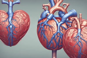
- The cardiovascular system consists of the heart, blood vessels and blood.
Heart Chambers
- The heart has four chambers: the left atrium, the right atrium, the left ventricle, and the right ventricle.
- The left and right atria are the superior chambers that receive blood and send it to the ventricles.
- The left and right ventricles are the inferior chambers that pump blood away.
- The left side of the heart has oxygenated blood, while the right side has deoxygenated blood
Heart Valves
- Heart valves prevent backflow to ensure one-way blood flow.
- Atrioventricular (AV) valves are located between an atrium and a ventricle on each side of the heart
- Semilunar valves are between a ventricle and an arterial trunk
- The right AV valve is also known as the tricuspid valve.
- The left AV valve is also known as the bicuspid or mitral valve.
System Circulations
- The cardiovascular system has two major circulations: pulmonary circulation, systemic circulation.
- Pulmonary circulation transports blood from the right side of the heart to the alveoli of the lungs for gas exchange and back to the left side.
- Systemic circulation transports blood from the left side of the heart to the systemic cells of the body for nutrient and gas exchange, and back to the right side of the heart.
Pulmonary Circulation Blood Flow
- Deoxygenated blood enters the right atrium from the venae cavae and coronary sinus, then passes through the right AV valve into the right ventricle.
- Blood passes through the pulmonary semilunar valve, entering the pulmonary trunk, and goes through the pulmonary arteries to both lungs.
- The blood enters pulmonary capillaries of both lungs for gas exchange.
- Oxygenated blood enters the right and left pulmonary veins and returns to the left atrium of the heart.
Systemic Circulation Blood Flow
- Oxygenated blood enters the left atrium
- Passes through the left AV valve (bicuspid or mitral valve)
- Enters the left ventricle
- Then passes through aortic semilunar valve
- Enters the aorta
- It proceeds to be distributed by the systemic arteries
- The enters systemic capillaries for nutrient and gas exchange
- Deoxygenated blood drains into the superior vena cava (SVC), inferior vena cava (IVC), and coronary sinus, and then finally enters the right atrium
Pericardium
- The pericardium surrounds the heart and has three layers: the fibrous pericardium, the parietal layer of serous pericardium, and the visceral layer of serous pericardium (epicardium).
- The pericardial cavity is located between the parietal and visceral layers of the serous pericardium and contains serous fluid.
Heart Wall Layers
- The heart wall varies in thickness, ventricles (pumping chambers) have thicker walls than atria.
- The left ventricle has a thicker wall than the right ventricle because the left ventricle must generate high pressure to force blood through systemic circulation, while the right only pumps to nearby lungs.
- There are three layers of the heart wall: epicardium, myocardium and endocardium.
Cardiac Muscle Metabolism and Properties
- Cardiac muscle has a high demand for energy, has an extensive blood supply, numerous mitochondria, and myoglobin and creatine kinase.
- Cardiac muscle can use different types of fuel molecules like fatty acids, glucose, lactic acid, amino acids, and ketone bodies.
- Cardiac muscle mostly relies on aerobic metabolism and is susceptible to failure when oxygen is low.
- Interference with blood flow to heart muscle can cause cell death.
Fibrous Skeleton
- The fibrous skeleton is made of dense irregular connective tissue.
- Provides structural support at the boundary of atria and ventricles.
- It forms fibrous rings to anchor valves, provides framework for attachment of cardiac muscle, and acts as an electrical insulator to prevent ventricles from contracting at the same time as atria.
Coronary Circulation
- Coronary circulation delivers blood to the heart wall.
- Coronary arteries transport oxygenated blood to the heart wall.
- Coronary veins transport deoxygenated blood away from the heart wall toward the right atrium.
Heart's Conduction System
- The conduction system initiates and conducts electrical events to ensure proper timing of contractions.
- It has specialized cardiac muscle cells that have action potentials but do not contract.
- Its activity is influenced by the autonomic nervous system.
Cardiac Center and Innervation
- The cardiac center is located in the medulla oblongata.
- It contains cardioacceleratory and cardioinhibitory centers, and sends signals via sympathetic and parasympathetic pathways.
- It receives signals from baroreceptors and chemoreceptors in the cardiovascular system.
- The cardiac center modifies cardiac activity but does not initiate it, however, it influences rate and force of heart's contractions.
Divisions of the Autonomic Nervous System
- Parasympathetic innervation decreases heart rate.
- Sympathetic innervation increases heart rate and force of contraction.
Contraction of the Heart
- Action potential must first be initiated in the SA node
- Is propagated throughout the atria and the conduction system
- The action potential is propagated across the sarcolemma of cardiac muscle cells, resulting in muscle contraction.
- Thin filaments slide past thick filaments and sarcomeres shorten within cardiac muscle cells.
SA Node Activity
- Resting membrane potential(RMP) of the SA node = -60mV.
- Slow voltage-gated Na+ channels open to cause inflow of Na+, which changes the membrane potential from -60mV to -40mV.
- This causes Fast voltage-gated Ca2+ channels open to cause calcium (Ca2+) inflow
- Causes membrane potential to change from -40 mV to just above 0 mV.
- Causes voltage-gated Ca2+ channels to close and Voltage-gated K+ channels to open.
- K+ outflow then allows membrane potential returns to RMP -60 mV and K+ channels close
Action Potential Through Cardiac Conduction System
- Generated at the sinoatrial (SA) node and spreads via gap junctions between cardiac muscle cells throughout the atria to atrioventricular (AV) node.
- There is a delay at the AV node before it passes to the AV bundle within the interventricular septum.
- The AV bundle conducts the action potential to the left and right bundle branches and then to the Purkinje fibers.
- The actin potential then is spread via gap junctions between cardiac muscle dells throughout the ventricles
Cardiac Muscle Cell at Rest
- At rest, a cardiac muscle cell has a resting membrane potential (RMP) of -90 mV, with all three voltage channels closed.
- Three phases: depolarization, plateau, and repolarization.
- Fast voltage-gated Na+ channels open, allowing Na+ to rapidly enter the cell, reversing the polarity from negative to positive (-90 mV to +30 mV).
- Then these channels then close voltage-gated, K+ channels open, causing K+ outflow of cardiac muscle cells.
- Slow voltage-gated, Ca2+ channels open, causing Ca2+ to enter the cell, with no electrical change and the depolarized state maintained.
- Voltage-gated, and Ca2+ channels then close
- Voltage-gated K+ channels remain open, and K+ moves out of the cell and is reversed from positive to negative (+30 mV to –90 mV).
ECG Reading
- Skin electrodes detect electrical signals of cardiac muscle cells.
- It is a Common diagnostic tool known as an electrocardiogram (ECG/EKG).
Waves and Segments of a Normal ECG
- The P wave reflects electrical changes of atrial depolarization originating in the SA node.
- The QRS complex refers to Electrical changes associated with ventricular depolarization, and the Atria also simultaneously repolarizing.
- The T wave refers to Electrical change associated with ventricular repolarization.
- P-Q segment is associated with atrial cells’ plateau (atria are contracting).
- S-T segment is associated with the ventricular plateau (ventricles are contracting).
Intervals of a Normal ECG
- P-R interval is time from beginning of the P wave to beginning of the QRS deflection- which is from atrial depolarization to beginning of ventricular depolarization
- Time needed to transmit action potential through entire conduction system
- Q-T interval is time from the beginning of the QRS to the end of the T wave and reflects the time of ventricular action potentials as well as length that depends upon heart rate
Cardiac Cycle
- Encompasses all events in the heart from the start of one beat to the start of the next
- Includes both systole (contraction) and diastole (relaxation)
- Contraction increases pressure; relaxation decreases it
- Blood moves down its pressure gradient (high to low) where all
- Valves ensure that flow is forward (closure prevents backflow)
Ventricular Activity
- Is the most important driving force to raise ventricular pressure
- AV valves are pushed closed
- Semilunar valves pushed open and blood ejected to artery
- Ventricular relaxation lowers ventricular pressure where semilunar valves close and have No pressure from below keeping them open, and AV valves are open where there is No pressure pushing them closed
Cardiac Cycle Phases
- Includes all events in the heart from the start of one beat to the start of the next and there are 5 phases:
- Atrial Contraction and Ventricular filling - atria contract, ventricles relax, ventricle pressure is less than atria and arterial trunk. All valves open except atrial and ventricular trunk
- Isometric Contraction - Atria relax, Ventricles contract, valves closed, and has EDV of 130mL
- Ventricle Ejection - Atria relax, Ventricles contract, valves closed, and as SV of 70mL
- Isometric Relaxation
- Atrial Relaxation and Ventricular Filling
Cardiac Output
- The amount of blood pumped by a single ventricle in one minute and is measured in liters per minute
- Measure of the effectiveness of the cardiovascular system
- It increases in healthy individuals during exercise
- Is determined by heart rate (beats per minute) and stroke volume (amount of blood ejected per beat)
- calculated using formula: HR × SV = CO and is typically 75 beats/min × 70 ml/beat = 5,250 ml/min = 5.25 L/min
Stroke Volume
-
Amount of blood ejected in one beat from one ventricle and in influenced by:
- Venous return: volume of blood returned to the heart where there Is a direct relation to SV which Determines amount of ventricular blood prior to contraction
- preload- pressure stretching heart wall before shortening
- Inotropic agents:
- Afterload is
- Venous return: volume of blood returned to the heart where there Is a direct relation to SV which Determines amount of ventricular blood prior to contraction
-
Volume determines preload, where pressure stretches heart wall before shortening
-
The Frank-Starling law (Starling's Law) states When EDV increases, the greater stretch of heart wall results in more optimal overlap of thick and thin filaments where the heart then contracts more forcefully when filled with more blood, so SV increases The venous return may also be increased by increased venous pressure or increased time to fill where venous pressure increases during exercise as muscles squeeze veins and the time available to fill increases with slower heart rate such as Athletes
Cardiac Output
- Increases with heart rate and inotropy and decreases as afterload increases
Studying That Suits You
Use AI to generate personalized quizzes and flashcards to suit your learning preferences.