Podcast
Questions and Answers
What is the location of the SA Node?
What is the location of the SA Node?
- In the area where the pulmonary trunk enters the RV
- In the area where the IVC enters the RA
- In the area where the aorta enters the LV
- In the area where the SVC enters the RA (correct)
What is the thickness of the RV myocardium compared to the LV?
What is the thickness of the RV myocardium compared to the LV?
- 2x thicker
- Equal in thickness
- Half the thickness
- 1/3 the thickness (correct)
What is the function of papillary muscles?
What is the function of papillary muscles?
- To control the tricuspid valve (correct)
- To control the mitral valve
- To pump blood from the heart
- To regulate the heart rate
What is the location of the left atrium?
What is the location of the left atrium?
What is the nerve supply to the heart responsible for?
What is the nerve supply to the heart responsible for?
What is the outflow part of the RV?
What is the outflow part of the RV?
What is the location of the oesophagus in relation to the left atrium?
What is the location of the oesophagus in relation to the left atrium?
What is the function of the trabeculae carnae?
What is the function of the trabeculae carnae?
What is the function of the sympathetic innervation in the heart?
What is the function of the sympathetic innervation in the heart?
Which artery supplies oxygenated blood to the myocardium?
Which artery supplies oxygenated blood to the myocardium?
What is the function of the tunica media in the walls of arteries?
What is the function of the tunica media in the walls of arteries?
What is the difference between superficial and deep veins?
What is the difference between superficial and deep veins?
What is the function of the lymphatic system in the cardiovascular system?
What is the function of the lymphatic system in the cardiovascular system?
What is the term for the reduction of blood supply to the heart muscle?
What is the term for the reduction of blood supply to the heart muscle?
What is the purpose of the coronary sinus?
What is the purpose of the coronary sinus?
What is the function of the vasoconstriction and vasodilation?
What is the function of the vasoconstriction and vasodilation?
What is a bronchopulmonary segment?
What is a bronchopulmonary segment?
What is the function of type II cells in the alveoli?
What is the function of type II cells in the alveoli?
What is the name of the passageways that connect respiratory bronchioles to alveoli?
What is the name of the passageways that connect respiratory bronchioles to alveoli?
What is the name of the veins that drain lung tissues?
What is the name of the veins that drain lung tissues?
What is the name of the lymphatic plexus that drains lung tissue and visceral pleura?
What is the name of the lymphatic plexus that drains lung tissue and visceral pleura?
What is the origin of the left bronchial artery?
What is the origin of the left bronchial artery?
What is the name of the cells that form the respiratory membrane?
What is the name of the cells that form the respiratory membrane?
What is the number of alveoli in each lung?
What is the number of alveoli in each lung?
What is the function of the pulmonary ligament?
What is the function of the pulmonary ligament?
What is the main function of the diaphragm during respiration?
What is the main function of the diaphragm during respiration?
What is the name of the potential space between the two layers of the pleural sac?
What is the name of the potential space between the two layers of the pleural sac?
Which of the following muscles is involved in inspiration?
Which of the following muscles is involved in inspiration?
What is the function of the visceral pleura?
What is the function of the visceral pleura?
What is the name of the thick median partition that separates the right and left pleural cavities?
What is the name of the thick median partition that separates the right and left pleural cavities?
What is the term for the physical movement of air into and out of the bronchial tree?
What is the term for the physical movement of air into and out of the bronchial tree?
What is the shape of each lung?
What is the shape of each lung?
What are the two parts of the pericardium?
What are the two parts of the pericardium?
Which surface of the heart faces inferiorly and rests on the diaphragm?
Which surface of the heart faces inferiorly and rests on the diaphragm?
What is the shape of the heart?
What is the shape of the heart?
How many chambers does the heart have?
How many chambers does the heart have?
What is the function of the right atrium?
What is the function of the right atrium?
What is the name of the valve between the right atrium and right ventricle?
What is the name of the valve between the right atrium and right ventricle?
What is the name of the ridge in the right atrium?
What is the name of the ridge in the right atrium?
What is the groove that separates the atria from the ventricles?
What is the groove that separates the atria from the ventricles?
Flashcards are hidden until you start studying
Study Notes
Cardiac Plexus and Synapse
- The cardiac plexus is found in the walls of the atria and decreases heart rate, force of contraction, and constricts coronary arteries.
- Sympathetic innervation, on the other hand, increases heart rate and force of contraction, and dilates coronary arteries.
Coronary Arteries
- The coronary arteries supply blood to the heart muscle.
- The right coronary artery and left coronary artery are the two main branches of the coronary arteries.
- The circumflex artery, marginal artery, anterior interventricular artery, and posterior interventricular artery are some of the smaller branches.
Coronary Sinus
- The coronary sinus is the largest vein of the heart and opens into the right atrium.
- It receives tributaries from the great cardiac vein, small cardiac vein, middle cardiac vein, and others.
Blood Flow
- The blood flows from the right atrium to the right ventricle, then to the lungs, and then to the left atrium, left ventricle, and finally to the rest of the body.
Arteries
- Arteries have elastic tissue in their walls, which enables them to recover their original diameter after expansion.
- This recoil propels blood during diastole and is responsible for filling the coronary arteries.
- Arteries divide into smaller arteries with less elastic tissue and more muscular tissue, which eventually divide into arterioles.
- Arterioles are responsible for maintaining blood pressure by providing peripheral resistance.
Arterial Structure
- The walls of arteries have three coats: tunica intima, tunica media, and tunica adventitia.
- Vasoconstriction decreases the size of the lumen, while vasodilation increases it.
Veins
- Veins have less elastic tissue than arteries and are more distensible.
- They receive tributaries that enter the heart and have low pressure.
- Valves are found in the lower half of the body to ensure the flow of blood to the heart.
- Veins above the heart do not require valves.
Types of Veins
- Superficial veins are deep to the skin and drain into the deep veins.
- Deep veins are deep to the muscle and have a more direct route to the heart.
Superior Vena Cava
- The superior vena cava drains blood from the head, neck, upper limb, and thorax.
- It receives blood from the internal jugular vein, subclavian vein, and brachiocephalic veins.
Lymphatics
- The lymphatic system carries clear fluid called lymph.
- Lymphoid tissue is found in many organs, lymph nodes, and lymphoid follicles.
- The lymphatic system drains from tissue spaces protein-containing fluid that escapes from the blood capillaries and transports it back into the bloodstream.
- It also transports fats from the gastrointestinal tract to the bloodstream and produces lymphocytes.
Heart
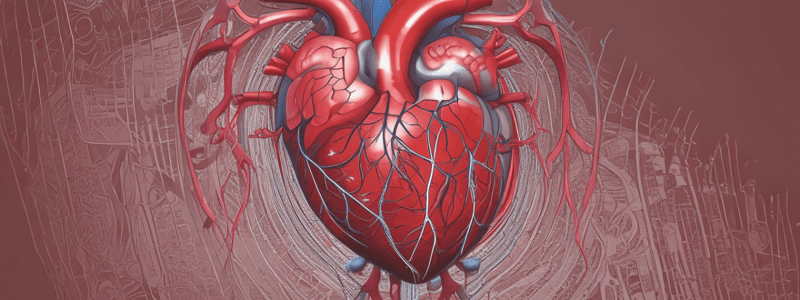
- The heart is a 4-chambered organ with a fibroserous sac enclosing it.
- The pericardium has two parts: the fibrous pericardium and the serous pericardium.
- The heart has an atrial surface, a ventricular surface, a base, and an apex.
- The right atrium receives venous blood from the whole body and sends it to the right ventricle.
- The right ventricle pumps blood to the lungs, and the left ventricle pumps blood to the rest of the body.
Right Atrium
- The right atrium receives blood from the superior vena cava, inferior vena cava, and coronary sinus.
- The crista terminalis and musculi pectinati are prominent features of the right atrium.
- The fossa ovalis is a thumbprint impression above the inferior vena cava opening.
Right Ventricle
- The right ventricle pumps blood to the lungs.
- The outflow part of the right ventricle is the infundibulum.
- The trabeculae carnae and papillary muscles are found in the right ventricle.
Left Atrium
- The left atrium receives blood from the four pulmonary veins.
- The interior of the left atrium is smooth-walled.
Left Ventricle
- The left ventricle pumps blood to the rest of the body.
- The outflow from the left ventricle is the aorta.
- The interior of the left ventricle has trabeculae carnae, papillary muscles, and chordae tendinae.
Nerve Supply
- The heart is supplied by the autonomic nervous system, which regulates heart rate, force of contraction, and cardiac output.
- Parasympathetic innervation decreases heart rate, while sympathetic innervation increases it.
Bronchopulmonary Segments
- Each lobe of the lungs can be divided into smaller units called bronchopulmonary segments.
- Each segment has a segmental bronchus and a tertiary branch of the pulmonary artery.
- The segments are supplied by the pulmonary veins and are surgically resectable.
Respiratory System
- The respiratory system consists of the bronchi, bronchioles, alveolar ducts, and alveoli.
- The alveoli are the site of gas exchange, and the type II cells produce surfactant to reduce surface tension.
Arterial Supply of Lungs
- The bronchial arteries arise from the systemic circulation and supply the lung and its associated tissues with nutrients.
- The left bronchial artery arises from the descending thoracic aorta, and the right bronchial artery arises from the 3rd posterior intercostal artery.
Venous Drainage of Lungs
- The bronchial veins drain the lung tissues and drain into the azygos vein or the accessory hemiazygos vein.
- The pulmonary veins drain the oxygenated blood from the lungs and drain into the left atrium.
Lymphatic Drainage of Lungs
- The superficial lymphatic plexus drains the lung tissue and visceral pleura.
- The bronchopulmonary (hilar) lymph nodes drain the lung and its associated tissues.
Pleura
- The pleura is a double-layered sac that surrounds the lung and lines the pulmonary cavity.
- The visceral pleura covers the lung and fissures, and the parietal pleura lines the pulmonary cavity.
- The pulmonary ligament is a fold of parietal pleura that provides space for the pulmonary veins to expand.
Thoracic Cavity
- The thoracic cavity contains the right and left pleural cavities, which are completely invaginated and occupied by the lungs.
- The mediastinum is a thick median partition that separates the right and left pleural cavities.
- The heart lies in the mediastinum.
Pulmonary Ventilation
- Pulmonary ventilation refers to the physical movement of air into and out of the bronchial tree.
- The respiratory muscles, including the diaphragm, intercostal muscles, and others, are responsible for pulmonary ventilation.
Studying That Suits You
Use AI to generate personalized quizzes and flashcards to suit your learning preferences.


