Podcast
Questions and Answers
What is the main function of the cardiovascular system?
What is the main function of the cardiovascular system?
- To regulate body temperature
- To digest food and absorb nutrients
- To transport blood throughout the body (correct)
- To filter waste products from the blood
Which of the following is a component of the cardiovascular system?
Which of the following is a component of the cardiovascular system?
- Kidneys
- Lungs
- Brain
- Heart (correct)
What type of blood vessels carry blood away from the heart?
What type of blood vessels carry blood away from the heart?
- Veins
- Capillaries
- Venules
- Arteries (correct)
Which of the following describes the location of the heart?
Which of the following describes the location of the heart?
Into how many chambers is the heart divided?
Into how many chambers is the heart divided?
What are the upper chambers of the heart called?
What are the upper chambers of the heart called?
What is the function of the right atrium?
What is the function of the right atrium?
Which valve does blood pass through when moving from the right atrium to the right ventricle?
Which valve does blood pass through when moving from the right atrium to the right ventricle?
What type of blood does the left atrium receive?
What type of blood does the left atrium receive?
What is the function of the left ventricle?
What is the function of the left ventricle?
What is the name of the valve that the left ventricle pumps blood through?
What is the name of the valve that the left ventricle pumps blood through?
Which of the following is NOT a valve in the heart?
Which of the following is NOT a valve in the heart?
What is the outer covering of the heart called?
What is the outer covering of the heart called?
How does the heart receive its blood supply?
How does the heart receive its blood supply?
What is the medical term for a reduced blood supply to the heart, potentially leading to chest pain?
What is the medical term for a reduced blood supply to the heart, potentially leading to chest pain?
What condition can result from the occlusion of main arteries in the heart?
What condition can result from the occlusion of main arteries in the heart?
What happens to the heart during systemic circulation?
What happens to the heart during systemic circulation?
What happens during the pulmonary circulation?
What happens during the pulmonary circulation?
Which chamber of the heart initiates pulmonary circulation?
Which chamber of the heart initiates pulmonary circulation?
Which structure does the systemic circulation start in?
Which structure does the systemic circulation start in?
Which great blood vessel feeds the tissues of the body during systematic circulation?
Which great blood vessel feeds the tissues of the body during systematic circulation?
In portal circulation, where is venous blood collected from?
In portal circulation, where is venous blood collected from?
What organ does the portal vein deliver blood to?
What organ does the portal vein deliver blood to?
Where does the portal circulation pathway terminate?
Where does the portal circulation pathway terminate?
Which heart valve is located behind the left border of the sternum, opposite the 4th costal cartilage?
Which heart valve is located behind the left border of the sternum, opposite the 4th costal cartilage?
Which heart valve is located behind the body of the sternum, opposite the 4th intercostal space?
Which heart valve is located behind the body of the sternum, opposite the 4th intercostal space?
How many points is the heart border outlined by?
How many points is the heart border outlined by?
What point is the (apex of the heart) located at?
What point is the (apex of the heart) located at?
Which structure is the heart not composed of?
Which structure is the heart not composed of?
What occurs when main arteries of the heart are occluded?
What occurs when main arteries of the heart are occluded?
What is the approximate location of the heart in accordance to the ribs?
What is the approximate location of the heart in accordance to the ribs?
What is the rough ratio from left to right in correspondence to the heart in the median plane?
What is the rough ratio from left to right in correspondence to the heart in the median plane?
What shape is the heart?
What shape is the heart?
Which of the following is delivered to the tissues of the body during systemic circulation?
Which of the following is delivered to the tissues of the body during systemic circulation?
Which choice is not a component of the cardiovascular system?
Which choice is not a component of the cardiovascular system?
Which of these elevated cardia enzymes are associated with Myocardial Infarction (MI)?
Which of these elevated cardia enzymes are associated with Myocardial Infarction (MI)?
How many borders does the heart have?
How many borders does the heart have?
Which great blood vessel are the right coronary and left coronary a branch of?
Which great blood vessel are the right coronary and left coronary a branch of?
Which of the following is a type of blood vessel?
Which of the following is a type of blood vessel?
Which of the following is the valve that deoxygenated blood passes throught to enter the right ventricle?
Which of the following is the valve that deoxygenated blood passes throught to enter the right ventricle?
What is the approximate ratio of the heart's location in relation to the median plane?
What is the approximate ratio of the heart's location in relation to the median plane?
Which great blood vessel do the right and left coronary arteries branch from?
Which great blood vessel do the right and left coronary arteries branch from?
Which of the following best describes the location of the pulmonary valve?
Which of the following best describes the location of the pulmonary valve?
Flashcards
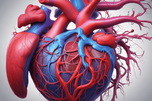
Cardiovascular system
Cardiovascular system
The cardiovascular system consists of the heart and blood vessels (arteries, veins, and capillaries).
Arteries
Arteries
Vessels carrying blood away from the heart.
Veins
Veins
Vessels carrying blood back to the heart.
Capillaries
Capillaries
Signup and view all the flashcards
The Heart
The Heart
Signup and view all the flashcards
Atria
Atria
Signup and view all the flashcards
Ventricles
Ventricles
Signup and view all the flashcards
Borders of the Heart
Borders of the Heart
Signup and view all the flashcards
Surfaces of the Heart
Surfaces of the Heart
Signup and view all the flashcards
Base of the Heart
Base of the Heart
Signup and view all the flashcards
Apex of the Heart
Apex of the Heart
Signup and view all the flashcards
Right Atrium
Right Atrium
Signup and view all the flashcards
Tricuspid Valve
Tricuspid Valve
Signup and view all the flashcards
Left Atrium
Left Atrium
Signup and view all the flashcards
Mitral Valve
Mitral Valve
Signup and view all the flashcards
Ventricles
Ventricles
Signup and view all the flashcards
Right Ventricle
Right Ventricle
Signup and view all the flashcards
Left Ventricle
Left Ventricle
Signup and view all the flashcards
Heart Valves
Heart Valves
Signup and view all the flashcards
Pericardium
Pericardium
Signup and view all the flashcards
Heart's Blood Supply
Heart's Blood Supply
Signup and view all the flashcards
Ischemic Heart Disease
Ischemic Heart Disease
Signup and view all the flashcards
Occlusion of Main Arteries
Occlusion of Main Arteries
Signup and view all the flashcards
Surface Anatomy of the Heart
Surface Anatomy of the Heart
Signup and view all the flashcards
Point A
Point A
Signup and view all the flashcards
Point B
Point B
Signup and view all the flashcards
Point C
Point C
Signup and view all the flashcards
Point D
Point D
Signup and view all the flashcards
Upper Border of Heart
Upper Border of Heart
Signup and view all the flashcards
Right Border of Heart
Right Border of Heart
Signup and view all the flashcards
Lower Border of Heart
Lower Border of Heart
Signup and view all the flashcards
Left Border of Heart
Left Border of Heart
Signup and view all the flashcards
Location of Pulmonary Valve
Location of Pulmonary Valve
Signup and view all the flashcards
Location of Aortic Valve
Location of Aortic Valve
Signup and view all the flashcards
Location of Mitral Valve
Location of Mitral Valve
Signup and view all the flashcards
Location of Tricuspid Valve
Location of Tricuspid Valve
Signup and view all the flashcards
Systemic Circulation
Systemic Circulation
Signup and view all the flashcards
Pulmonary Circulation
Pulmonary Circulation
Signup and view all the flashcards
Portal Circulation
Portal Circulation
Signup and view all the flashcards
Study Notes
- CVS is the Cardiovascular System
Cardiovascular System Components
- The cardiovascular system consists of the heart and blood vessels.
- Blood vessels include arteries, veins, and capillaries.
Heart Location and Description
- The heart is a conical muscular pump located behind the sternum and costal cartilages.
- It extends from the 2nd to the 6th costal cartilages.
- Approximately 2/3 of the heart lies to the left, and 1/3 to the right, of the median plane.
Heart Anatomy
- The heart has 4 chambers: 2 atria (right and left) and 2 ventricles (right and left).
- It has 4 borders: upper, lower, right, and left.
- The heart has 4 surfaces: anterior, posterior, right, and left.
- The base of the heart is directed backward, and the apex is directed downward and to the left.
Heart Chambers
- The right atrium receives deoxygenated blood from all body parts via the superior and inferior vena cava.
- The right atrium sends blood to the right ventricle through the tricuspid valve.
- The left atrium receives oxygenated blood from the lungs through 4 pulmonary veins.
- The left atrium pumps blood to the left ventricle via the mitral valve.
- The right ventricle sends deoxygenated blood to the pulmonary artery (via the pulmonary valve), which then divides into two branches for each lung, where oxygenation occurs.
- The two ventricles are separated by the interventricular septum.
- The left ventricle pumps oxygenated blood to the body through the aortic valve, into the aorta and its branches.
Heart Valves and Coverings
- The heart contains 4 valves: tricuspid, pulmonary, mitral, and aortic.
- The heart is covered by the fibrous pericardium and serous pericardium.
Blood Supply to the Heart
- The heart is supplied by 2 arteries: the right coronary and left coronary arteries.
- These arteries branch from the ascending aorta.
Ischemic Heart Disease
- Narrowing of small coronary artery branches due to atherosclerosis leads to angina pectoris.
- Occlusion of main arteries leads to myocardial infarction (MI).
- Myocardial infarction results in elevated cardiac enzymes, specifically CK and troponin.
Surface Anatomy of the Heart
- Borders of the heart can be outlined on the body's surface using 4 points.
- Point A: Left 2nd costal cartilage, 4 cm from the median plane.
- Point B: Right 3rd costal cartilage, 3 cm from the median plane.
- Point C: Right 6th costal cartilage, 3 cm from the median plane.
- Point D: (apex of the heart) Left 5th intercostal space, 9 cm from the median plane.
Borders based on surface anatomy
- Upper border: lies between points A and B.
- Right border: lies between points B and C.
- Lower border: lies between points C and D.
- Left border: lies between points D and A.
Surface Anatomy of Heart Valves
- Pulmonary valve: Located behind the left border of the sternum, opposite the 3rd costal cartilage
- Aortic valve: Located behind the left border of the sternum, opposite the 3rd intercostal space
- Mitral valve: Located behind the left border of the sternum, opposite the 4th costal cartilage
- Tricuspid valve: Located behind the body of the sternum, opposite the 4th intercostal space
Systemic Circulation
- Begins in the left ventricle where oxygenated blood is pumped through the aorta and its branches.
- Blood reaches all body tissues, where gas and material exchange occurs.
- Deoxygenated blood is collected by small veins, then large veins, and finally enters the right atrium via the superior and inferior vena cava.
- Blood flows from the right atrium to the right ventricle and this completes systemic circulation with a new cycle starting.
Pulmonary Circulation
- Initiates in the right ventricle where venous blood flows through the pulmonary artery and its two branches to the lungs.
- Gas exchange happens in the lungs.
- Oxygenated blood returns to the left atrium via the 4 pulmonary veins, proceeding to the left ventricle, and initiating a new cycle.
Portal Circulation
- Venous blood from the stomach, spleen, pancreas, and intestines is collected into the portal vein.
- The portal vein enters the liver (through the porta hepatis) and divides into branches that end in liver sinusoids.
- Blood exits liver sinusoids through hepatic veins that drain into the inferior vena cava, then to the right atrium.
Studying That Suits You
Use AI to generate personalized quizzes and flashcards to suit your learning preferences.