Podcast
Questions and Answers
What is the primary function of the cardiovascular system?
What is the primary function of the cardiovascular system?
- To circulate blood and transport nutrients and waste. (correct)
- To protect the body from infections.
- To facilitate gas exchange in the lungs.
- To regulate body temperature.
Which part of the heart is primarily responsible for pumping blood to the lungs?
Which part of the heart is primarily responsible for pumping blood to the lungs?
- Right atrium
- Left ventricle
- Left atrium
- Right ventricle (correct)
In a cardiac assessment, which of the following techniques is NOT typically used?
In a cardiac assessment, which of the following techniques is NOT typically used?
- Transfusion (correct)
- Percussion
- Auscultation
- Palpation
What anatomical feature does the heart extend from in terms of intercostal spaces?
What anatomical feature does the heart extend from in terms of intercostal spaces?
Which cardiac structure primarily prevents backflow of blood?
Which cardiac structure primarily prevents backflow of blood?
What measurement reflects the size of a normal human heart?
What measurement reflects the size of a normal human heart?
Which of the following best describes the location of the heart within the thoracic cavity?
Which of the following best describes the location of the heart within the thoracic cavity?
What is a potential consequence of problems within the cardiovascular system?
What is a potential consequence of problems within the cardiovascular system?
What layer of the heart wall is primarily responsible for the heart's contraction?
What layer of the heart wall is primarily responsible for the heart's contraction?
Which heart sound is associated with the closing of the aortic and pulmonic valves?
Which heart sound is associated with the closing of the aortic and pulmonic valves?
In which phase of the cardiac cycle do the AV valves close?
In which phase of the cardiac cycle do the AV valves close?
Where is the S3 heart sound best heard?
Where is the S3 heart sound best heard?
What is the primary function of the pericardial cavity?
What is the primary function of the pericardial cavity?
Which layer of the pericardium is fused to the heart?
Which layer of the pericardium is fused to the heart?
What event marks the end of diastole in the cardiac cycle?
What event marks the end of diastole in the cardiac cycle?
During which phase is the S4 heart sound typically produced?
During which phase is the S4 heart sound typically produced?
Where is the carotid artery pulse best felt?
Where is the carotid artery pulse best felt?
Which position is recommended for optimal auscultation at the cardiac apex?
Which position is recommended for optimal auscultation at the cardiac apex?
What are the two sets of jugular veins mentioned?
What are the two sets of jugular veins mentioned?
What should be checked during a physical assessment of the cardiovascular system?
What should be checked during a physical assessment of the cardiovascular system?
What describes Cheyne-Stokes respirations?
What describes Cheyne-Stokes respirations?
Which of the following is NOT a subjective data symptom of cardiovascular problems?
Which of the following is NOT a subjective data symptom of cardiovascular problems?
What technique is used to estimate heart size most accurately?
What technique is used to estimate heart size most accurately?
What lifestyle factor could contribute to heart disease?
What lifestyle factor could contribute to heart disease?
What does the presence of pallor and cyanosis indicate during cardiovascular assessment?
What does the presence of pallor and cyanosis indicate during cardiovascular assessment?
Which position should the patient be in for the inspection of jugular venous pressure (JVP)?
Which position should the patient be in for the inspection of jugular venous pressure (JVP)?
What is considered an abnormal finding during the inspection of the chest?
What is considered an abnormal finding during the inspection of the chest?
During the examination of JVP, which lighting technique enhances the visualization of venous pulsations?
During the examination of JVP, which lighting technique enhances the visualization of venous pulsations?
What should the examiner look for during the visual inspection of the internal jugular veins?
What should the examiner look for during the visual inspection of the internal jugular veins?
What is the normal range of elevation above the sternal angle when measuring JVP?
What is the normal range of elevation above the sternal angle when measuring JVP?
Which of the following is NOT a focus area during cardiovascular inspection?
Which of the following is NOT a focus area during cardiovascular inspection?
What does an earlobe crease indicate in the context of cardiovascular assessment?
What does an earlobe crease indicate in the context of cardiovascular assessment?
What is the correct position for the patient during the assessment of Jugular Venous Pressure (JVP)?
What is the correct position for the patient during the assessment of Jugular Venous Pressure (JVP)?
Which area corresponds to the Aortic area during chest palpation?
Which area corresponds to the Aortic area during chest palpation?
What could be a potential consequence of bilateral carotid palpation?
What could be a potential consequence of bilateral carotid palpation?
Which technique is recommended for palpating the apical pulse?
Which technique is recommended for palpating the apical pulse?
What should be avoided when palpating carotid arteries?
What should be avoided when palpating carotid arteries?
In which area is the tricuspid valve palpation typically assessed?
In which area is the tricuspid valve palpation typically assessed?
Why may the apical impulse be difficult to palpate in certain patients?
Why may the apical impulse be difficult to palpate in certain patients?
During the inspection phase, what general assessments are recommended?
During the inspection phase, what general assessments are recommended?
What change may affect the palpation of the apical impulse in older patients?
What change may affect the palpation of the apical impulse in older patients?
Which area is associated with S1 being louder than S2 during auscultation?
Which area is associated with S1 being louder than S2 during auscultation?
Which technique should be used to auscultate carotid arteries effectively?
Which technique should be used to auscultate carotid arteries effectively?
What is the purpose of asking the patient to hold their breath during carotid auscultation?
What is the purpose of asking the patient to hold their breath during carotid auscultation?
Which position is best to auscultate for aortic valve disease?
Which position is best to auscultate for aortic valve disease?
During auscultation at Erb's point, how are S1 and S2 perceived?
During auscultation at Erb's point, how are S1 and S2 perceived?
Why should a light palpation be performed if an occlusion is noted during carotid auscultation?
Why should a light palpation be performed if an occlusion is noted during carotid auscultation?
What finding indicates the apical impulse is difficult to palpate due to anatomical changes in older patients?
What finding indicates the apical impulse is difficult to palpate due to anatomical changes in older patients?
Flashcards
Cardiovascular System
Cardiovascular System
The system of the heart and blood vessels that circulates blood throughout the body, delivering oxygen and nutrients.
Heart Chambers
Heart Chambers
The four hollow spaces (atria and ventricles) within the heart that pump blood.
Cardiac Muscle
Cardiac Muscle
Specialized muscle tissue that forms the walls of the heart.
Heart Valves
Heart Valves
Signup and view all the flashcards
Heart Landmarks
Heart Landmarks
Signup and view all the flashcards
Cardiac Assessment
Cardiac Assessment
Signup and view all the flashcards
Heart Size
Heart Size
Signup and view all the flashcards
Mediastimun
Mediastimun
Signup and view all the flashcards
Fibrous Pericardium
Fibrous Pericardium
Signup and view all the flashcards
Serous Pericardium
Serous Pericardium
Signup and view all the flashcards
Cardiac Cycle
Cardiac Cycle
Signup and view all the flashcards
Systole
Systole
Signup and view all the flashcards
Diastole
Diastole
Signup and view all the flashcards
S1 Heart Sound
S1 Heart Sound
Signup and view all the flashcards
S2 Heart Sound
S2 Heart Sound
Signup and view all the flashcards
S3 Heart Sound
S3 Heart Sound
Signup and view all the flashcards
Carotid Artery Pulse Location
Carotid Artery Pulse Location
Signup and view all the flashcards
Jugular Venous Pressure (JVP)
Jugular Venous Pressure (JVP)
Signup and view all the flashcards
Inspecting JVP
Inspecting JVP
Signup and view all the flashcards
Jugular Veins Types
Jugular Veins Types
Signup and view all the flashcards
Cardiac Apex Location
Cardiac Apex Location
Signup and view all the flashcards
Normal JVP Elevation
Normal JVP Elevation
Signup and view all the flashcards
Tangential Lighting
Tangential Lighting
Signup and view all the flashcards
Cardiac Assessment Techniques
Cardiac Assessment Techniques
Signup and view all the flashcards
Patient Preparation (Cardiac)
Patient Preparation (Cardiac)
Signup and view all the flashcards
Pulsations
Pulsations
Signup and view all the flashcards
Abnormal Pulsations (Heves/Lifts)
Abnormal Pulsations (Heves/Lifts)
Signup and view all the flashcards
Cheyne-Stokes Respirations
Cheyne-Stokes Respirations
Signup and view all the flashcards
Physical Assessment Equipment
Physical Assessment Equipment
Signup and view all the flashcards
Skin Color Changes
Skin Color Changes
Signup and view all the flashcards
Subjective Data in Cardiac History
Subjective Data in Cardiac History
Signup and view all the flashcards
Splinter Hemorrhage
Splinter Hemorrhage
Signup and view all the flashcards
JVP Measurement
JVP Measurement
Signup and view all the flashcards
Carotid Pulse Palpation
Carotid Pulse Palpation
Signup and view all the flashcards
Precordial Assessment
Precordial Assessment
Signup and view all the flashcards
Apical Pulse
Apical Pulse
Signup and view all the flashcards
Chest Palpation for Thrills
Chest Palpation for Thrills
Signup and view all the flashcards
Carotid Artery Landmarks
Carotid Artery Landmarks
Signup and view all the flashcards
Jugular Venous Pressure (JVP)
Jugular Venous Pressure (JVP)
Signup and view all the flashcards
Precordial Landmarks
Precordial Landmarks
Signup and view all the flashcards
Palpating the Apical Impulse
Palpating the Apical Impulse
Signup and view all the flashcards
Auscultating Heart Sounds
Auscultating Heart Sounds
Signup and view all the flashcards
S1 Heart Sound
S1 Heart Sound
Signup and view all the flashcards
S2 Heart Sound
S2 Heart Sound
Signup and view all the flashcards
Carotid Artery Auscultation
Carotid Artery Auscultation
Signup and view all the flashcards
Auscultation Positions
Auscultation Positions
Signup and view all the flashcards
Palpating Thoracic Area
Palpating Thoracic Area
Signup and view all the flashcards
Percussion of the Chest
Percussion of the Chest
Signup and view all the flashcards
Study Notes
Cardiovascular System Assessment
- System function: The cardiovascular system continuously circulates blood, providing oxygen and nutrients to the body's organs and tissues, and removing waste.
- Components: It's a complex system of the heart (pump) and blood vessels (plumbing).
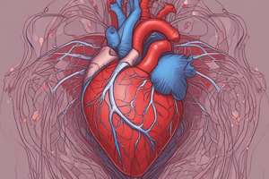
- Heart structure: The heart is a hollow, muscular, four-chambered organ in the mediastinum. It's roughly the size of a clenched fist and weighs approximately 255g in women and 310g in men.
- Heart position: The heart extends vertically from the second to the fifth intercostal space and horizontally from the right edge of the sternum to the left midclavicular line. The upper portion (near the second intercostal space) is the base, and the lower portion (near the fifth intercostal space and left midclavicular line) is the apex.
- Heart coverings and walls: The heart has three layers: epicardium (outermost), myocardium (thickest layer of contractile cardiac muscle cells), and endocardium. It's encased in a pericardium (double-layered sac), with lubricating fluid in the pericardial cavity.
- Cardiac cycle: The cardiac cycle involves filling and emptying of heart chambers. Systole is ventricular contraction (AV valves close first, then aortic & pulmonic valves). Diastole is ventricular relaxation (aortic & pulmonic valves close first, then AV valves).
- Heart sounds: The "lub" (S₁) is from mitral and tricuspid valve closure (beginning of systole). The "dub" (S₂) is from aortic and pulmonary valve closure (beginning of diastole). S₁ is best heard at the apex, and S₂ at the base of the heart.
- Additional heart sounds: S₃ (extra heart sound in early diastole) and S₄ (extra heart sound in late diastole) may be heard, using the stethoscope's bell.
- Neck vessels: Carotid arteries supply the neck and head with oxygenated blood. They are located in groove between trachea and sternocleidomastoid muscles, slightly below the mandible. Carotid artery pulse is assessed with the examiner palpating with their fingers.
- Jugular veins: Jugular veins return deoxygenated blood from the head and neck to the heart via the superior vena cava. Internal jugular veins are deep, while external jugular veins are more superficial.
- Subjective data and history: Medical history and lifestyle factors are crucial (hypertension, cholesterol levels, smoking, diet, etc.) in cardiovascular assessments. Patient reporting of symptoms (pain, breathlessness, etc.) is also key.
- Respiratory problems: Respiratory problems like tachypnea, Cheyne-Stokes respirations, hemoptysis, and cough could result from a heart disorder.
- Physical assessment tools: Inspection, palpation, percussion, and auscultation are used. Diagram shows this assessment procedure.
- Patient preparation: Explain the procedure, expose the anterior chest, and position the patient appropriately for examination.
- Equipment: Examination gown, drape, stethoscope, ruler, pillow, penlight/light source, and a watch with a second hand.
- Inspection techniques: Inspect face, lips, ears, scalp, jugular veins, carotid arteries, hands and fingers paying attention to color, movement, creases, distention. Inspect the chest, abdomen, legs, and skeletal structure to identify pulsations/heaves/lifts.
- Palpation techniques: Palpating carotid pulse, thrills, apical pulse.
- Palpating heart locations: Assess the aortic, pulmonic, Erb's point, mitral, and tricuspid areas.
- Percussion techniques: Percussion of the chest to determine heart size and borders.
- Auscultation techniques: Listen to the heart sounds in various locations (aorta, pulmonic, Erb's point, tricuspid, mitral areas) and for any bruits in the carotid arteries. Diagram indicates areas for auscultation.
- Measuring JVP: Determine the jugular venous pressure and position of the venous pulsations in the neck to determine central venous pressure. Palpate and listen for any abnormal sounds in the neck.
Landmarks for Cardiac Assessment
- Sternum
- Clavicles
- Ribs
- 2nd to 5th intercostal spaces
- Midsternal line
- Midclavicular lines
- Anterior axillary line
Studying That Suits You
Use AI to generate personalized quizzes and flashcards to suit your learning preferences.